

- •Copyright
- •Contents
- •Dedication
- •Preface
- •Acknowledgments
- •Contributors
- •Contributors to the Previous Edition
- •Review Test
- •Answers and Explanations
- •Review Test
- •Answers and Explanations
- •Review Test
- •Answers and Explanations
- •Review Test
- •Answers and Explanations
- •Review Test
- •Answers and Explanations
- •Review Test
- •Answers and Explanations
- •Review Test
- •Answers and Explanations
- •Review Test
- •Answers and Explanations
- •Review Test
- •Answers and Explanations
- •Review Test
- •Answers and Explanations
- •IV. Hypertension
- •VI. Nephrotic Syndrome (NS)
- •VII. Hemolytic Uremic Syndrome (HUS)
- •VIII. Hereditary Renal Diseases
- •IX. Renal Tubular Acidosis (RTA)
- •XI. Chronic Kidney Disease (CKD) and End-Stage Renal Disease (ESRD)
- •XII. Structural and Urologic Abnormalities
- •XIII. Urolithiasis
- •XIV. Urinary Tract Infection (UTI)
- •Review Test
- •Answers and Explanations
- •Review Test
- •Answers and Explanations
- •Review Test
- •Answers and Explanations
- •Review Test
- •Answers and Explanations
- •IV. Food Allergy
- •VI. Urticaria (Hives)
- •VII. Drug Allergy
- •VIII. Asthma
- •IX. Immunology Overview
- •X. Disorders of Lymphocytes (Figure 15-2)
- •XI. Disorders of Granulocytes (Figure 15-3)
- •XII. Disorders of the Complement System
- •Review Test
- •Answers and Explanations
- •Review Test
- •Answers and Explanations
- •Review Test
- •Answers and Explanations
- •Review Test
- •Answers and Explanations
- •Review Test
- •Answers and Explanations
- •Review Test
- •Answers and Explanations
- •Comprehensive Examination
- •Index
Answers and Explanations
1.The answer is B [I.A.2]. The hemoglobin of a healthy full-term infant is high at birth and decreases during the next several months, reaching its nadir, or physiologic lowest point, by 2–3 months of age. The hemoglobin of a preterm infant is at its physiologic low point at 1– 2 months of age. Iron-deficiency anemia does not generally appear until 9–24 months of age, as a result of inadequate iron intake and depletion of iron stores acquired during fetal life.
Fetal hemoglobin, a major constituent of red blood cells during early postnatal life, gradually declines and disappears by 6–9 months of age. Macrocytic anemia, caused most commonly by folic acid or vitamin B12 deficiency, would not normally occur at 2 months of age.
2.The answer is C [V.B.2]. This patient’s clinical presentation and age are most consistent with chronic benign neutropenia (CBN) of childhood. This noncyclic neutropenia is most common in children younger than 4 years of age. CBN is characterized by normal appearance and growth and a history of mild infections, such as sinusitis, cellulitis, and otitis media. Absolute neutrophil count (ANC) and white blood cell counts are low. Chédiak–Higashi and Kostmann syndromes are characterized by more severe infections, and Chédiak–Higashi syndrome is also notable for the presence of oculocutaneous albinism. Kostmann syndrome (severe congenital agranulocytosis) is an autosomal recessive disorder with frequent severe infections and a very low ANC. Shwachman–Diamond syndrome is characterized by poor growth, pancreatic insufficiency, and metaphyseal chondrodysplasia. Cyclic neutropenia, as its name suggests, is characterized by regular cycles of neutropenia occurring on average every 21 days.
3.The answer is B [IV.E.2.b.(1) and Table 13-6]. The most likely diagnosis is immune thrombocytopenic purpura (ITP) based on the acuteness of the presentation and the classic history of signs and symptoms after a viral infection. Spontaneous recovery is the rule, occurring in 70–80% of patients. Clinical features of ITP most commonly include cutaneous or mucous membrane bleeding, rather than bleeding into joints (hemarthroses). Platelet transfusions are generally not recommended because transfused platelets will be destroyed by the patient’s antibodies. Patients with ITP have low platelet counts but normal activated partial thromboplastin time and prothrombin time. Treatment includes supportive care, intravenous immune globulin, or corticosteroids, and sometimes anti-D immunoglobulin. Chronic ITP occurs more commonly in patients older than 10 years.
4.The answer is D [I.D.1]. The history of excessive intake of iron-poor cow’s milk and the presence of a microcytic, hypochromic anemia at 15 months of age are both consistent with iron-deficiency anemia. In iron-deficiency anemia, findings include increased free erythrocyte protoporphyrin, increased transferrin, and decreased transferrin saturation. Significant physical and intellectual effects may occur in iron-deficiency anemia and include poor weight gain, diminished attention, and diminished abilities to learn. One of the earliest laboratory findings is a low serum ferritin. Reticulocyte count is often elevated, reflecting increased bone marrow activity.
5.The answer is E [I.F.3.d]. The presence of both hemoglobin (Hgb) A and Hgb S indicates that this patient has sickle cell trait (all children also have Hgb F at birth). The presence of Hgb A excludes sickle cell disease; affected patients with sickle cell disease have only Hgb S and Hgb F. Patients with sickle cell trait are generally asymptomatic, although during adolescence they may have an inability to concentrate the urine or hematuria. Patients do not have anemia; they are generally free of crises, unless they have severe hypoxemia, and they have normal splenic function. Prophylactic penicillin is unnecessary given the normal splenic function.
6.The answer is D [Table 13-3]. Extremity pain in patients with sickle cell disease may be caused by trauma, osteomyelitis, or a vaso-occlusive crisis. Because there is no trauma or fever, a painful bone crisis (a type of vaso-occlusive crisis) is most likely. Appropriate management
520
includes pain control with a narcotic and a nonsteroidal anti-inflammatory agent. There is no need to obtain a blood culture because the child has no fever. Red blood cell transfusion to a hemoglobin level of 14 g/dL is inappropriate because it will lead to increased blood viscosity that may result in increased vaso-occlusion. If the pain is not well controlled with analgesia and improved hydration, a partial exchange transfusion might be indicated. Because osteomyelitis is less likely given the acute onset of the pain and the absence of fever, magnetic resonance imaging would not be an appropriate initial management step.
7.The answer is E [IV.B.3]. von Willebrand disease is characterized by mild to moderate bleeding in most patients. Common signs and symptoms include bruising, epistaxis, menorrhagia (i.e., prolonged or excessive uterine bleeding occurring at regular intervals), and bleeding after surgical procedures or dental extraction. Hemarthroses are unusual and are more typical in hemophilia A and B, in which deep soft tissue bleeding occurs. In addition, these bleeding disorders occur in males; they have X-linked inheritance. Vitamin K deficiency in an adolescent would likely be caused by medications or disorders that cause diminished vitamin K absorption, such as pancreatic insufficiency, biliary obstruction, or prolonged diarrhea. Symptoms would also likely be more severe. Immune thrombocytopenic purpura may present with petechiae, bruising, or nosebleeds. However, the onset of symptoms is generally acute.
8.The answer is C [II.B.1]. The patient most likely has Fanconi anemia, or congenital aplastic anemia, on the basis of pancytopenia, her age at presentation, and her history of absence (hypoplasia may also occur) of the thumb and radius. Fanconi anemia is an inherited lifelong disorder that results in bone marrow failure. Almost all patients have short stature, and many also have skin hyperpigmentation and kidney anomalies. Management includes transfusions and bone marrow transplant. Because Fanconi anemia is characterized by bone marrow failure, the reticulocyte count would be expected to be very low. Because the hemoglobin and white blood cell count are also low, this girl does not have immune thrombocytopenic purpura.
9.The answer is B [I.D.2.d]. This patient’s anemia and physical features suggest β-thalassemia. β-Thalassemia occurs most commonly in patients of Mediterranean background and is caused by deletion of the β-globin chain. If untreated, β-thalassemia results in bone marrow hyperplasia (often noted within the facial bones), delays in growth and puberty, and hepatosplenomegaly. Many children suffer from hemochromatosis (iron overload) as a complication, and therefore iron is contraindicated in these patients. The presence of both hemoglobin S and hemoglobin F is not consistent with β-thalassemia and would instead
suggest sickle cell anemia. Spherocytes are not present on blood smear.
10.The answer is E [I.E.1]. Feedings exclusively with goat’s milk as a sole source of milk protein can lead to folic acid deficiency and a macrocytic anemia. Dietary folic acid is the treatment. Spoon-shaped nails are seen in iron-deficiency anemia, and a smooth red tongue is seen in vitamin B12 deficiency.
11.The answers are F, C, and A, respectively [IV.E.2.b.(4), IV.E.2.a.(1).(a), and IV.E.2.a.(1).(b)]. The 2-year-old boy has a large hemangioma that sequesters and destroys platelets, which is termed Kasabach–Merritt syndrome. The 5-year-old boy has Wiskott–Aldrich syndrome, which is characterized by eczema, defects in T- and B-cell immunity, and low platelet counts. The newborn girl has thrombocytopenia–absent radius (TAR) syndrome, which is characterized by thrombocytopenia, at times cardiac and renal disease, and absence of the radius. The thumb is present in TAR syndrome, in contrast to Fanconi anemia (pancytopenia with hypoplasia or absence of the radius and thumb).
12.The answers are B and A, respectively [Table 13-5]. The 6-month-old infant has Diamond– Blackfan anemia, which is characterized by rapid onset of anemia within the first year of life and physical abnormalities, including triphalangeal thumbs, short stature, and cardiac and
521
renal anomalies, in one-fourth to one-third of patients. Treatment includes transfusions and corticosteroids. The 2-year-old girl has transient erythroblastopenia of childhood, which is characterized by the slow onset of anemia after the first year of life. The cause is likely a postviral autoimmune reaction, and no treatment is generally required. Parvovirus B19 red blood cell aplasia generally results in no symptoms of anemia in healthy children. Associated features may include a “slapped cheek” red facial rash and upper respiratory symptoms.
13.The answers are C, D, A, and E, respectively [Table 13-6]. The clinical characteristics and laboratory abnormalities can differentiate the causes of bleeding in pediatric patients. The 6- year-old boy has vitamin K deficiency, which can occur with pancreatic insufficiency, biliary obstruction, and prolonged diarrhea. Vitamin K deficiency affects the vitamin K–dependent coagulation factors (II, VII, IX, and X) and therefore results in hemarthroses, a prolonged prothrombin time (PT) and activated partial thromboplastin time (aPTT), and normal bleeding times. The 2-year-old boy has disseminated intravascular coagulation (DIC), which may be caused by malignancy, sepsis, snakebite, heat stroke, and burns. DIC is characterized by abnormalities in all coagulation constituents, including low platelet counts and prolonged aPTT, PT, and bleeding time. Both petechiae and hemarthroses may be present. The 5-year-old boy has hemophilia B, or factor IX deficiency. Hemophilia B is characterized by hemarthroses and prolonged aPTT, but normal PT and bleeding time. The 8-year-old boy has von Willebrand disease, which can present with epistaxis, menorrhagia, and bleeding after tonsillectomy or dental surgery. von Willebrand disease is characterized by prolonged aPTT and prolonged bleeding time, but normal PT. Hemarthroses may sometimes occur.
522

C H A P T E R 1 4
523

Oncology
Carol Diamond
524

I.General Considerations
A.Incidence. Approximately 6000–7000 children between 1 and 15 years of age develop cancer each year.
1.Cancer is the leading cause of death from disease in childhood.
2.Unlike adult cancers, most childhood cancers are not carcinomas. The most common childhood cancers, in order of declining incidence, are leukemia, brain tumors, lymphoma, neuroblastoma, soft tissue sarcomas, Wilms tumor, and bone tumors.
B.Etiology. The cause of childhood cancers is often unknown. However, genetic disorders, immunodeficiency diseases, infections, and environmental factors may predispose to certain cancers.
1.Ten to fifteen percent of cancers have a familial association or are associated with a genetic disorder. Table 14-1 lists common genetic syndromes and their associated cancer(s).
2.Immunodeficiency diseases may predispose to cancer, including the following examples:
a.Wiskott–Aldrich syndrome, characterized by B- and T-cell dysfunction, atopic dermatitis, and thrombocytopenia, is associated with lymphoma and leukemia.
b.X-linked lymphoproliferative disease, associated with Epstein–Barr virus (EBV) infection, may result in lymphoma.
3.Infectious diseases, such as EBV infection, which can be associated with Burkitt lymphoma and Hodgkin disease, human papillomavirus (HPV) which can cause cervical, orophayngeal and penile cancer, and human immunodeficiency virus (HIV) infection, which can predispose to Kaposi sarcoma
4.Environmental factors, such as prior chemotherapy and ionizing radiation, may result in malignancy.
C.Typical presenting features of childhood cancer
1.Persistent fever, especially if associated with weight loss or night sweats, may be associated with leukemia, lymphoma, and other cancers.
2.Palpable or visible mass
a.Abdominal mass should be considered malignant until proven otherwise. Wilms tumor and neuroblastoma are the two most common malignant abdominal tumors that may present with an abdominal mass.
b.Mass on the trunk or extremities may be caused by rhabdomyosarcoma or bone tumor.
3.Bone pain may reflect metastatic cancer, primary tumors of bone or connective tissue, or leukemic infiltration of bone marrow.
4.Supraclavicular lymphadenopathy, nontender, firm lymph nodes, or enlarging lymph nodes may be caused by leukemia, lymphoma, or metastatic disease.
5.Early morning headache with vomiting, or change in gait, may be caused by a spaceoccupying tumor within the central nervous system (CNS).
6.Bruising, petechiae, and pallor may be caused by tumor infiltration of the bone marrow.
7.Leukocoria (i.e., white reflex in the pupillary area) may be caused by retinoblastoma (see Chapter 18, section VIII.B).
8.Hypertension may be caused by neuroblastoma, Wilms tumor, or pheochromocytoma.
Table 14-1
Genetic Disorders and Their Association with Childhood Cancer
Genetic Disorder |
Type of Cancer |
|
|
525
Down syndrome |
Leukemia (ALL or AML) |
Turner syndrome |
Gonadoblastoma |
Trisomy 13 |
Leukemia, teratoma |
Trisomy 18 |
Wilms tumor, neurogenic tumors |
Klinefelter syndrome |
Leukemia, germ cell tumors, breast cancer |
Fanconi anemia |
Leukemia |
Xeroderma pigmentosa |
Basal and squamous cell carcinoma, melanoma |
Ataxia telangiectasia |
Hodgkin and non-Hodgkin lymphoma, leukemia, sarcomas |
Bloom syndrome |
Leukemia, lymphomas, gastrointestinal malignancies, solid tumors |
Beckwith–Wiedemann syndrome |
Wilms tumor, hepatoblastoma, rhabdomyosarcoma, adrenocortical carcinoma |
Neurofibromatosis type 1 |
Brain tumors, lymphoma, leukemia, malignant schwannoma |
Neurofibromatosis type 2 |
Acoustic neuroma |
ALL = acute lymphocytic leukemia; AML = acute myelogenous leukemia.
526
II.Leukemias
A.Acute lymphocytic leukemia (ALL) [acute lymphoblastic leukemia]
1.Epidemiology
a.ALL accounts for 30% of all pediatric cancers.
b.ALL represents 80–85% of childhood leukemias.
c.Peak incidence occurs at 2–6 years of age. ALL is more common in males and in Caucasians.
2.Etiology. The cause is generally unknown; however, ALL may be associated with ionizing radiation, chemotherapy, genetic syndromes (e.g., Down syndrome, Bloom syndrome), chemical agents, and immunodeficiency diseases (e.g., ataxia telangiectasia).
3.Classification is based on the cell of origin, immunophenotype, and cytogenetic characteristics of the leukemic cells (i.e., lymphoblasts). The immunophenotype classification is as follows:
a.T-cell phenotype: 20%
b.Mature B-cell phenotype: <5%
c.Precursor B-cell phenotype: 70–80%. Pre–B-cell ALL may be further subdivided on the basis of the presence of common acute lymphocytic leukemia antigen (CALLA).
1.CALLA-positive (70%)
2.CALLA-negative (30%)
4.Clinical features
a.Fever and bone or joint pain are the most common symptoms. Bone or joint pain often manifests as refusal to bear weight.
b.Pallor, bruising, hepatosplenomegaly, and lymphadenopathy are the most common signs.
c.Epistaxis, anorexia, fatigue, testicular pain and swelling, and abdominal pain may also be present.
5.Diagnosis
a.ALL is suggested by a complete blood count (CBC) that demonstrates anemia and thrombocytopenia. The white blood cell (WBC) count is variable.
1.WBC is high in one-third of cases (>50,000 cells/mm3), normal in one-third of cases, and low in one-third of cases (<10,000 cells/mm3).
2.Leukemic blasts (i.e., lymphoblasts) are often seen.
3.Note: A normal CBC does not rule out leukemia.
b.Confirmation is by bone marrow evaluation demonstrating marrow replacement by lymphoblasts. Other normal marrow elements are decreased or absent. Cytogenetics to evaluate for translocations and immunophenotyping must be performed. Spinal fluid must also be obtained to determine the presence of CNS involvement.
c.Prognostic factors for ALL at the time of diagnosis are listed in Table 14-2. Most patients have disseminated disease at presentation, so there is no staging system for ALL.
6.Management. The best treatment for ALL remains under investigation, and patients should be encouraged to participate in a national clinical trial. Treatment is stratified by risk and initial response to therapy. Management involves four stages: induction, consolidation, delayed intensification, and maintenance.
a.Induction aims to destroy as many cancer cells as possible to induce remission.
1.Drugs vary based on study protocol but typically include corticosteroids, vincristine, and l-asparaginase. Intrathecal methotrexate is given to all
527
children during induction. Other agents are added on the basis of expected prognosis.
2.Remission is induced in 95% of patients.
3.Minimal residual disease (MRD), refers to the small number of leukemic cells that remain in the patient during or after treatment. It is assessed in peripheral blood and marrow by immunophenotyping at the end of induction, is a very powerful indicator of prognosis and guides further therapy.
b.Consolidation involves a continuation of systemic chemotherapeutic agents and prophylactic regimens to prevent CNS involvement, because systemic chemotherapy poorly penetrates the blood–brain barrier.
1.Intrathecal methotrexate is continued during consolidation.
2.Cranial irradiation may be given to high-risk children. Radiation should generally be avoided in children younger than 5 years, if possible, because of the risk of subsequent neuropsychological effects.
c.Delayed intensification is commonly used during the first few months of maintenance. It is comprised of regimens similar to those used during induction. Children with higher risk disease may receive longer periods of intensification.
d.Maintenance therapy involves daily and periodic chemotherapy during remission for up to 3 years. Chemotherapy is discontinued after 2–3 years if the patient remains disease-free.
e.Bone marrow transplant may be performed for very high–risk children and for those who have relapsed or were slow to enter remission after the first several months of therapy. (The bone marrow is the most common site of relapse.)
f.Complications during treatment often occur. Supportive care is important and includes management of anemia and thrombocytopenia with appropriate blood products and therapy for the following common complications:
1.Infection associated with neutropenia is potentially life-threatening. Children with fever and severe neutropenia (absolute neutrophil
count < 500 cells/mm3) must be assumed to have a serious bacterial infection, such as sepsis, until proven otherwise. Common infectious agents include
Staphylococcus aureus, Staphylococcus epidermidis, Pseudomonas aeruginosa, and
Escherichia coli. It is necessary to give empiric treatment with intravenous broad-spectrum antibiotics after appropriate cultures of blood and urine and any other noticeable sources of infection are obtained.
2.Opportunistic infections with organisms, such as herpes simplex virus,
Pneumocystis jiroveci (i.e., P. jiroveci pneumonia [PCP]), and fungi (Candida albicans, Aspergillus), may occur as a result of immunosuppression associated with chemotherapy. Fungal infection should be considered in patients with fever lasting longer than 1 week while on intravenous antibiotics. Prophylaxis with trimethoprim–sulfamethoxazole is generally effective in preventing PCP infection.
3.Metabolic complications from spontaneous or therapy-induced cell lysis (tumor lysis syndrome)
a.Hyperuricemia may result in renal insufficiency.
b.Hyperkalemia may result in cardiac dysrhythmias.
c.Hyperphosphatemia may result in hypocalcemia with tetany.
4.Other complications include medication-induced pancreatitis (l-asparaginase and corticosteroids), cardiomyopathy (doxorubicin), and cystitis (cyclophosphamide). Cranial irradiation may result in cognitive impairment, stroke, endocrine problems (e.g., growth delay, hypothyroidism,
528
hypopituitarism), and secondary malignancy.
7.Prognosis. The outlook for patients with ALL is generally good. Overall long-term survival occurs in 85% of patients.
B.Acute myelogenous leukemia (AML)
1.Epidemiology. AML represents 15–20% of childhood leukemias.
2.Etiology. The cause of AML is unknown. AML is associated with Down syndrome,
Fanconi anemia, Kostmann syndrome, and neurofibromatosis. It may also be associated with ionizing radiation and occur as a secondary malignancy resulting from chemotherapy.
3.Classification is based on cell of origin, immunophenotype, and cytogenetic characteristics.
4.Clinical features are similar to those of ALL.
a.Symptoms and signs include fever, hepatosplenomegaly, bruising and bleeding, gingival hypertrophy, and bone pain. Lymphadenopathy and testicular involvement are uncommon.
b.Laboratory findings may include pancytopenia or leukocytosis and disseminated intravascular coagulation (DIC).
5.Diagnosis is suggested by clinical features and blood smear demonstrating leukemic myeloblasts. Blasts containing Auer rods are consistent with myeloid leukemia. Confirmation is by immunophenotyping and cytogenetics of cells obtained by bone marrow evaluation.
6.Management. AML, unlike ALL, requires very intensive myeloablative therapy to induce remission. Bone marrow transplant is recommended for most patients in remission, if they have a human leukocyte antigen (HLA)–matched donor.
7.Prognosis. Aggressive chemotherapy is effective in 60% of patients. Bone marrow transplant from a matched sibling is curative in 70% of patients. Megakaryoblastic AML associated with Down syndrome is unusually responsive to chemotherapy and carries an excellent prognosis.
C.Chronic myelogenous leukemia (CML)
1.Epidemiology. CML is the least common type of leukemia in children, representing 3– 5% of childhood leukemia. Males are more commonly affected.
2.Classification. Two forms of CML occur in children.
a.Adult-type CML
1.Twice as common as the juvenile form
2.Occurs predominantly in older children and adolescents
3.Characterized by the presence of the Philadelphia chromosome (reciprocal translocation between the long arms of chromosomes 9 and 22, leading to the fusion gene BCR/ABL1 that produces the BCR-ABL fusion protein)
b.Juvenile chronic myelogenous leukemia (juvenile myelomonocytic leukemia [JMML])
1.Occurs predominantly in infants and children younger than 2 years
2.Sometimes characterized by abnormalities of chromosome 7 or 8. The Philadelphia chromosome is absent.
3.Clinical features. Signs and symptoms vary based on the type of CML.
a.Adult-type CML
1.Nonspecific symptoms such as fatigue, weight loss, and abdominal discomfort
2.Massive splenomegaly leading to abdominal distension. CML is typically discovered after an incidental finding of splenomegaly on examination.
3.Extremely high WBC (often >100,000 cells/mm3)
b.JMML
529

1.Fever and malaise
2.Chronic eczema-like facial rash
3.Suppurative lymphadenopathy
4.Petechiae and purpura
5.Moderate leukocytosis (<100,000 cells/mm3), anemia, and thrombocytopenia
4.Management. The first line of therapy for CML is a tyrosine kinase inhibitor (imatinib mesylate), which induces sustained remission in most patients. Bone marrow transplantation (BMT), with either HLA-matched (ideally) or unmatched donor, is reserved for those who do not have a rapid response, fall out of remission, or progress to acute leukemia. Radiation therapy is not effective.
5.Prognosis
a.Adult-type CML. With a tyrosine kinase inhibitor, the majority of patients can achieve and maintain a sustained remission, but BMT is still considered curative.
b.JMML is often fatal. Relapse occurs in 60% of cases, even with BMT.
Table 14-2
Prognostic Factors for ALL at Time of Diagnosis
Prognostic Factor |
Favorable |
Unfavorable |
Age |
1–9 years of age |
<1 or >9 years of age |
Sex |
Female |
Male |
Race |
White |
Black |
WBC |
<50,000 cells/mm3 |
>50,000 cells/mm3 |
Ploidy |
Hyperploidy (more than 53 chromosomes |
Low ploidy (fewer than 53 chromosomes within leukemic |
|
within leukemic cells) |
cells) or extreme hyperploidy |
Organ |
None |
Organomegaly, central nervous system involvement, |
involvement |
|
mediastinal mass |
Immunophenotype |
CALLA-positive |
CALLA-negative |
ALL = acute lymphocytic leukemia; CALLA = common acute lymphocytic leukemia antigen; WBC = white blood cell count.
530
III. Lymphomas
Lymphomas account for 10–15% of childhood cancers.
A.Hodgkin disease is a cancer of the B-cell lineage.
1.Epidemiology
a.Hodgkin disease can be associated with EBV infection. Patients with EBVassociated mononucleosis have a two to four times greater risk of developing Hodgkin disease later in life.
b.Hodgkin disease is more common in older children and adolescents.
2.Clinical features. Most children with Hodgkin disease present with painless lymphadenopathy, most commonly in the supraclavicular or cervical regions. Signs and symptoms of Hodgkin disease are listed in Table 14-3.
3.Diagnosis. The basis of diagnosis is histologic review of tissue obtained by lymph node biopsy. The hallmark histologic feature is the Reed–Sternberg cell, a large multinucleated cell with abundant cytoplasm.
4.Staging. Classification by the Ann Arbor system is the basis for treatment and provides prognostic information. There are four basic stages, and each stage is subclassified into “A” or “B,” reflecting clinical symptoms. A refers to lack of systemic symptoms. B refers to the presence of systemic symptoms, such as fever, night sweats, or >10% weight loss.
a.Stage I: involvement of a single lymph node or extralymphatic site
b.Stage II: involvement of two or more lymph node regions on the same side of the diaphragm, or extension to an extralymphatic site and one or more lymph node regions on the same side of the diaphragm
c.Stage III: involvement of lymph nodes on both sides of the diaphragm (in this case, the spleen is considered a lymph node)
d.Stage IV: diffuse or disseminated involvement of one or more extralymphatic organs or tissues
5.Management. Treatment is based on the child’s age, disease stage, and in some protocols, initial treatment response. Treatment most commonly includes chemotherapy and radiation therapy. Late complications of therapy include the following:
a.Growth retardation as a result of radiation therapy
b.Secondary malignancies, including breast cancer, AML, non-Hodgkin lymphoma, and thyroid and skin cancers.
c.Hypothyroidism
d.Impaired fertility
6.Prognosis. Overall, prognosis of stages I and II disease is excellent, with ≥90% long-term survival. More advanced disease carries a long-term survival rate of about 80%.
B.Non-Hodgkin lymphoma is an aggressive cancer and is 1.5 times more common than Hodgkin disease.
1.Epidemiology
a.Male predominance
b.Associated with immunodeficiency states, such as HIV infection, Wiskott–Aldrich syndrome, ataxia telangiectasia syndrome, and prior EBV infection.
c.Increasing incidence after 5 years of age
2.Classification. There are three major categories of non-Hodgkin lymphoma. (The more uncommon categories include anaplastic large cell and large cell, noncleaved immunoblastic lymphoma.)
a.Lymphoblastic lymphoma is histologically similar to the lymphoblast of ALL. It is generally T cell in origin. This accounts for about 30% of non-Hodgkin lymphoma.
531

b.Small, noncleaved cell lymphoma includes Burkitt lymphoma, the most common lymphoma in childhood. Burkitt lymphoma is B cell in origin. This accounts for about 40% of all non-Hodgkin lymphoma.
c.Large cell lymphoma is generally B cell in origin.
3.Clinical features (see Table 14-3). Painless lymphadenopathy is the most common presenting feature.
a.Lymphoblastic lymphoma commonly presents with an anterior mediastinal mass, and the patient may develop superior vena cava syndrome or airway obstruction as a result.
b.Small, noncleaved cell lymphoma
1.Intussusception, abdominal pain, or mass can be the presenting signs or symptoms. Lymphoma must be considered as a possible cause (lead point) in any child older than 3 years presenting with intussusception.
2.Burkitt lymphoma is endemic in Africa, where it presents as a jaw mass and is most often associated with EBV.
c.Large cell lymphoma commonly presents as enlargement of lymphoid tissue in the tonsils, adenoids, or Peyer patches.
4.Diagnosis and staging. The basis of diagnosis is on immunophenotyping, cytogenetics, and morphologic appearance of tissue. Evaluation for dissemination is the basis of staging. This evaluation often includes chest radiograph or chest computed tomography (CT) scan, abdominal and pelvis CT scan, positron emission tomography (PET) scan, bone marrow aspirate and biopsy, and cerebrospinal fluid analysis.
5.Management. Treatment must be rapid because of the aggressiveness of this cancer. Management includes surgery to remove or debulk the tumor, chemotherapy specific for the tumor type, prophylaxis for CNS disease, and treatment of tumor lysis syndrome, should it occur. Lymphoblastic lymphoma is treated in a similar way to ALL with at least 2 years of therapy, whereas the other non-Hodgkin lymphomas are treated with a shorter duration of therapy.
6.Prognosis. Outlook is best for localized lymphoma, with a cure rate >90%. Prognosis is poorest for patients with disseminated disease and for those who do not respond to therapy well initially, but overall greater than 70% of patients are cured with the current regimens used.
Table 14-3
Clinical Features of Hodgkin Disease and Non-Hodgkin Lymphoma
Clinical Feature |
Hodgkin Disease |
Non-Hodgkin Lymphoma |
Symptom onset |
Slow, indolent |
Rapid |
Common location |
Cervical and supraclavicular nodes |
Abdominal, mediastinal, and supraclavicular nodes |
Systemic symptoms* |
Relatively common (30%) |
Uncommon |
Abdominal findings† |
Rare |
Common |
Painless adenopathy |
Common |
Common |
SVC syndrome |
Rare |
Common |
Airway compression |
Rare |
Common |
*Systemic symptoms include fever, drenching night sweats, and weight loss.
†Abdominal findings include abdominal pain, intussusception, abdominal mass, and obstruction.
SVC = superior vena cava.
532
IV. Brain Tumors
A.Epidemiology
1.Brain tumors are the second most common childhood cancer after leukemia and are the most common solid tumors.
2.They account for 20% of all childhood cancers.
3.Brain tumors may be associated with underlying diseases such as neurofibromatosis, tuberous sclerosis, and von Hippel–Lindau disease.
B.Classification is by histology, grade, location, and molecular signature.
1.Histology
a.Glial cell tumors are most common (40–60% of brain tumors) and include astrocytomas. High-grade (i.e., aggressive) tumors often arise in the supratentorial region, and low-grade (i.e., less aggressive) tumors arise in the infratentorial region.
b.Primitive neuroectodermal tumors (PNETs) are the second most common tumor and include medulloblastomas arising from the cerebellum.
c.Ependymomas are the third most common tumor.
d.Craniopharyngiomas are the fourth most common tumor.
2.Grade. The grade of the tumor refers to its aggressiveness.
a.High grade: aggressive, proliferative cells
b.Low grade: less aggressive, more-differentiated cells
3.Location. Infratentorial tumors are more common than supratentorial tumors, except at age extremes of <1 or >12 years.
a.Medulloblastoma is the most common infratentorial tumor, followed by cerebellar astrocytoma and brainstem glioma.
b.Astrocytoma is the most common supratentorial tumor.
C.Clinical features. Signs and symptoms are typically based on the location of the tumor and the child’s age.
1.Key point: Even benign tumors can be lethal if their location interferes with brain function.
2.Initial nonspecific symptoms are caused by increased intracranial pressure (and are often worse during sleep or on awakening). Symptoms commonly subside during the day as venous return from the head improves with upright posture.
a.Headache: diffuse, occipital, or frontal
b.Vomiting: often resolves the accompanying headache
c.Drowsiness or irritability
d.Abnormal behavior
e.Ataxia: associated with cerebellar tumors
f.Seizure: associated with supratentorial tumors
g.Head tilt (torticollis)
3.Physical examination findings
a.Enlarged or bulging fontanelle in infants, or enlarged head circumference
b.Nystagmus
c.Papilledema
d.Cranial nerve abnormalities, especially sixth nerve palsy
e.Lethargy or irritability
4.Features associated with specific tumors
a.Optic glioma is associated with diminished vision, visual field deficits, and strabismus.
b.Craniopharyngioma is associated with growth retardation, delayed puberty, visual
533
changes, diabetes insipidus, and other hormonal problems because of involvement of the hypothalamic–pituitary axis.
D.Diagnosis
1.Neuroimaging by magnetic resonance imaging (MRI) is critical for diagnosis and management.
2.Cerebrospinal fluid obtained at surgery is useful for staging and assessment of tumor markers (i.e., α-fetoprotein or β-human chorionic gonadotropin for germ cell tumors; homovanillic acid [HVA], vanillylmandelic acid [VMA], and polyamines for medulloblastoma).
E.Management
1.Surgery. Resection or debulking of the tumor is the principal treatment.
2.Radiation therapy. Almost all brain tumors are radiosensitive. However, radiation should be reserved, if possible, for children older than 5 years because of the risk of adverse effects [see section II.A.6.f.(4)].
3.Chemotherapy. This method is effective for many tumors and is often used together with radiation therapy and surgery. Stem cell transplant is used, particularly for the younger child, to avoid radiation and thus avoid the late adverse effects of radiation.
F.Prognosis. Outlook depends on tumor grade, size, type, and resectability.
1.Astrocytomas. Low-grade, completely resectable astrocytomas have a good prognosis (>80% survival). High-grade astrocytomas have a poor prognosis (35% survival at
3 years) because of their infiltrative nature.
2.PNETs. Survival is >80% if the majority of the tumor can be resected and there are no metastases or extension. Prognosis is worse in children younger than 4 years.
3.Brainstem gliomas. The prognosis is poorest for intrinsic brainstem gliomas, which can respond temporarily to radiation, but are typically fatal.
534
V.Renal and Suprarenal Tumors
A.Neuroblastoma. This malignant tumor of neural crest cells may arise anywhere along the sympathetic ganglia chain and within the adrenal medulla.
1.Epidemiology
a.Neuroblastomas are the second most common solid tumors, after brain tumors.
b.Neuroblastomas are responsible for 8–10% of all childhood cancers.
c.Peak incidence is in the first 5 years of life. Median age at time of diagnosis is 2 years.
d.Approximately 75% occur in the abdomen or pelvis, 20% occur in the posterior mediastinum, and 5% occur in the neck.
2.Etiology. The cause is unknown; however, chromosomal abnormalities have been detected.
3.Clinical features. Signs and symptoms are presented in Figure 14-1.
4.Diagnosis
a.Urine excretion of excessive catecholamines, including VMA and HVA, is characteristic (found in 90% of patients). Definitive diagnosis is by tissue biopsy.
b.Staging is achieved with bone marrow biopsy, MRI or CT, and MIBG (metaiodobenzylguanidine) or PET scanning.
5.Staging. The most commonly used staging system is the International Neuroblastoma Staging System.
a.Stage 1: localized tumor with complete gross excision, with or without microscopic residual disease; representative ipsilateral lymph nodes are microscopically negative for tumor
b.Stage 2A: localized tumor with incomplete gross excision; representative ipsilateral nonadherent lymph nodes are microscopically negative for tumor
c.Stage 2B: localized tumor with or without complete gross resection, with ipsilateral nonadherent lymph nodes microscopically positive for tumor. Enlarged contralateral nodes must be negative for tumor.
d.Stage 3: unresectable unilateral tumor infiltrating across the midline, with or without regional lymph node involvement, or localized unilateral tumor with contralateral regional lymph node involvement, or midline tumor with bilateral extension by infiltration or by lymph node involvement
e.Stage 4: any primary tumor with dissemination to distant lymph nodes, bone, bone marrow, liver, or skin
f.Stage 4S: localized primary tumor with dissemination limited to the skin, liver, and marrow. Stage 4S is limited to infants.
6.Management. In addition to staging, the biologic features of the tumor, which include ploidy, morphology, and n-myc amplification, and the patient’s age determine treatment. Except for stage 4S, more aggressive biologic features generally correlate with the disease extent at diagnosis.
a.Surgery alone may be curative for stage 1 and 2 disease.
b.Chemotherapy and surgery are used for intermediate-risk disease (stage 3).
c.Advanced disease (stage 4) requires chemotherapy, surgery, stem cell transplant, radiation, and immunotherapy.
7.Prognosis
a.Excellent prognosis occurs in children younger than 1 year and in patients with stage 1 and 2 disease. Spontaneous regression without treatment may occur in young infants with stage 4S disease.
535
b.Prognosis is good with stage 3 disease, but for stage 4 disease, despite very aggressive treatment, including immunotherapy, only about 65% have long-term survival.
B.Wilms tumor (nephroblastoma) is a tumor of the kidney.
1.Epidemiology
a.Wilms tumor is the most common childhood renal tumor. It is responsible for 7% of childhood cancers.
b.Seventy-five percent of cases occur in children younger than 5 years (median age at diagnosis is 3 years).
c.Associated genetic findings or syndromes include Beckwith–Wiedemann syndrome (hemihypertrophy, macroglossia, and visceromegaly), deletion of the short arm of chromosome 11, and WAGR syndrome (Wilms tumor, aniridia, genitourinary abnormalities, and mental retardation).
2.Clinical features
a.Abdominal mass, generally found on routine evaluation, is the most common presentation. The mass is smooth and firm and sometimes crosses the midline.
b.Abdominal pain (50% of patients) with or without vomiting
c.Hematuria (25% of patients)
d.Hypertension (25% of patients) secondary to pressure on the renal artery or increased renin secretion by the tumor
e.Nonspecific findings include fever (rare), anorexia, weight loss, and constipation.
f.Associated congenital anomalies in 15% of cases
1.Genitourinary malformations
2.Hemihypertrophy
3.Sporadic aniridia
3.Diagnosis. Wilms tumor should be considered in any child who presents with hematuria or abdominal mass. Wilms tumor is bilateral in 5% of cases. Confirmation is by imaging with abdominal CT or MRI scan and by histologic evaluation of tissue.
4.Staging. The National Wilms Tumor Study Group classification is used to stage Wilms tumor.
a.Stage I: tumor limited to the kidney and completely excised intact without rupture
b.Stage II: tumor extends locally but can still be completely excised without residual disease
c.Stage III: residual tumor remains in the abdomen or spillage of tumor occurs during resection
d.Stage IV: distant metastasis to the lung (most common), liver, bone, and brain
e.Stage V: bilateral renal involvement
5.Management. Treatment includes prompt surgery for staging and to remove as much tumor as possible. Chemotherapy is used for all stages. Radiation therapy is also used for advanced disease (stages III and IV).
6.Prognosis. Outcome is usually excellent, with an overall cure rate >90%. Prognosis is dependent on staging and histology. Favorable histology and stage I, II, or III disease result in a 2-year survival rate >95%. Unfavorable histology accounts for 12% of cases but 90% of deaths.
536
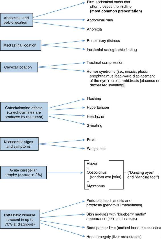
FIGURE 14.1 Clinical features of neuroblastoma.
537
538
VI. Soft Tissue Tumors
Rhabdomyosarcoma is the most common soft tissue sarcoma in childhood, and is a malignant tumor of the same embryonic mesenchyme that gives rise to skeletal muscle.
A.Epidemiology. Two-thirds of rhabdomyosarcomas occur in children younger than 10 years.
B.Etiology. The cause is generally unknown. Patients with neurofibromatosis are at higher risk.
C.Clinical features. Signs and symptoms depend on the site of involvement. Any part of the body may be affected. The initial presentation is usually a painless soft tissue mass.
1.Head and neck, including the orbit, are the most common sites of involvement (40% of cases).
a.Orbital tumors typically present with proptosis, chemosis (i.e., conjunctival edema), eyelid swelling, and cranial nerve palsies.
b.Nasopharyngeal tumors typically present with epistaxis, airway obstruction, and chronic sinusitis.
c.Laryngeal tumors typically present with hoarseness.
2.Genitourinary tract is the second most common site of involvement (20% of cases). Tumors in this location typically present with hematuria, urinary tract obstruction, vaginal bleeding, and/or an abdominal mass.
3.The extremities are the third most common site of involvement (18% of cases). Tumors in this location present with a painless growing mass.
4.Other sites of involvement include the trunk, retroperitoneum, mediastinum, and paratesticular and perianal regions.
D.Diagnosis. Rhabdomyosarcoma should be considered in any patient presenting with a painless enlarging mass. Imaging studies (CT or MRI) are performed to determine the extent of local extension and to prepare for surgical excision. Immunohistochemistry and cytogenetic evaluation of tissue obtained by biopsy provides a definitive diagnosis.
E.Management. Treatment requires chemotherapy and either surgery or surgery and radiation for local control.
F.Prognosis. Tumors of the head and neck and the genitourinary tract have the best prognosis, with a cure rate >90% if localized. Poor prognosis is associated with metastases (20% have metastatic disease at time of diagnosis) and with recurrence. Tumors at sites other than the head and neck or the genitourinary tract carry an intermediate prognosis with survival about 70%.
539
VII. Bone Tumors
A.Osteogenic sarcoma is a malignant tumor that forms osteoid or new bone.
1.Epidemiology
a.Osteogenic sarcoma is the most common malignant bone tumor in children.
b.The incidence of this tumor peaks during the rapid growth spurt of adolescence. Males are more commonly affected.
2.Etiology. The cause is unknown; however, it is associated with previous retinoblastoma, Paget disease of bone, radiation therapy for cancer, and fibrous dysplasia.
3.Clinical features. About 50% of tumors occur in the distal femur or proximal tibia. Other signs and symptoms are listed in Table 14-4, which compares osteogenic sarcoma with Ewing sarcoma, the second most common malignancy of bone in children.
4.Diagnosis. Diagnosis is suggested by findings on radiographs and MRI. Definitive diagnosis is by tissue biopsy. Bone scan and chest CT scan are performed to evaluate for metastatic disease.
5.Management
a.Surgery to remove the primary tumor is performed by limb amputation or limb salvage procedures after initial chemotherapy.
b.Chemotherapy includes high-dose methotrexate, cisplatin, and doxorubicin.
c.Pulmonary metastases identified at the time of diagnosis are resected if still evident after initial chemotherapy.
6.Prognosis. For nonmetastatic disease, about 70% of patients are cured, and if metastatic at diagnosis, about 50% are cured. Prognosis for those who do not respond to chemotherapy or relapse is very poor.
B.Ewing sarcoma is a sarcoma characterized as a small, round, blue cell tumor (undifferentiated, monomorphous cell appearance).
1.Epidemiology
a.Ewing sarcoma is the second most common malignant bone tumor.
b.It most commonly occurs during adolescence. Males are more commonly affected.
c.It is rare in Asians and African Americans.
2.Etiology. The cause is unknown. However, 95% have a chromosomal translocation between chromosomes 11 and 21 (a similar translocation to that noted in PNET brain tumors).
3.Clinical features. Signs and symptoms are listed in Table 14-4, which compares Ewing sarcoma and osteogenic sarcoma. Ewing sarcoma may occasionally develop in soft tissue instead of bone.
4.Diagnosis
a.Diagnosis is suggested by radiographic findings; however, similar findings are found in osteomyelitis, lymphoma, osteogenic sarcoma, and Langerhans cell histiocytosis (LCH).
b.MRI of affected bone can better delineate the tumor and its local extension.
c.Definitive diagnosis is by histologic evaluation of tissue obtained by open biopsy.
d.Bone scan, chest CT scan, and bone marrow aspiration and biopsy are performed to assess for metastatic disease.
5.Management. Treatment includes multiagent chemotherapy followed by surgical excision, when possible. If the lesion is not surgically resectable, radiation therapy is used for local control. Late complications of radiation therapy include pathologic fractures at the tumor site, retarded bone growth, limb length discrepancy, functional impairment, and secondary malignancy.
540
6.Prognosis. Outcome is good for local disease, with a 3- to 5-year survival rate of 80%. Prognosis is especially poor if metastases are present at diagnosis.
Table 14-4
Clinical and Radiographic Features of Osteogenic Sarcoma and Ewing Sarcoma
Feature |
Osteogenic Sarcoma |
Ewing Sarcoma |
Site |
Metaphysis of tubular long bones |
Flat bones and diaphysis of tubular bones; |
|
50% occur near the knee |
occasionally extraosseous |
|
Most common sites (in order): distal femur, proximal |
Most common sites (in order): axial skeleton |
|
tibia, proximal humerus, proximal femur |
(especially pelvis), humerus, femur |
Local and |
Pain, swelling, and soft tissue massSystemic symptoms |
Pain, swelling, and soft tissue mass |
systemic |
uncommon |
Fever, malaise, and weight loss |
findings |
|
Leukocytosis and elevated ESR |
Radiographic |
Periosteal reaction with “sunburst” appearance |
Periosteal reaction with “onion skin” |
findings |
|
appearance |
|
Lytic; or mixed lytic and destructive changes |
Destructive changes |
Metastases |
Occurs in 15% at presentation |
Occurs in 25% at presentation |
|
Lungs (90%) and bone (10%) |
Lungs (50%), bone (25%), and bone marrow |
|
|
(25%) |
ESR = erythrocyte sedimentation rate.
541
VIII. Liver Tumors
Liver tumors include hepatoblastoma and hepatocellular carcinoma.
A.Epidemiology
1.Hepatoblastoma is the most common type of liver tumor in childhood. It almost always occurs in children younger than 3 years of age. It is also associated with Beckwith– Wiedemann syndrome and familial polyposis, and is more common in children born prematurely.
2.Hepatocellular carcinoma may occur in both young children and in adolescents. It is associated with chronic active hepatitis B infection, biliary atresia, glycogen storage disease type I, α1-antitrypsin deficiency, and hereditary tyrosinemia.
B.Clinical features. Signs and symptoms are similar in liver tumors and include presentation with a right upper abdominal mass, loss of appetite, and weight loss. Jaundice is generally absent, and quite often patients are asymptomatic.
C.Diagnosis. Diagnosis is made by abdominal imaging with CT or MRI scan and finding elevation of the serum tumor marker α-fetoprotein.
D.Management. Treatment includes surgical resection, if possible, and chemotherapy. Chemotherapy may convert a previously unresectable tumor to one that is amenable to surgery.
E.Prognosis. Outcome depends on surgical resectability and chemosensitivity. Nonmetastatic hepatoblastoma carries a cure rate of about 75%, whereas hepatocellular carcinoma carries a cure rate of about 40%.
542
IX. Retinoblastoma
See Chapter 18, section VIII.B.
543
X. Germ Cell Tumors (Germinomas)
Germ cell tumors are rare malignancies derived from the cellular precursors of sperm and eggs.
A.Classification is by location and degree of cell differentiation. Germ cell tumors may be located in the gonadal region (i.e., testis, ovary) or in extragonadal regions (i.e., anterior mediastinum, sacrococcygeal area, pineal gland or suprasellar region of the brain, retroperitoneum, neck). Types of germ cell tumors include seminoma (in males), dysgerminoma (in females), teratoma, yolk sac tumor, embryonal cell carcinoma, and choriocarcinoma.
B.Specific tumors
1.Teratomas are tumors containing more than one of the three primary germ cell layers (i.e., ectoderm, mesoderm, and endoderm). Mature teratomas often contain skin, hair, or teeth, whereas immature teratomas contain fetalor embryonal-type structures.
Teratomas may be benign or malignant. Malignant potential is based on the amount of immature tissue and the presence or absence of other germ cell tumor cells within the
teratoma.
a.Sacrococcygeal teratoma is the most common teratoma during the first year of life. The majority (75%) occur in females. The tumor arises from the coccyx and presents as a soft tissue mass. Almost all (95%) are benign. Treatment includes surgical excision of both the tumor and the coccyx to prevent recurrence.
b.Anterior mediastinal teratomas are generally benign and may present with signs and symptoms of airway obstruction.
c.Ovarian teratomas are the most common ovarian tumor and are generally benign.
The teratoma is suggested by the presence of calcium within the tumor on an abdominal radiograph.
2.Testicular tumors may be derived from germ cells or stromal cells; 70% of childhood testicular tumors are germinomas.
a.Epidemiology
1.The most common of these germinomas are yolk sac tumors (60%), followed by teratomas (15%) and, rarely, seminomas and embryonal carcinoma. (See also Chapter 3, section IX.C.1.)
2.Peak ages are younger than 5 years and during adolescence.
3.There is an association with cryptorchid testes.
4.One-third of testicular tumors in childhood are benign, unlike in adults, in whom almost all testicular tumors are malignant.
5.Germ cell tumors are more common in patients with sex chromosome abnormalities.
b.Clinical features. Signs and symptoms include a solid, firm, painless testicular mass or generalized testicular swelling. Serum α-fetoprotein is elevated in yolk sac tumors. Malignant tumors may extend locally or may metastasize to retroperitoneal lymph nodes, lung, or liver.
c.Management. Treatment is based on the tumor type and size. Treatment of yolk sac tumors involves radical orchiectomy and, if necessary, retroperitoneal lymph node dissection.
3.Ovarian tumors most commonly include yolk sac tumors, teratomas, and dysgerminomas.
a.Epidemiology
1.One-third are malignant. The younger the child, the more likely it is that the tumor will be malignant.
544
2.Tumors increase in frequency during puberty.
b.Clinical features. Signs and symptoms include abdominal mass, abdominal pain caused by torsion of the tumor together with the ovary, and vaginal bleeding. As with males, serum α-fetoprotein is elevated in yolk sac tumors in females.
c.Management. Treatment is based on tumor type and typically includes surgical resection, chemotherapy, and, sometimes, radiation therapy.
545
XI. Langerhans Cell Histiocytosis (LCH)
A.Definition. LCH is characterized by infiltration of organs with Langerhans cells.
B.Etiology. The cause is unknown. It appears that some cases represent clonal cellular proliferation, whereas others represent immunologic dysregulation.
C.Clinical features are highly variable.
1.Skeletal involvement occurs in 80% of patients.
a.The skull is most commonly involved.
b.Single or multiple bony lesions may be present and may be painful, palpable, and associated with swelling.
c.Pathologic fractures may occur.
d.Chronic draining ears may indicate LCH involving the mastoid.
2.Skin involvement occurs in 50% of patients. It typically manifests as seborrheic dermatitis of the diaper area and scalp (it mimics cradle cap) and is most common in intertriginous areas.
3.Pituitary or hypothalamic involvement may lead to growth retardation, diabetes insipidus, hypogonadism, and panhypopituitarism.
4.Other features include lymphadenopathy, hepatosplenomegaly, exophthalmos, anemia, and pulmonary infiltrates.
5.Nonspecific systemic features are very uncommon and include weight loss, fatigue, fever, and failure to thrive.
D.Diagnosis is by identifying the typical histologic features on biopsy of skin or bone lesions. There are characteristic morphologic features and immunohistochemical markings.
E.Management
1.Therapy is geared to abort organ destruction or dissemination.
2.Corticosteroids and chemotherapeutics are considered standard of care. If a single lesion or organ is involved, local curettage may be used.
F.Prognosis varies with the extent and location of disease. Single lesions may spontaneously resolve after curettage. Response rate to current treatments is high. Long-term complications include growth impairment, learning problems, hearing loss, orthopedic deformities, and chronic lung disease.
546