

- •Copyright
- •Contents
- •Dedication
- •Preface
- •Acknowledgments
- •Contributors
- •Contributors to the Previous Edition
- •Review Test
- •Answers and Explanations
- •Review Test
- •Answers and Explanations
- •Review Test
- •Answers and Explanations
- •Review Test
- •Answers and Explanations
- •Review Test
- •Answers and Explanations
- •Review Test
- •Answers and Explanations
- •Review Test
- •Answers and Explanations
- •Review Test
- •Answers and Explanations
- •Review Test
- •Answers and Explanations
- •Review Test
- •Answers and Explanations
- •IV. Hypertension
- •VI. Nephrotic Syndrome (NS)
- •VII. Hemolytic Uremic Syndrome (HUS)
- •VIII. Hereditary Renal Diseases
- •IX. Renal Tubular Acidosis (RTA)
- •XI. Chronic Kidney Disease (CKD) and End-Stage Renal Disease (ESRD)
- •XII. Structural and Urologic Abnormalities
- •XIII. Urolithiasis
- •XIV. Urinary Tract Infection (UTI)
- •Review Test
- •Answers and Explanations
- •Review Test
- •Answers and Explanations
- •Review Test
- •Answers and Explanations
- •Review Test
- •Answers and Explanations
- •IV. Food Allergy
- •VI. Urticaria (Hives)
- •VII. Drug Allergy
- •VIII. Asthma
- •IX. Immunology Overview
- •X. Disorders of Lymphocytes (Figure 15-2)
- •XI. Disorders of Granulocytes (Figure 15-3)
- •XII. Disorders of the Complement System
- •Review Test
- •Answers and Explanations
- •Review Test
- •Answers and Explanations
- •Review Test
- •Answers and Explanations
- •Review Test
- •Answers and Explanations
- •Review Test
- •Answers and Explanations
- •Review Test
- •Answers and Explanations
- •Comprehensive Examination
- •Index
Review Test
1.You are called to the nursery to examine a “floppy” female infant born within the past
24 hours. The neonate is hypotonic with diminished deep tendon reflexes. There are no tongue fasciculations. When you greet the baby’s mother, she is anxious and has difficulty releasing her grip after shaking your hand. Which of the following is the most likely diagnosis?
A.Muscular dystrophy
B.Congenital myotonic dystrophy
C.Neonatal myasthenia gravis
D.Spinal muscular atrophy, type 1
E.Infantile botulism
2.A 10-year-old girl is being evaluated for “weakness” of 3 days’ duration. Medical history is significant for a 4-day episode of diarrhea 2 weeks before her current presentation. She is otherwise well with no chronic medical conditions and is taking no medications. Physical examination reveals symmetric weakness at the ankles and knees, with normal strength at the hip joints. Deep tendon reflexes are absent in the distal lower extremities. Sensory examination is normal. Which of the following statements is most consistent with the most likely diagnosis?
A.This patient is most likely to have had a prodromal gastroenteritis with Salmonella typhi.
B.Electromyography would be expected to show normal nerve conduction.
C.Management should include intravenous immune globulin.
D.This patient is likely to have elevated antistreptolysin O or anti-DNase B titers.
E.The prognosis for complete recovery is poor.
3.A 6-month-old male infant is evaluated for lethargy and poor feeding. Recent dietary changes include the introduction of cereals, fruits, and herbal tea with honey. Physical examination reveals an afebrile infant with normal vital signs. Neurologic examination is notable for decreased muscle tone and a weak suck. Which of the following statements regarding this infant’s most likely diagnosis is most accurate?
A.Infants typically present with ascending paralysis.
B.Antibiotics should be administered immediately.
C.Infants present with brisk deep tendon reflexes.
D.Electromyography is not helpful in making the diagnosis.
E.Constipation is often the initial symptom in infants.
4.An 8-year-old girl is noted by her teacher to have brief staring spells throughout the day. She is referred to you for further evaluation. Neurologic examination is normal. You order an electroencephalogram, which shows a generalized 3-Hz spike-and-wave discharge pattern arising from both hemispheres. Which of the following statements regarding the most likely diagnosis is most accurate?
A.Further questioning would probably reveal that this patient loses control of her bladder during the event.
B.This patient’s condition is inherited in an autosomal recessive pattern.
C.This patient’s staring spells would be expected to last less than 10 seconds each.
D.This patient is likely to have prolonged postictal periods.
E.Phenobarbital is the drug of choice for this patient’s condition.
5.A 4-month-old female infant is brought to your office by her parents, who are concerned about some behaviors they have witnessed. They note that during the past week, she has had brief jerking episodes, lasting 1–2 seconds each, with sudden arm extension followed by flexion of the head. You order an electroencephalogram, which reveals a highly disorganized pattern of high-amplitude spike and waves in both cerebral hemispheres, consistent with a hypsarrhythmia pattern. Which of the following is the most commonly identified cause of the
484
patient’s disorder?
A.Prior episode of bacterial meningitis
B.Perinatal asphyxia
C.Shaken baby syndrome
D.Tuberous sclerosis
E.Neurofibromatosis type 1
6.A 6-year-old boy complains of a 4-day history of low-back pain and difficulty walking. On examination, you note weakness in his lower extremities and absent lower extremity deep tendon reflexes. Sensation in the lower extremities is intact. A magnetic resonance imaging scan of the spine is normal. Which of the following is the most likely diagnosis?
A.Duchenne muscular dystrophy
B.Myasthenia gravis
C.Acute cerebellar ataxia of childhood
D.Guillain–Barré syndrome
E.Becker muscular dystrophy
7.A 2-year-old boy is brought to the office by his parents, who note that their son has had weakness in his legs for the past several months that appears to be getting worse. On examination, you note that the calf muscles are enlarged. Which of the following statements regarding this patient’s condition is correct?
A.DNA testing is normal.
B.Dystrophin levels are normal.
C.Progressive weakness leads to loss of ambulation before 12 years of age.
D.Electromyography reveals delayed nerve conduction.
E.Treatment with intravenous immune globulin can transiently improve symptoms.
8.Two hours ago a 2-year-old boy had a 5-minute episode of whole body shaking associated with a temperature of 39.4°C (103°F). The boy’s parents state that their son has had runny nose and cough for the past 24 hours. He now acts normal except for his cold symptoms, according to his parents. The parents remind you that this is his second febrile seizure. Physical examination is normal, revealing no focal neurologic signs. Which of the following would be the most appropriate recommendation at this time?
A.Order a stat head computed tomography scan.
B.Order an electroencephalogram.
C.Begin treatment with phenobarbital.
D.Reassure the parents that no workup or medications are necessary at this point.
E.Admit the patient to the hospital for overnight observation.
9.A 12-year-old girl is brought to see you for evaluation of frequent headaches. She has had headaches three times per week for the past several months, and her parents are concerned that she may be having migraines. Which of the following statements would support this diagnosis?
A.The headaches are bilateral, dull, and achy, but mild.
B.The neurologic examination is abnormal during the headache.
C.The headaches awaken the patient in the early morning hours.
D.The duration of the headache is >1 hour.
E.This patient is likely to have an aura with her headache.
10.Examination of a comatose 13-year-old boy in the emergency department is significant for bilaterally nonreactive and dilated pupils. An oculocephalic maneuver is negative. Which of the following is the most likely diagnosis?
A.Postictal state from status epilepticus
B.Brainstem injury
C.Subdural hematoma with herniation
485
D.Opiate overdose
E.Alcohol intoxication
11.A 2-month-old boy is brought to your office for evaluation with a 2-day history of poor feeding and vomiting. Parents deny fever, cold symptoms, or diarrhea. On physical examination, the child is well hydrated but has a large anterior and posterior fontanelle and a persistent downward deviation of both eyes. This patient’s presentation is most consistent with which of the following diagnoses?
A.Cerebral infarct
B.Congenital myotonic dystrophy
C.Myasthenia gravis
D.Becker muscular dystrophy
E.Infantile hydrocephalus
The response items for statements 12–14 are the same. You will be required to select one answer for each statement in the following set.
A.Dandy–Walker malformation
B.X-linked hydrocephalus
C.Infantile botulism
D.Spinal muscular atrophy type 1 (Werdnig–Hoffman disease)
E.Juvenile myasthenia gravis
F.Congenital myotonic dystrophy
For each patient, select the most likely diagnosis.
1.A 4-month-old male infant has generalized weakness, hypotonia, areflexia, and tongue fasciculations.
2.An 8-month-old female infant with a 5-day history of constipation presents with a weak cry, hyporeflexia, ophthalmoplegia, and loss of the ability to sit without support.
3.A 6-month-old male has had hypotonia, facial weakness, areflexia, and a history of feeding problems since birth.
486
Answers and Explanations
1.The answer is B [I.F.3]. This patient most likely has congenital myotonic dystrophy. Patients present with hypotonia and often have feeding and respiratory problems. Facial weakness and hyporeflexia are common. Infants acquire the disorder through autosomal dominant inheritance, most commonly from an affected mother. Mothers of infants with congenital myotonic dystrophy have myotonia, an inability to relax contracted muscles, which manifests as difficulty releasing a hand grip during a firm handshake. Muscular dystrophy rarely presents during infancy. Infants with botulism have constipation, hypotonia, problems with suck and swallow, and progressive weakness that may lead to paralysis. Neonatal myasthenia is a transient muscle disorder caused by the transplacental passage of acetylcholine receptor antibodies. Weakness and hypotonia may be present, but deep tendon reflexes are preserved. Infants with spinal muscular atrophy have hypotonia but also have characteristic tongue fasciculations.
2.The answer is C [VII.D]. This patient’s presentation is most consistent with Guillain–Barré syndrome. The diagnosis of Guillain–Barré syndrome should be considered in any child with ascending symmetric weakness or paralysis, absence of deep tendon reflexes, and a normal sensory examination. Management should be initiated as soon as the diagnosis is established because of the risk of respiratory muscle paralysis. Intravenous immune globulin is the preferred treatment in children. Many infectious agents have been associated with Guillain– Barré syndrome, but the most common infectious agent is Campylobacter jejuni, which causes a prodromal gastroenteritis. Electromyography would be expected to demonstrate decreased nerve conduction velocity or conduction block. There is no known association between Guillain–Barré syndrome and prior group A β-hemolytic streptococcal infection. The prognosis for children with Guillain–Barré syndrome is excellent, and complete recovery is likely.
3.The answer is E [I.F.2]. Infantile botulism is caused by the ingestion of Clostridium botulinum spores and the release of botulinum toxin within the intestine. The toxin prevents the release of acetylcholine at peripheral cholinergic synapses, initially causing constipation, which is followed by a weak suck and swallow, cranial nerve palsies, and weakness. Patients with infantile botulism have a symmetric descending paralysis. Contaminated honey is a common source of the toxin. Physical examination is notable for diffuse weakness, hypotonia, and hyporeflexia (i.e., diminished deep tendon reflexes). The diagnosis is suggested by the history and physical examination findings and confirmed by the identification of the toxin or bacteria within the stool. Electromyography may also be helpful in diagnosis and may show brief, small-amplitude muscle potentials with an incremental response during high-frequency stimulation. Treatment includes supportive care and botulism immune globulin. Antibiotics are not helpful.
4.The answer is C [V.L.4]. This patient’s clinical presentation and electroencephalogram (EEG) are consistent with absence epilepsy of childhood. Patients usually present with multiple absence seizures, which are brief staring spells that occur without warning and are not followed by postictal drowsiness. Urinary continence and loss of posture are also not seen in absence seizures. Absence seizures last less than 10 seconds and have a very characteristic EEG pattern, showing a generalized 3-Hz spike-and-wave abnormality. Absence epilepsy of childhood is inherited in an autosomal dominant pattern with age-dependent penetrance. The antiepileptic medications usually used include ethosuximide, valproic acid, or lamotrigine.
5.The answer is D [V.L.3]. This clinical presentation is consistent with infantile spasms. Patients
with infantile spasms typically present with brief, myoclonic jerks, lasting 1–2 seconds each, occurring in clusters of 5–10 seizures spread out over 3–5 minutes. Patients may have sudden
487
extension of the arms and sudden flexion of the head (jackknife or salaam seizures). A variety of different prenatal, perinatal, and postnatal insults to the central nervous system may result in infantile spasms. Tuberous sclerosis is the most commonly identified cause of this disorder. Perinatal asphyxia, intraventricular hemorrhage, and meningitis are other causes of infantile spasms. Neurofibromatosis type 1 is not typically associated with infantile spasms.
6.The answer is D [VII.D]. Guillain–Barré syndrome (acute inflammatory demyelinating polyneuropathy) typically presents with ascending paralysis without sensory loss. Despite this finding, about 50% of children complain of low-back pain or discomfort in their legs. Deep tendon reflexes are absent, and spinal magnetic resonance imaging is normal. The diagnosis is based on the findings of albuminocytologic dissociation in the cerebrospinal fluid and by decreased nerve conduction velocity on electrophysiologic studies. In both Duchenne and Becker muscular dystrophy, the onset of weakness is slow and progressive. Myasthenia gravis, which is more common in girls, presents with weakness that increases during the day and normal deep tendon reflexes. Acute cerebellar ataxia of childhood presents with ataxia (unsteady gait or truncal unsteadiness) rather than weakness.
7.The answer is C [IX.B, IX.E, and IX.I.1]. This patient’s presentation with increasing weakness in the lower extremities and calf enlargement is consistent with Duchenne muscular dystrophy, an X-linked progressive degenerative muscle disorder for which there is no cure. Children typically present between 2 and 5 years of age with gait problems and weakness, and they are often wheelchair dependent by around their 12th birthday. On examination, patients have enlarged calf muscles (pseudohypertrophy) as a result of fatty infiltration of the degenerating muscles, and laboratory studies reveal elevated creatine kinase levels. The diagnosis can be made by DNA testing of the dystrophin gene, which shows a deletion in more than 90% of patients. Electromyography shows small, polyphasic muscle potentials but normal nerve conduction velocities. Intravenous immune globulin has no role in the management of Duchenne muscular dystrophy.
8.The answer is D [V.K.7]. Febrile seizures are defined as any seizure that accompanies a fever owing to a non-CNS (central nervous system) cause in patients between the ages of 6 months and 6 years. Febrile seizures are benign events that are not generally associated with serious acute or long-term neurologic sequelae. A computed tomography scan is not indicated in the absence of papilledema or focal neurologic deficits. An electroencephalogram is useful when the diagnosis of epilepsy is being considered but not in recurrent febrile seizures. A complex febrile seizure is distinguished from a simple febrile seizure if it lasts more than 15 minutes, has focal features, or recurs within 24 hours. For simple febrile seizures, anticonvulsant treatment is usually not initiated until a patient has multiple febrile seizures. Febrile seizures do not require inpatient observation.
9.The answer is D [VI.B and VI.C]. Migraines are characterized by unilateral, or sometimes bilateral, frontal throbbing headaches that last for at least 1 hour. The neurologic examination of patients with migraines is usually normal. Headaches that occur on awakening in the morning are more characteristic of headaches resulting from increased intracranial pressure.
Migraine without aura is the most common form of migraine in children.
10.The answer is B [IV.C.6]. The physical examination of this patient’s eyes, including both the negative oculocephalic test and bilateral dilated nonreactive pupils, is most consistent with brainstem injury. The oculocephalic maneuver is also termed doll’s eyes, and an abnormal response (when the head is turned, the eyes follow the head and continue to look straight ahead rather than drifting to midline) suggests a damaged vestibular system. Pupils may also be dilated during and immediately after a seizure or after topical ophthalmic application of a dilating agent, but the vestibular nerve is usually unaffected in these situations (the oculocephalic maneuver would be positive). An enlarging subdural hematoma can cause uncal herniation with a unilateral dilated, nonreactive pupil. Opiate ingestion causes
488
constricted pupils. Alcohol intoxication less frequently causes coma and more often leads to somnolence. Alcohol intoxication can cause pupils to be slow to react but pupils would not be nonreactive.
11.The answer is E [II.D]. This patient’s eye findings are termed the setting-sun sign, or a tonic downward deviation of the eyes. The setting-sun sign is suggestive of hydrocephalus. Increased pressure in the third ventricle from noncommunicating hydrocephalus injures the upward gaze center in the midbrain, causing this downward deviation of the eyes. In contrast, patients with myasthenia commonly present with ptosis. There are no specific eye abnormalities in congenital myotonic dystrophy, muscular dystrophy, or cerebral infarcts.
12.The answers are D, C, and F, respectively [I.F.1–3]. The 4-month-old infant has spinal muscular atrophy type 1 (Werdnig–Hoffman disease), an autosomal recessive disorder that presents with hypotonia, weakness, problems with suck and swallow, and tongue fasciculations. It presents within the first 6 months of life. The 8-month-old infant has infantile botulism, an environmentally acquired disorder of the neuromuscular junction in which botulinum neurotoxin blocks release of acetylcholine at the neuromuscular junction. The toxin is released from the spores of Clostridium botulinum, which are found in contaminated honey or soil. Constipation is often the presenting symptom, followed by weakness, cranial nerve findings, poor abilities to feed, and paralysis. The 6-month-old infant has congenital myotonic dystrophy, an autosomal dominant disorder presenting in infancy with hypotonia, facial weakness, and feeding problems. The diagnosis may be missed if the mother of the patient is not seen or examined because the typical myotonia (i.e., inability to relax contracted muscles) is most apparent in adults.
489

C H A P T E R 1 3
490
Hematology
Carol Diamond
491
I.Anemia
A.General concepts
1.Definition. Anemia is a reduction in the red blood cell (RBC) number or in the hemoglobin (Hgb) concentration to a level more than 2 standard deviations below the mean.
2.Hgb and age
a.The Hgb is high at birth in most newborns and then declines, reaching the physiologic lowest point (nadir) between 2 and 3 months of age in the term infant and between 1 and 2 months of age in the preterm infant. Hgb values reach adult levels after puberty.
b.Fetal hemoglobin (Hgb F) is a major constituent of Hgb during fetal and early postnatal life. It declines and gradually disappears by 6–9 months of age.
3.Epidemiology. Anemia is one of the most common laboratory abnormalities during childhood. Approximately 20% of all children in the United States and 80% of children in developing nations have anemia at some time during childhood.
B.Classification
1.Classification is made on the basis of the mean corpuscular volume (MCV) and the morphologic appearance of the RBC (i.e., size, color, and shape). Terms used include the suffix -cytic, referring to size, and the suffix -chromic, referring to color. Primary classifications include the following:
a.Microcytic, hypochromic anemia (small, pale RBCs; low MCV)
b.Macrocytic anemia (large RBCs; high MCV)
c.Normocytic, normochromic anemia (normal RBCs in size, color, and shape; normal MCV)
2.Classification based on reticulocyte count is also helpful. The reticulocyte count reflects the number of immature RBCs in the circulation and, therefore, the activity of the bone marrow in producing RBCs. The usual percentage of RBCs that are reticulocytes is 1% (normal absolute count = 50,000 cells/mm3). In the steady state, when a patient has a normal Hgb level, the reticulocytes should constitute 1% of all RBCs. In most anemias, reticulocyte counts should rise. A low reticulocyte count indicates bone marrow failure or diminished hematopoiesis.
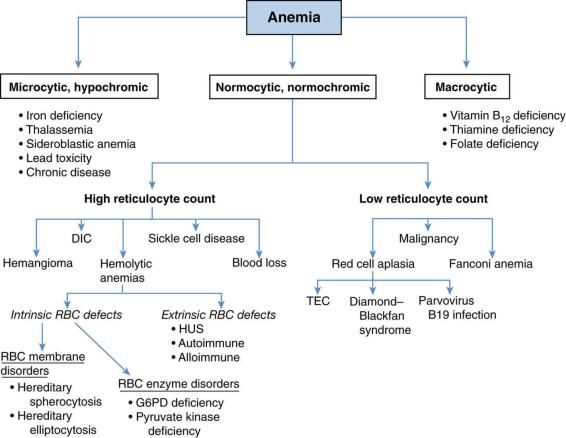
3.Figure 13-1 presents the differential diagnosis of anemia on the basis of the previously discussed classification schemes. Descriptions of the more common forms of anemia in childhood follow.
C.Clinical features of anemia (Table 13-1)
D.Microcytic, hypochromic anemias. The two most common types of microcytic, hypochromic anemia during childhood are iron-deficiency anemia and β-thalassemia minor.
1.Iron-deficiency anemia is the most common blood disease during infancy and childhood.
a.Etiology. The majority of cases are caused by inadequate iron intake.
1.Nutritional iron deficiency is most common in two age groups.
a.Nine to twenty-four months of age: owing to inadequate intake and inadequate iron stores (which are typically depleted by 4–6 months of age). Blood loss during birth may contribute to the anemia. The typical toddler’s diet consists of large quantities of iron-poor cow’s milk. Ironrich foods (e.g., iron-fortified cereal, meats, legumes) or iron supplementation is therefore recommended beginning at 4–6 months of age to prevent anemia.
492
b.Adolescent girls: owing to poor diet, rapid growth, and loss of iron in menstrual blood
2.Occult blood loss with resultant iron deficiency may be secondary to polyps, Meckel diverticulum, inflammatory bowel disease (IBD), peptic ulcer disease, celiac disease, and the early ingestion of whole cow’s milk before 1 year of age.
b.Clinical features. The signs and symptoms of anemia are listed in Table 13-1.
c.Laboratory findings
1.Because iron stores disappear first, an early finding of iron-deficiency anemia is low serum ferritin. Ferritin can be a useful assessment of iron stores; however, because ferritin is also an acute-phase reactant, it may be increased in infection, disease states, and stress, therefore appearing normal.
2.As serum iron decreases, iron-binding capacity increases, manifested as increased transferrin and decreased transferrin saturation.
3.Increased free erythrocyte protoporphyrin may be noted.
4.Other findings include a normal or increased reticulocyte count.
d.Management
1.Elemental iron (4–6 mg/kg/day) is prescribed orally for mild to moderate anemia. Iron is given with vitamin C (e.g., orange juice) to enhance intestinal iron absorption.
2.Dietary counseling may be required to increase nutritional iron through ironrich foods (e.g., meats, legumes, prunes, iron-fortified cereals).
3.RBC transfusion may be required for severe anemia associated with cardiovascular compromise. Not only will this improve oxygen-carrying capacity, but this will contribute to iron repletion.
4.Further evaluation to rule out other causes of anemia is necessary in patients with anemia unresponsive to iron.
2.α-Thalassemia and β-thalassemia syndromes
a.Definition. Thalassemia is a group of inherited anemias characterized by defective synthesis of one of the Hgb chains.
b.Pathophysiology
1.Normally, the major Hgb in RBCs is hemoglobin A1, a tetramer of two α- chains and two β-chains. HgbA2 and Hgb F may also be present in small amounts.
2.α-Thalassemia results from defective α-globin chain synthesis, and β- thalassemia results from defective β-globin chain synthesis.
3.Both types of thalassemia result in hemolysis that leads to increased bone marrow activity. As marrow activity increases, the marrow spaces enlarge, increasing the size of bones in the face, skull, and other bones if severe and untreated.
c.α-Thalassemia is the result of deletions of the α-globin chain and occurs predominantly in Southeast Asians. There are four states categorized on the basis of the number of α-globin genes deleted (normally there are four α-globin genes per diploid cell).
1.Silent carrier. One α-globin gene is deleted. Patients have no anemia and are asymptomatic.
2.α-Thalassemia minor. Two α-globin genes are deleted. Patients have mild microcytic anemia.
3.Hgb H disease. Three α-globin genes are deleted. Patients have moderate to severe anemia at birth with an elevated Hgb Bart’s (which is a type of Hgb made up of four gamma-globins that bind oxygen very strongly and do not
493
release it to tissue). Anemia is lifelong.
4.Fetal hydrops. Four α-globin genes are deleted. Only Hgb Bart’s are formed, and in utero, this causes profound anemia, congestive heart failure (CHF), and death if not identified early enough for intrauterine transfusion to occur.
d.β-Thalassemia is the result of mutations of the β-globin chain. Because there are only two β-globin genes in each cell, there are only two states:
1.β-Thalassemia major (Cooley anemia or homozygous β-thalassemia) may be caused by either total absence of the β-globin chains or deficient β-globin chain production.
a.Epidemiology. β-Thalassemia major occurs predominantly among patients of Mediterranean background.
b.Clinical features. Clinical findings include profound hemolytic anemia beginning in infancy, marked hepatosplenomegaly, and, if untreated, bone marrow hyperplasia in sites that result in a characteristic “thalassemia facies” appearance (frontal bossing, maxillary hyperplasia with prominent cheekbones, and skull deformities). Delayed growth and puberty may also be present.
c.Laboratory findings. Studies show severe hypochromia and microcytosis, elevated reticulocyte count, target cells and poikilocytes (abnormally shaped red blood cells) on the blood smear, and elevated unconjugated bilirubin, serum iron, and lactate dehydrogenase (LDH). Electrophoresis demonstrates low or absent Hgb A and elevated Hgb F.
d.Management. Treatment includes lifelong transfusions, chelation therapy to avoid iron overload and often splenectomy. Bone marrow transplant is curative and is the therapy of choice.
e.Complications. Hemochromatosis (iron accumulation within the heart, liver, lungs, pancreas, and skin) is a major complication and is caused by increased iron absorption from the intestine and from iron in transfused RBCs. Chelation of iron with the intravenous agent deferoxamine and/or the oral agent deferasirox promotes iron excretion and may help prevent or delay hemochromatosis.
2.β-Thalassemia minor (heterozygous β-thalassemia or β-thalassemia trait) causes a mild asymptomatic anemia with Hgb levels 2–3 g/dL below ageappropriate norms.
a.Laboratory findings include hypochromia and microcytosis with target cells and anisocytosis (excessive variation of size of RBCs) on smear.
b.No treatment is required.
c.It is important to note that patients with β-thalassemia minor may be very easily misdiagnosed as having iron-deficiency anemia and treated inappropriately with iron. However, the iron level in β-thalassemia minor is normal or elevated. Patients with thalassemia minor have normal to elevated RBC counts as opposed to iron deficiency in which the RBC count is low to normal.
3.Sideroblastic anemia is a group of anemias characterized by the presence of ring sideroblasts in the bone marrow. Ring sideroblasts result from the accumulation of iron in the mitochondria of RBC precursors. Sideroblastic anemia may be inherited or may be acquired as a result of drugs or toxins (e.g., isoniazid, alcohol, lead poisoning, chloramphenicol).
4.Lead poisoning (see Chapter 1, section IV.J) and chronic diseases, such as malignancy, infections, and kidney disease (termed anemia of inflammation), may present with a
494
microcytic, hypochromic anemia.
E.Macrocytic (megaloblastic) anemias. These anemias are characterized by large RBCs with
MCV > 95. The two major causes in children are folic acid and vitamin B12 deficiencies. In addition, rare but important causes of macrocytic anemia include bone marrow failure syndromes [e.g., Fanconi anemia, see section II.B.1].
1.Folic acid deficiency
a.Etiology. Causes include decreased folic acid intake (i.e., from a diet lacking uncooked fresh fruits and vegetables or from exclusive feedings with goat’s milk as the sole source of milk protein) and decreased intestinal absorption of folic acid
(i.e., from diseases affecting the small intestine, such as celiac disease, chronic infectious enteritis, Crohn disease, or medications, such as anticonvulsants and oral contraceptives).
b.Clinical features. In addition to the characteristic signs and symptoms of anemia, patients may have failure to thrive, chronic diarrhea, and irritability.
c.Diagnosis. Documentation of low serum folic acid is diagnostic.
d.Management. Treatment includes dietary folic acid and identification and treatment of the underlying cause.
2.Vitamin B12 deficiency
a.Normal physiology. To be absorbed, dietary vitamin B12 must first combine with a glycoprotein (intrinsic factor) secreted by the gastric parietal cells. Absorption then occurs in the terminal ileum.
b.Etiology. Causes include inadequate dietary intake (e.g., from a strict vegetarian [vegan] diet), an inherited inability to secrete intrinsic factor (juvenile pernicious anemia), or an inability to absorb vitamin B12 (e.g., Crohn disease, short gut syndrome).
c.Clinical features. In addition to the characteristic features of anemia, patients may also have anorexia, a smooth red tongue, and neurologic manifestations (such as ataxia, hyporeflexia, and positive Babinski responses).
d.Diagnosis. Documentation of low serum vitamin B12 level is diagnostic.
e.Management. Treatment is by monthly intramuscular vitamin B12 injections.
F.Normocytic, normochromic anemias. These anemias are characterized by normal size (normal MCV) and shape of the RBCs.
1.General concepts
a.Common causes include hemolytic anemias (premature destruction of RBCs), some RBC aplasias, and sickle cell (SS) anemia.
b.Reticulocyte count may be used to differentiate among the disorders [see Figure 13-1].
1.Low reticulocyte count reflects bone marrow suppression or failure and can be seen with RBC aplasias, viral suppression, medication effect, and pancytopenia associated with aplastic anemia.
2.High reticulocyte count reflects high bone marrow production of RBCs as seen in hemolytic anemias, recent acute hemorrhage, or any other condition associated with shortened RBC life span.
2.Hemolytic anemias
a.Intrinsic RBC defects that cause hemolysis include RBC membrane disorders and RBC enzyme disorders. Examples of RBC membrane disorders include hereditary spherocytosis and hereditary elliptocytosis, and examples of RBC enzyme disorders include glucose-6-phosphate dehydrogenase (G6PD) deficiency and pyruvate kinase (PK) deficiency.
1.Hereditary spherocytosis is the most common inherited abnormality of the
495
RBC membrane and occurs predominantly in persons of Northern European ancestry. There is a large spectrum of phenotypes, with some patients who are largely asymptomatic and others who are transfusion-dependent starting in infancy.
a.Etiology. There is a deficiency or abnormality of the structural RBC membrane protein spectrin, causing the RBC to assume its spherical shape. Inheritance is usually autosomal dominant.
b.Clinical features. Clinical findings are related to extravascular hemolysis. Infants may present with jaundice and anemia. By 2–3 years of age, patients develop pallor, weakness, and splenomegaly, as spherocytes are trapped in the spleen and destroyed. Other complications include aplastic crises, most commonly associated with parvovirus B19 infection, and pigmentary gallstones.
c.Laboratory findings. Studies show an elevated reticulocyte count, hyperbilirubinemia, spherocytes on blood smear, increased MCHC (mean corpuscular hemoglobin concentration), and abnormal RBC fragility with osmotic fragility studies.
d.Management. Treatment includes transfusions. Splenectomy cures the disorder, but to decrease the incidence of invasive disease caused by encapsulated bacteria, splenectomy is generally delayed until after
5 years of age.
2.Hereditary elliptocytosis is another autosomal dominant defect in the structure of spectrin that may or may not result in hemolysis. Clinical features are more variable than in hereditary spherocytosis. The majority of patients are asymptomatic, although 10% have jaundice at birth, and later may develop splenomegaly and gallstones. Elliptical RBCs are found on blood smear in older children. Treatment includes splenectomy for patients with severe chronic hemolysis. No treatment is needed for patients who have wellcompensated hemolysis.
3.Glycolytic enzymatic defects of RBCs include glucose-6-phosphate dehydrogenase deficiency and PK deficiency.
a.Glucose-6-phosphate dehydrogenase deficiency (G6PD) is the most common RBC enzymatic defect. It may occur as an acute hemolytic disease, induced by infection or medications, or as a chronic hemolytic disease. The epidemiology, etiology, clinical findings, and treatment are described in Table 13-2.
b.PK deficiency is an autosomal recessive disorder that results in decreased production of PK isoenzyme leading to ATP (adenosine triphosphate) depletion and decreased RBC survival.
1.Clinical features include pallor, jaundice, and splenomegaly. Kernicterus has been reported in neonates.
2.Laboratory findings include varying degrees of anemia and a blood smear showing polychromatic RBCs.
3.Diagnosis is by finding decreased PK activity in the RBCs.
4.Management includes transfusions and splenectomy for severe disease.
b.Defects extrinsic to the RBC that cause hemolysis
1.Autoimmune hemolytic anemia (AIHA) occurs when antibodies are misdirected against the RBCs.
a.Etiology
496
1.Primary AIHA is generally idiopathic in which no underlying disease is identified. Viral infections and occasionally drugs may be causal in some patients.
2.Secondary AIHA is associated with an underlying disease process, such as lymphoma, systemic lupus erythematosus (SLE), or immunodeficiency.
b.Clinical features
1.Fulminant acute-type AIHA occurs in infants and young children and is preceded by a respiratory infection. Presenting features include the acute onset of pallor, jaundice, hemoglobinuria, and splenomegaly. A complete recovery is expected.
2.Prolonged-type AIHA is characterized by a protracted course and high mortality. Underlying disease is frequently present.
c.Laboratory findings. Studies show severe anemia, spherocytes on blood smear, prominent reticulocytosis, and leukocytosis. A direct Coombs test is positive (detects coating of antibodies on the surface of RBCs or complement).
d.Management. Treatment may include transfusions that unfortunately may provide only transient benefit. Corticosteroids are often used for severe anemia and are continued until hemolysis diminishes. The acute form responds well to steroids.
2.Alloimmune hemolytic anemia occurs when antibodies from someone else are directed at the patients’ RBCs and is most commonly caused by newborn Rh and ABO hemolytic diseases.
a.Rh hemolytic disease occurs when the mother, who has no Rh antigen (maternal Rh negative), produces antibodies to the Rh antigen on her fetus’s RBCs (fetal Rh positive). In subsequent pregnancies, antibodies pass from the mother to the fetus causing hemolysis that presents as severe jaundice (which can lead to kernicterus), anemia, hepatosplenomegaly, and hydrops fetalis. A direct Coombs test is strongly positive.
b.ABO hemolytic disease occurs when the mother is blood group O and her fetus is blood group A, B, or AB. The mother produces antibodies to either the A or B blood group antigen that then pass to the fetus, causing hemolysis with resultant jaundice. A direct Coombs test is weakly positive. Of note, ABO disease can occur in the first pregnancy, unlike
Rh hemolytic disease.
c.Management. Treatment may include phototherapy for mild to moderate jaundice and exchange transfusion for severe jaundice.
3.Microangiopathic hemolytic anemia
a.Definition. This form of anemia results from mechanical damage to RBCs caused by passage through an injured vascular endothelium.
b.Etiology. Causes include severe hypertension, hemolytic uremic syndrome (HUS), artificial heart valves, a giant hemangioma, and disseminated intravascular coagulation (DIC).
c.Clinical features. Signs and symptoms are those characteristic of anemia and thrombocytopenia.
d.Laboratory findings. Studies show RBC fragmentation seen as “burr” cells, “target” cells, and irregularly shaped cells on the blood smear, along with thrombocytopenia.
497
e.Management. Therapy includes supportive care and treatment of the underlying cause.
3.SS hemoglobinopathies
a.Epidemiology. SS disease occurs in 1 in 800 black newborns in the United States. Eight percent have S trait. Compound heterozygotic disease can occur with Hgb C or β-thalassemia, leading to Hgb SC or sickle β-thalassemia disease, respectively.
b.Etiology and pathophysiology
1.SS disease is caused by a single amino acid substitution of valine for glutamic acid on the number 6 position of the β-globin chain of Hgb.
2.The mutation results in polymerization of Hgb within the RBC membrane when the RBC is exposed to low oxygen or acidosis.
3.Polymerization of Hgb results in a distorted RBC shape (sickled) that leads to decreased RBC life span (hemolysis) and occlusion of small vessels, resulting in distal ischemia, infarction, and organ dysfunction.
4.SS disease is the result of having two genes for Hgb S (homozygous).
5.S trait is defined as having only one gene for Hgb S (heterozygous). Persons with S trait have Hgb A (50–60%), Hgb S (35–45%), and a small percentage of Hgb F. Patients are asymptomatic without anemia unless exposed to severe hypoxemia. Some patients have an inability to concentrate the urine or may present with hematuria (5%) during adolescence.
c.Diagnosis. Diagnosis of SS disease is now usually made at birth through state newborn screening programs. Hgb electrophoresis is a highly sensitive and specific test that demonstrates Hgb S and Hgb F (fetal hemoglobin) in the newborn with SS disease.
d.Clinical features. Clinical characteristics are not generally present until protective Hgb F declines (by 6 months of age). Clinical episodes are often termed crises because they occur suddenly. Table 13-3 describes the clinical features and management of the common SS disease crises.
e.Laboratory findings (Table 13-4)
f.Management Historically, infection was the leading cause of death due to impaired splenic function.
a.Patients are at risk for infection with encapsulated bacteria (i.e.,
Haemophilus influenzae type b, Streptococcus pneumoniae, Salmonella, Neisseria meningitidis).
b.Fever in any patient with SS disease is managed with urgent assessment and appropriate cultures (blood and urine), chest radiograph to rule out pneumonia, and parenteral antibiotics until bacterial infection can be safely excluded.
c.Osteomyelitis may occur and may mimic a painful bone crisis.
Infection is most commonly caused by Salmonella species acquired through the gastrointestinal (GI) tract, although Staphylococcus aureus may also cause osteomyelitis. Clinical features include fever and pain, induration, tenderness, warmth, and erythema of the involved area. Treatment includes appropriate intravenous antibiotics.
g.Preventive care
1.Hydroxyurea, a chemotherapeutic agent that increases Hgb F, has been shown to decrease the incidence of vaso-occlusive crises.
2.Daily oral penicillin prophylaxis is started in the first few months of life to decrease the risk of S. pneumoniae infection.
498
3.Daily folic acid is given to prevent folic acid deficiency.
4.Routine immunizations and also yearly influenza vaccination, 23-valent polysaccharide pneumococcal vaccine at 2 years of age, and meningococcal vaccine should all be given.
5.Serial transcranial Doppler ultrasound or magnetic resonance angiography is recommended beginning at 2 years of age to identify patients at increased risk for stroke.
6.Bone marrow transplant is curative and is considered for children with severe manifestations.
h.Prognosis
1.Median life expectancy is in the 50s.
2.Long-term complications include delayed growth and puberty, cardiomegaly, hemochromatosis, cor pulmonale, pulmonary hypertension, renal insufficiency, gallstones, poor wound healing, avascular necrosis of the femoral and humeral heads, osteopenia, retinopathy, and diminished cognitive and school performance.
i.Other SS diseases include sickle cell–thalassemia disease (with clinical features similar to SS disease) and sickle cell–hemoglobin C disease (Hgb SC disease) caused by the inheritance of both Hgb S and Hgb C genes. Clinical features of Hgb SC disease are less severe than SS disease.
4.RBC aplasias are a group of congenital or acquired blood disorders characterized by anemia, reticulocytopenia, and a paucity of RBC precursors in the bone marrow. The clinical features of the three most common disorders occurring in childhood, congenital hypoplastic anemia (Diamond–Blackfan anemia), transient erythroblastopenia of childhood, and parvovirus B19–associated RBC aplasia, are presented in Table 13-5.
499

FIGURE 13.1 Classification and differential diagnosis of anemia. DIC = disseminated intravascular coagulation; HUS = hemolytic uremic syndrome; TEC = transient erythroblastopenia of childhood;
G6PD = glucose-6-phosphate dehydrogenase; RBC = red blood cell.
Table 13-1
Clinical Features of Anemia
Mild
Pallor (noted especially on skin and on mucous membranes)
Diminished attention
Moderate
Weakness and fatigue
Decreased exercise tolerance
Irritability
Tachycardia
Tachypnea
Anorexia
Systolic heart murmur
Severe
Congestive heart failure
Cardiac dilation
Shortness of breath
Hepatosplenomegaly
Spoon-shaped nails
Table 13-2
Features of Glucose-6-Phospate Dehydrogenase (G6PD) Deficiency
Epidemiology
Mediterranean, Arabic, Asian, and African ethnic groups
Pathophysiology
G6PD enzyme is critical for protecting the RBC from oxidative stress; deficiency results in RBC damage when the RBC is exposed to oxidants
Triggers of
hemolysis Infection Fava beans
Drugs (e.g., sulfa, salicylates, antimalarials)
Clinical features
Symptoms occur 24–48 hours after exposure to oxidant
Hemolysis occurs, resulting in abdominal pain, V/D, fever, and hemoglobinuria followed by jaundice; HSM may be present
Laboratory
findings Hemoglobinuria
Elevated reticulocyte count
500
Smear shows “bite” cells, “hemighosts” and Heinz bodies
Diagnosis
Low levels of G6PD in RBCs
Treatment
Transfusions as needed; splenectomy is not beneficial
V/D = vomiting, diarrhea; RBC = red blood cell; HSM = hepatosplenomegaly.
Table 13-3
Clinical Features and Management of Crises Occurring in Sickle Cell Disease
Crisis |
Clinical Features |
Management |
|
Vaso-occlusive crisis |
|
||
Painful bone |
Most common crisis |
Pain control |
|
crisis |
|||
Ischemia/infarction of bone or marrow |
Intravenous fluids at 1–1.5 × |
||
|
|||
|
Deep, gnawing, or throbbing pain lasting 3–7 days |
maintenance |
|
|
Subtype: acute dactylitis—painful swelling of digits of |
Incentive spirometry to decrease the |
|
|
the hands and feet |
risk of acute chest syndrome |
|
|
|
Severe, unremitting pain may respond |
|
|
|
to partial exchange transfusion. (Simple |
|
|
|
transfusion of RBCs is not indicated. It |
|
|
|
increases viscosity of blood and may |
|
|
|
worsen crisis.) |
|
|
|
|
|
Acute |
Abdominal pain and distension |
Low threshold for imaging the |
|
abdominal |
|||
Often caused by sickling within mesenteric artery |
abdomen |
||
crisis |
|||
|
Same management as for painful bone |
||
|
|
||
|
|
crisis |
|
|
|
|
|
Stroke |
Dysarthria and hemiplegia, but may be asymptomatic |
Same management as for painful bone |
|
|
|||
|
Occurs in up to 11% of patients (subclinical stroke |
crisis |
|
|
occurs in up to 20% of patients) |
Urgent exchange transfusion |
|
|
|
Patient should be started on chronic |
|
|
|
transfusion program to prevent |
|
|
|
recurrence, which occurs in 60–90% |
|
|
|
|
|
Priapism |
Painful, sustained erection |
Same management as for painful bone |
|
|
|||
|
Always consider SS disease in any patient presenting |
crisis |
|
|
with priapism |
|
|
|
|
|
|
Acute chest syndrome (ACS) |
|
||
|
New pulmonary infiltrate associated with respiratory |
Careful hydration and pain |
|
|
symptoms (e.g., cough, shortness of breath, chest pain) |
management |
|
|
Hypoxemia |
Oxygen |
|
|
May be severe and may cause up to 25% of deaths in |
Appropriate antibiotics (usually |
|
|
patients with SS disease |
cefuroxime and azithromycin) |
|
|
Causes include infection (e.g., viral, Mycoplasma |
Incentive spirometry |
|
|
pneumoniae, Chlamydia pneumoniae, Streptococcus |
Early use of partial exchange |
|
|
pneumoniae), sickling, atelectasis, fat embolism, painful |
transfusion in a patient who does not |
|
|
bone crisis involving the ribs, and pulmonary edema |
improve rapidly |
|
|
from fluid overload |
|
|
|
|
|
|
Sequestration |
Rapid accumulation of blood in spleen (or less |
Supportive care |
|
crisis |
|||
commonly, liver) |
Transfusion of RBCs |
||
|
|||
|
Occurs in patients <6 years of age |
Splenectomy recommended by some |
|
|
Abdominal distension, abdominal pain, shortness of |
practitioners because recurrence occurs |
|
|
breath, tachycardia, pallor, fatigue, and shock; |
in up to 50% |
|
|
mortality can be high |
|
|
|
Lab findings: low Hgb; elevated reticulocytes |
|
|
|
|
|
|
|
|
|
|
501
Aplastic crisis |
|
|
Temporary cessation of RBC production often caused |
Supportive care |
|||||
|
|
|
by parvovirus B19 or other infectious agent |
Transfusion of RBCs |
|||||
|
|
|
Pallor, fatigue, tachycardia |
|
|
||||
|
|
|
Lab findings: low Hgb; low reticulocytes |
|
|
||||
|
|
|
|
|
|
|
|
|
|
Hyperhemolytic |
|
|
Rapid hemolysis; often occurs in patients with other |
Supportive care |
|||||
crisis |
|
|
|||||||
|
|
hemolytic diseases (e.g., G6PD deficiency) |
Transfusion of RBCs |
||||||
|
|
|
|||||||
|
|
|
Pallor, fatigue, tachycardia, jaundice |
|
|
||||
|
|
|
Lab findings: low Hgb; elevated reticulocytes; elevated |
|
|
||||
|
|
|
bilirubin |
|
|
|
|
|
|
|
|
|
|
|
|
|
|
|
|
RBCs = red blood cells; SS = sickle cell; G6PD = glucose-6-phosphate dehydrogenase; Hgb = hemoglobin. |
|||||||||
Table 13-4 |
|
|
|
|
|
|
|
||
Usual Laboratory Findings in Sickle Cell Anemia |
|
|
|||||||
|
|
|
|
|
|
|
|
||
|
|
|
|
|
|
|
|
||
Red blood cell life span |
10–50 days |
|
|
|
|
|
|||
Hemoglobin |
|
6–9 g/dL |
|
|
|
|
|
||
Hematocrit |
|
18–27% |
|
|
|
|
|
||
Reticulocyte count |
|
5–15% |
|
|
|
|
|
||
White blood cell count |
12,000–20,000 cells/mm3 |
|
|
|
|
||||
Platelet count |
|
Increased, often > 500,000 platelets/µL |
|
|
|
|
|||
Bilirubin |
|
Increased |
|
|
|
|
|
||
Blood smear |
|
Sickled cells, target cells, Howell–Jolly bodies |
|
|
|
|
|||
Bone marrow |
|
Erythroid hyperplasia |
|
|
|
|
|||
Table 13-5 |
|
|
|
|
|
|
|
||
Characteristics of the Red Blood Cell Aplasias |
|
|
|||||||
|
|
|
|
|
|
|
|||
|
|
|
|
|
|
|
|||
Aplasia |
|
Etiology |
Clinical Features |
|
Laboratory Findings |
Treatment |
|||
Congenital |
|
|
Unknown |
Anemia within the first year |
|
↓Hgb |
RBC transfusion |
||
hypoplastic |
|
|
|
||||||
|
|
Autosomal |
of life |
|
↓Reticulocytes |
Corticosteroids |
|||
anemia |
|
|
|
||||||
|
|
recessive or |
Rapid onset |
|
↑Hgb F |
(up to 70% |
|||
(Diamond– |
|
|
|
||||||
|
|
autosomal |
One-fourth to one-third have |
|
↓or normal |
respond) |
|||
Blackfan |
|
|
|
||||||
|
|
dominant |
physical findings: |
|
platelet count |
Bone marrow |
|||
anemia) |
|
|
|
||||||
|
|
inheritance |
craniofacial, renal, cardiac |
|
Marrow: |
transplant if no |
|||
|
|
|
|
|
|||||
|
|
|
|
|
anomalies; short stature; |
|
↓RBC |
response to |
|
|
|
|
|
|
triphalangeal thumbs |
|
precursors; |
corticosteroids |
|
|
|
|
|
|
Signs and symptoms of |
|
other marrow |
|
|
|
|
|
|
|
anemia |
|
elements |
|
|
|
|
|
|
|
|
|
|
normal |
|
|
|
|
|
|
|
|
|
|
|
TEC |
|
|
Unknown |
Anemia begins >1 year of age |
|
↓Hgb |
Spontaneous |
||
|
|
|
|
|
|||||
|
|
|
|
Possible |
Slow in onset |
|
↓Reticulocytes |
recovery within |
|
|
|
|
|
postviral |
Signs and symptoms of |
|
Normal |
several weeks |
|
|
|
|
|
autoimmune |
anemia |
|
platelet count |
No treatment |
|
|
|
|
|
reaction |
|
|
|
Marrow: |
required |
|
|
|
|
|
|
|
|
↓RBC |
|
|
|
|
|
|
|
|
|
precursors |
|
|
|
|
|
|
|
|
|
|
|
Parvovirus B19– |
|
|
Parvovirus |
Anemia generally not |
|
↓Hgb |
Spontaneous |
||
associated pure |
|
|
|
||||||
|
|
B19 |
symptomatic |
|
↓Reticulocytes |
recovery within |
|||
RBC aplasia* |
|
|
|
||||||
|
|
|
|
infection |
May have associated URI |
|
Normal |
2 weeks |
|
|
|
|
|
|
symptoms and facial rash |
|
platelet count |
RBC transfusions |
|
|
|
|
|
|
(“slapped cheeks”) of fifth |
|
|
may be required |
|
|
|
|
|
|
disease |
|
|
for patients with |
|
|
|
|
|
|
Aplastic crisis in patients |
|
|
aplastic crisis |
|
|
|
|
|
|
with SS disease |
|
|
associated with |
|
|
|
|
|
|
|
|
|
|
SS disease |
|
|
|
|
|
|
|
|
|
|
*Note that Epstein–Barr virus, cytomegalovirus, human immunodeficiency virus (HIV), and drugs (e.g., chloramphenicol) may cause an acquired RBC aplasia similar to parvovirus B19.
502
TEC = transient erythroblastopenia of childhood; SS = sickle cell; URI = upper respiratory infection; Hgb = hemoglobin; RBCs = red blood cells.
503
II.Pancytopenia and Aplastic Anemia
A.Definition. Pancytopenia is defined as low white blood cells (WBCs), RBCs, and platelets and implies bone marrow failure.
B.Pancytopenia may be congenital or acquired.
1.Congenital aplastic anemia is also known as Fanconi anemia.
a.Etiology. Inheritance is autosomal recessive.
b.Clinical features
1.Onset of bone marrow failure occurs at a mean age of 7 years. Typical presentation is with ecchymosis and petechiae.
2.Skeletal abnormalities, which include short stature in almost all patients, and absence or hypoplasia of the thumb and radius
3.Skin hyperpigmentation
4.Renal abnormalities, including renal tubular acidosis
c.Laboratory findings. Studies show pancytopenia, RBC macrocytosis, low reticulocyte count, elevated Hgb F, and bone marrow hypocellularity.
d.Management. Treatment includes transfusions of RBCs and platelets as needed, and bone marrow transplant from an HLA-compatible donor, if available. Immunosuppressive therapy (e.g., corticosteroids, cyclosporin) may also help.
2.Acquired aplastic anemia
a.Etiology. Causes include drugs (e.g., sulfonamides, anticonvulsants, chloramphenicol), infections (e.g., human immunodeficiency virus [HIV], Epstein– Barr virus [EBV], cytomegalovirus [CMV], hepatitis), chemicals, and radiation. These all may damage bone marrow stem cells directly or may induce autoimmune destruction. Acquired aplastic anemia is most often idiopathic.
b.Clinical features. Signs and symptoms include bruising, petechiae, pallor, and fatigue, or serious infection as a result of neutropenia.
c.Laboratory findings. Studies show pancytopenia, low reticulocyte count, and hypocellular bone marrow.
d.Management. Treatment includes identifying and stopping the causative agent, transfusions as needed, bone marrow transplant, and immunosuppressive therapy.
504
III.Polycythemias
A.Definition. Polycythemia is defined as an increase in RBCs relative to total blood volume. It may also be defined as a hematocrit (Hct) > 60% or as an Hgb or Hct more than 2 standard deviations above normal values for age.
B.Primary polycythemia (polycythemia vera) is an extremely rare cause of polycythemia during childhood. This is a myeloproliferative disorder seen typically in older adults.
C.Secondary polycythemia is caused by increased erythropoietin production. Production may be appropriate or inappropriate.
1.Appropriate polycythemia may be caused by chronic hypoxemia as a result of cyanotic congenital heart disease (the most common cause of polycythemia in childhood), pulmonary disease, or living at high altitudes.
2.Inappropriate polycythemia may be caused by benign and malignant tumors of the kidney, cerebellum, ovary, liver, and adrenal gland; excess hormone production (e.g., corticosteroids, growth hormone, androgens), and kidney disease.
3.Clinical features include a ruddy facial complexion with a normal-sized liver and spleen.
4.Laboratory findings reveal elevated Hgb and Hct but normal platelet and WBC counts. Erythropoietin levels are high.
5.Management is directed toward identifying and treating the underlying cause. Phlebotomy is also used to keep the Hct < 60%.
D.Relative polycythemia refers to an apparent increase in RBC mass caused by a decrease in plasma volume. The most common cause is dehydration, and this should be considered in every patient with a high Hgb or Hct. Appropriate fluid management normalizes the Hct.
E.Complications of polycythemia include thrombosis (vaso-occlusive crisis, stroke, myocardial infarction) and bleeding.
505
IV. Disorders of Hemostasis
A.General concepts
1.Hemostasis requires normal function of three important elements: blood vessels, platelets, and soluble clotting factors. Hemorrhage may result from deficiency or dysfunction of any of these elements. Thrombosis may also occur but is rare during childhood.
2.The clotting cascade is depicted in Figure 13-2.
3.Clinical features suggesting abnormal hemostasis include the following:
a.Cutaneous bleeding (e.g., ecchymoses, petechiae)
b.Spontaneous epistaxis that is severe and recurrent without an obvious cause
c.Prolonged bleeding after simple surgical procedures, circumcision, trauma, or dental extraction
d.Recurrent hemarthroses
e.Deep venous thrombosis, pulmonary embolism, or stroke
4.Diagnostic studies. Evaluation for clotting abnormality typically includes these screening tests:
a.Complete blood count (CBC)
b.Platelet count
c.Blood smear to evaluate platelet morphology
d.Activated partial thromboplastin time (aPTT)
e.Prothrombin time (PT)
f.Platelet function assay
g.The laboratory and clinical findings of coagulation disorders are summarized in
Table 13-6.
5.The differential diagnosis of disorders of hemostasis is summarized in Figure 13-3.
B.Congenital clotting factor disorders. These disorders include deficiency of factor VIII and von Willebrand disease (both of which are factor VIII–related disorders) and deficiency of factor IX.
1.General considerations. Factor VIII disorders include two inherited disorders, hemophilia A and von Willebrand disease, which are described in more detail below. These two diseases involve different regions and different functions of the factor VIII molecule.
a.Hemophilia A represents a defect in factor VIII procoagulant activity (antihemophilic factor; factor VIII protein). Platelet function is normal.
b.In von Willebrand disease, factor VIII procoagulant activity is variable, but platelet function is defective because of a decrease or defect in von Willebrand factor, a protein required for platelet adhesion to blood vessel wall. It also functions as a carrier protein for factor VIII.
2.Factor VIII deficiency—hemophilia A
a.Inheritance is X-linked and occurs in 1 in 5000–10,000 male births. More than 200 different mutations or deletions have been identified in the factor VIII gene.
b.Clinical features
1.Hemarthroses (involving the knees, elbows, and ankles most commonly) and deep soft tissue bleeding are the hallmarks. Bleeding into the iliopsoas muscle may be especially severe as a result of delayed recognition of the bleeding and the potential for significant blood accumulation. Risk of serious and life-threatening hemorrhage is lifelong.
2.Severe, moderate, and mild forms exist based on the activity level of factor
506
VIII protein.
a.Severe: spontaneous bleeding (<1% factor VIII protein activity)
b.Moderate: bleeding only with trauma (1–5% factor VIII protein activity)
c.Mild: bleeding only after surgery or major trauma (>5% factor VIII protein activity)
3.Central nervous system (CNS) bleeding is the most dreaded complication and is usually the result of head trauma.
c.Laboratory findings
1.Prolonged aPTT (in mild form, aPTT may be normal)
2.Normal PT, platelet count, and platelet function assay
3.Low factor VIII protein activity in the presence of normal von Willebrand factor assay
d.Management. Treatment includes prevention of trauma and replacement of factor VIII. Desmopressin acetate (DDAVP) causes the release of stored factor VIII from the patient’s own cells and may be useful in mild hemophilia A.
3.von Willebrand disease. This group of disorders involves defects or deficiency in the von Willebrand factor (vWf) portion of the factor VIII complex and is the most common hereditary bleeding disorder.
a.Etiology. Inheritance is most commonly autosomal dominant.
b.Categories
1.Type I (classic type): mild quantitative deficiencies of vWf and factor VIII protein. It is the most common form.
2.Type II: qualitative abnormality in vWf
3.Type III: absence of vWf; the most severe type
c.Clinical features
1.Most patients have mild to moderate bleeding, usually involving mucocutaneous surfaces. More profound bleeding occurs in type III disease.
2.Common signs and symptoms include epistaxis, menorrhagia, bruising, and bleeding after dental extraction or tonsillectomy. Excessive bleeding after trauma may occur.
3.Hemarthroses are unusual.
d.Laboratory findings
1.Prolonged bleeding time and prolonged aPTT may be present, but not always (but they are always present in type III disease).
2.Quantitative assay for vWf antigen and activity (ristocetin cofactor assay) are diagnostic.
e.Management. DDAVP induces vWf release from endothelial cells and is used for mild to moderate bleeding and for prophylaxis before surgery. DDAVP is most useful in type I disease and is sometimes effective in type II disease. Cryoprecipitate, which contains intact vWf, may be used for serious bleeding, for extensive surgeries, or for type III disease.
4.Factor IX deficiency—hemophilia B (Christmas disease). This X-linked disorder has clinical features similar to those of hemophilia A and occurs in 1 in 50,000 males. aPTT is prolonged and low factor IX activity is found. PT and platelet count are normal. Management includes factor IX replacement.
C. Acquired clotting factor disorders
1.Vitamin K deficiency
a.Vitamin K, a fat-soluble vitamin, is essential for the synthesis of both procoagulant and anticoagulant factors, such as factors II, VII, IX, and X and proteins C and S.
b.Etiology
507
1.Dietary deficiency is unusual, except during early infancy.
2.Pancreatic insufficiency, biliary obstruction, and prolonged diarrhea may result in diminished ability to absorb vitamin K.
3.Medications may interfere with vitamin K metabolism (e.g., cephalosporins, rifampin, isoniazid, warfarin).
4.Hemorrhagic disease of the newborn is the result of vitamin K deficiency. It may occur early (within 24 hours after birth), within the first week of life (classic form), or late (1–3 months after birth).
c.Clinical features. Clinical manifestations include bruising, oozing from skin puncture wounds (e.g., previous blood draw sites), and bleeding into organs.
Hemorrhagic disease of the newborn is characterized by serious bleeding in the early and late forms, but classic disease generally presents only with cutaneous bleeding, hematemesis, and bleeding from the circumcision site or umbilical cord. CNS bleeding may occur occasionally.
d.Laboratory findings include prolonged aPTT and PT.
e.Management. Treatment includes administration of vitamin K. Intramuscular administration of vitamin K after birth prevents hemorrhagic disease of the newborn. In severe disease, fresh-frozen plasma (FFP) may be needed.
2.Liver disease
a.The liver is the major site of production of most coagulation factors. Therefore, with liver disease, synthesis of clotting factors is often diminished, with the vitamin K– dependent factors most severely affected [see section IV.C.1.a]. Consumption of clotting factors and platelets may also occur with liver disease.
b.Laboratory findings. Laboratory results are the same as those seen in DIC [see section IV.C.3], including prolonged PT and aPTT, increased fibrin degradation products, and thrombocytopenia.
c.Management. Treatment includes vitamin K, FFP, and platelets as needed.
3.DIC
a.Definition. DIC refers to a group of laboratory and clinical features indicative of both accelerated fibrinogenesis and fibrinolysis. The initiating event is clotting that leads to consumption of procoagulant factors and resultant hemorrhage.
b.Etiology. DIC is a secondary phenomenon that occurs in response to local factors (e.g., large hemangiomas as seen in Kasabach–Merritt syndrome) and systemic factors (e.g., sepsis, hypothermia, malignancy, heat stroke, snakebite, burns).
c.Clinical features. Signs include cutaneous and internal organ bleeding.
d.Laboratory findings. Studies show thrombocytopenia, prolongation of PT and aPTT, reduction in clotting factors (especially fibrinogen and factors II, V, and VIII), elevated fibrin degradation products (positive D-dimer assay), and fragmented and helmet-shaped RBCs on blood smear.
e.Management. Therapy includes treatment of the underlying cause and transfusions of fibrinogen, FFP, and platelets as needed. Heparin may be useful if the underlying defect cannot be corrected.
D.Disorders of blood vessels. These diseases affect the integrity of blood vessels and may present with bleeding.
1.Henoch–Schönlein purpura, an IgA-mediated vasculitis, presents with palpable purpura on the lower extremities and buttocks, renal insufficiency, arthritis, and abdominal pain. Platelet count is normal. (See also Chapter 16, section I.)
2.Hereditary hemorrhagic telangiectasia is an autosomal dominant disorder characterized by locally dilated and tortuous veins and capillaries of the skin and mucous membranes.
508
3.Scurvy is vitamin C deficiency and causes impaired collagen synthesis that results in weakened blood vessels.
4.Inherited disorders of collagen synthesis (e.g., Ehlers–Danlos syndrome) may result in capillary fragility.
5.Malnutrition and corticosteroids may weaken the collagen supporting vessels.
E.Platelet abnormalities
1.General concepts
a.Platelet abnormalities may be quantitative (i.e., decreased or increased in number) or qualitative (i.e., intrinsic abnormality in function).
b.Thrombocytopenia is defined as a decreased number of platelets, generally <100,000/µL. It is the most common cause of bleeding.
2.Quantitative disorders may be secondary to diminished platelet production or to increased platelet destruction or sequestration (within the spleen). They may also be congenital or acquired.
a.Decreased platelet production
1.Congenital disorders
a.Wiskott–Aldrich syndrome is an X-linked disorder characterized by thrombocytopenia with unusually small platelets, eczema, and defects in T- and B-cell immunity.
b.Thrombocytopenia–absent radius (TAR) syndrome is an autosomal recessive disorder characterized by thrombocytopenia and limb abnormalities, especially absence of the radius (note that the thumb is present, in contrast to Fanconi anemia, in which the thumb is absent; see section II.B.1). Cardiac and renal disease may be present. Thrombocytopenia improves in the second or third year of life.
2.Acquired disorders are generally those that cause pancytopenia, including infiltration of the bone marrow, infections, drugs, and aplastic anemia [see section II.B.2].
b.Increased platelet destruction
1.Immune-mediated thrombocytopenias
a.Immune thrombocytopenic purpura (ITP) is the most common acquired platelet abnormality in childhood.
1.Etiology. ITP may be viral, drug-induced, or idiopathic.
2.Pathophysiology. Because ITP often follows a viral infection, it is thought that the virus triggers antibodies that cross-react with platelets, causing their destruction and removal by the spleen.
3.Clinical features. Illness typically occurs 1–4 weeks after a viral infection. It begins abruptly with cutaneous bleeding (e.g., petechiae, bruising) or mucous membrane bleeding (e.g., epistaxis, gum bleeding). Internal bleeding into the brain (occurs in <1%), kidneys, or GI tract may occur but are rare.
4.Laboratory findings. Studies reveal thrombocytopenia and a blood smear showing few large “sticky” platelets.
5.Management. Treatment includes supportive care. Very low platelet counts (<10,000/µL) or active bleeding warrants treatment with intravenous immunoglobulin (IVIG) or corticosteroids. Anti-D immunoglobulin is a second-line agent that may also be effective. This immunoglobulin binds to erythrocyte D antigen (Rh) on RBCs (patients must be Rh-positive). These antibody-coated RBCs are cleared by the spleen, preferentially allowing platelets to escape
509
destruction. Platelet transfusions are generally avoided because transfused platelets are rapidly destroyed.
6.Prognosis. Most cases (70–80%) resolve spontaneously within months. Chronic ITP, which occurs in 10–20%, is diagnosed if ITP lasts >6 months. Chronic ITP results in long-lasting or relapsing thrombocytopenia and is more common in adults and in children older than 10 years. Splenectomy results in a normal platelet count in 75% of patients with chronic ITP, but because of the risk of infection after spleen removal, there is a reluctance to perform a splenectomy on children.
b.Neonatal immune-mediated thrombocytopenia
1.Passive autoimmune thrombocytopenia occurs when the mother has ITP, and antibodies against her own platelets cross the placenta and destroy the fetus’s platelets. The mother has thrombocytopenia.
2.Isoimmune thrombocytopenia occurs when the mother produces antibodies against her fetus’s platelets as a result of sensitization to an antigen that her own platelets lack. The mother’s platelet count is normal.
2.Drugs, DIC, and an enlarged spleen may all cause platelet destruction.
3.HUS is characterized by thrombocytopenia, in association with acute renal failure and hemolytic anemia (see Chapter 11, section VII).
4.Large hemangiomas may sequester and destroy platelets (e.g., Kasabach– Merritt syndrome characterized by an enlarging hemangioma, microangiopathic hemolytic anemia, thrombocytopenia, and consumptive coagulopathy).
3.Qualitative platelet disorders (i.e., defect in platelet function despite normal number) may be congenital or acquired.
a.Congenital disorders
1.Glanzmann thrombasthenia is an autosomal recessive disorder characterized by diminished ability of platelets to aggregate and form a clot as a result of deficient adhesive glycoprotein IIb/IIIa (receptor for fibrinogen) on the platelet cell membrane.
2.Bernard–Soulier syndrome is an autosomal recessive disorder characterized by decreased platelet adhesion as a result of absence of platelet membrane glycoprotein Ib (receptor for collagen). Severe hemorrhage may occur, and large unusual platelets are seen on blood smear.
b.Acquired disorders are usually caused by drugs (e.g., aspirin, valproic acid) that impair platelet function. Uremia and severe liver disease may also decrease platelet function.
F.Hypercoagulability
1.Inherited coagulation abnormalities leading to hypercoagulability most commonly include deficiencies of proteins C and S or antithrombin III, or mutations in factor V
(factor V Leiden) and prothrombin.
a.Protein C deficiency
1.Protein C is a vitamin K–dependent factor that is the most potent anticoagulant protein known. Homozygous and heterozygous deficiency states have been described, and inheritance may be either autosomal recessive or dominant.
2.Clinical features
510
a.Homozygotes usually have no protein C activity and are detected soon after birth. Purpura fulminans, a nonthrombocytopenic purpura, is often the initial presentation. It is characterized by fever, shock, and rapidly spreading skin bleeding and intravascular thrombosis.
b.Heterozygotes often present later with deep venous or CNS thrombosis.
3.Diagnosis is by careful family history and specific testing for protein C.
4.Management. Treatment may include heparin, FFP, and warfarin. Purified concentrates of protein C have been used.
b.Protein S and antithrombin III deficiencies, and factor V Leiden and prothrombin mutations present similarly to protein C deficiency. Specific testing for levels and function of each factor is diagnostic.
2.Disease states associated with thrombosis include SS disease, malignancy, inflammatory disease (e.g., ulcerative colitis), liver disease, kidney disease (e.g., nephrotic syndrome), dehydration, vasculitis (e.g., Kawasaki disease), diabetes mellitus, and homocystinuria. Pregnancy and contraceptive use may also be associated with thrombosis.
511
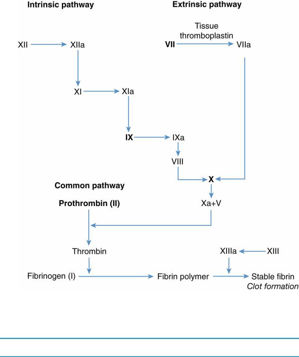
FIGURE 13.2 Coagulation cascade. Activated partial thromboplastin time measures the function of the intrinsic pathway and the extrinsic pathway, except for factor VII; the prothrombin time measures the function of the extrinsic pathway (factors VII, X, and V), fibrinogen, and prothrombin (factor II). Factors in bold type are vitamin K–dependent coagulation factors.
Table 13-6
Laboratory and Clinical Findings in Coagulation Disorders
Disorder |
aPTT |
PT |
Platelet Function Assay |
Platelet Count |
Petechiae |
Hemarthroses |
Factor VIII, IX deficiency |
Prolonged |
Normal |
Normal |
Normal |
No |
Yes |
von Willebrand |
Prolonged |
Normal |
Abnormal |
Normal |
No |
Rare |
Thrombocytopenia |
Normal |
Normal |
* |
Low |
Yes |
No |
|
||||||
Platelet function defect |
Normal |
Normal |
Abnormal |
Normal |
Yes |
No |
Vitamin K deficiency |
Prolonged |
Prolonged |
Normal |
Normal |
Yes |
Yes |
DIC |
Prolonged |
Prolonged |
Abnormal |
Low |
Yes |
Sometimes |
*Platelet function assays may be unreliable with thrombocytopenia and therefore are typically not indicated.
aPTT = activated partial thromboplastin time; PT = prothrombin time; DIC = disseminated intravascular coagulation.
512
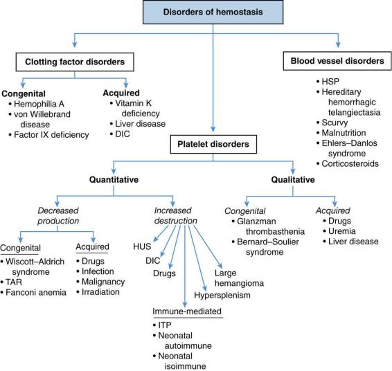
FIGURE 13.3 Overview of the differential diagnosis of disorders of hemostasis. DIC = disseminated intravascular coagulation; HUS = hemolytic uremic syndrome; HSP = Henoch–Schönlein purpura;
TAR = thrombocytopenia–absent radius syndrome; ITP = immune thrombocytopenic purpura.
513
V.Neutropenia
A.General concepts
1.Definition. Neutropenia is a low absolute number of neutrophils and is often expressed as the absolute neutrophil count (ANC; percentage of WBCs that are neutrophils, bands, and immature myeloid cells).
2.Risk of infection is directly related to the ANC.
a.Mild neutropenia is an ANC of 1000–1500 cells/mm3.
b.Moderate neutropenia is an ANC of 500–1000 cells/mm3. Infection generally involves the mucous membranes and skin (e.g., stomatitis, cellulitis, gingivitis).
c.Severe neutropenia is an ANC of <500 cells/mm3. Severe infections may result, such as pneumonia, sepsis, and meningitis. S. aureus and Gram-negative bacteria (e.g.,
Klebsiella, Serratia, Escherichia coli, and Pseudomonas) are typical organisms.
B.Neutropenia caused by decreased production
1.Infections are the most common cause of neutropenia during childhood. Viruses (e.g., HIV, EBV, CMV, hepatitis A and B, influenza A, parvovirus B19), bacteria (e.g., typhus, Rocky Mountain spotted fever), and protozoans (e.g., malaria) may all suppress the bone marrow, marginate neutrophils, or exhaust marrow reserves, resulting in neutropenia.
2.Chronic benign neutropenia (CBN) of childhood, a common cause of neutropenia in children younger than 4 years, refers to a group of acquired and inherited disorders with noncyclic neutropenia as the only abnormality. Its etiology is presumed to be autoimmune.
a.Clinical features. CBN has a variable course, with most children having an increased incidence of mild infections, such as otitis media, sinusitis, pharyngitis, and cellulitis. Severe infections may occur but are uncommon. Children are otherwise healthy, with normal appearance and growth.
b.Laboratory findings. Studies show a low ANC with a normal or slightly low WBC. Bone marrow demonstrates immature neutrophil precursors (development of mature neutrophils is arrested).
c.Prognosis. In most children, CBN resolves spontaneously within months to years.
3.Severe congenital agranulocytosis (Kostmann syndrome) is an autosomal recessive disorder with frequent and life-threatening pyogenic bacterial infections beginning in
infancy. ANC is usually <300 cells/mm3. These patients must be supported with granulocyte colony-stimulating factor or undergo bone marrow transplant for cure.
4.Cyclic neutropenia
a.Clinical features. Cyclic alterations in neutrophil counts result in regular episodes of neutropenia with resultant infections. Fever, oral ulcers, and stomatitis may occur during the neutropenia. Cycles last an average of 21 days in 70% of patients. Some cases are inherited in an autosomal dominant pattern.
b.Diagnosis is made by documenting the cyclic nature of the neutropenia by obtaining serial neutrophil counts during a 2- to 3-month period.
c.Treatment is with granulocyte colony–stimulating factor during periods of neutropenia, although some patients spontaneously remit during adolescence.
5.Genetic syndromes associated with neutropenia
a.Chédiak–Higashi syndrome is an autosomal recessive disorder characterized by oculocutaneous albinism, large blue-gray granules in the cytoplasm of neutrophils, neutropenia, and blond or brown hair with silver streaks. Patients are at high risk for serious infection.
b.Cartilage–hair hypoplasia syndrome is an autosomal recessive disorder
514
characterized by short stature, immunodeficiency, fine hair, and neutropenia.
6.Shwachman–Diamond syndrome is characterized by exocrine pancreatic insufficiency with malabsorption, short stature caused by metaphyseal chondrodysplasia, and neutropenia. Failure to thrive and recurrent infections (especially otitis media) are common.
7.Drugs (e.g., antibiotics, anticonvulsants, aspirin), environmental toxins, radiation, and chemotherapy may all cause neutropenia.
8.Metabolic diseases, such as hyperglycinemia, methylmalonic acidemia, and Gaucher disease, may result in neutropenia.
C.Neutropenia caused by increased destruction
1.Infections (especially viral)
2.Drugs
3.Hypersplenism
4.Autoimmune neutropenia describes a disorder in which antineutrophil antibodies are produced in response to infection (e.g., EBV), drugs, SLE, and juvenile rheumatoid arthritis (JRA), or for unknown reasons.
5.Isoimmune neutropenia describes the passive transfer of antineutrophil antibodies from the mother to her fetus after maternal sensitization by antigens on the fetal neutrophils. Infants are initially susceptible to infection, but neutropenia resolves by 8 weeks of life.
515