

- •Copyright
- •Contents
- •Dedication
- •Preface
- •Acknowledgments
- •Contributors
- •Contributors to the Previous Edition
- •Review Test
- •Answers and Explanations
- •Review Test
- •Answers and Explanations
- •Review Test
- •Answers and Explanations
- •Review Test
- •Answers and Explanations
- •Review Test
- •Answers and Explanations
- •Review Test
- •Answers and Explanations
- •Review Test
- •Answers and Explanations
- •Review Test
- •Answers and Explanations
- •Review Test
- •Answers and Explanations
- •Review Test
- •Answers and Explanations
- •IV. Hypertension
- •VI. Nephrotic Syndrome (NS)
- •VII. Hemolytic Uremic Syndrome (HUS)
- •VIII. Hereditary Renal Diseases
- •IX. Renal Tubular Acidosis (RTA)
- •XI. Chronic Kidney Disease (CKD) and End-Stage Renal Disease (ESRD)
- •XII. Structural and Urologic Abnormalities
- •XIII. Urolithiasis
- •XIV. Urinary Tract Infection (UTI)
- •Review Test
- •Answers and Explanations
- •Review Test
- •Answers and Explanations
- •Review Test
- •Answers and Explanations
- •Review Test
- •Answers and Explanations
- •IV. Food Allergy
- •VI. Urticaria (Hives)
- •VII. Drug Allergy
- •VIII. Asthma
- •IX. Immunology Overview
- •X. Disorders of Lymphocytes (Figure 15-2)
- •XI. Disorders of Granulocytes (Figure 15-3)
- •XII. Disorders of the Complement System
- •Review Test
- •Answers and Explanations
- •Review Test
- •Answers and Explanations
- •Review Test
- •Answers and Explanations
- •Review Test
- •Answers and Explanations
- •Review Test
- •Answers and Explanations
- •Review Test
- •Answers and Explanations
- •Comprehensive Examination
- •Index
Review Test
1.A 10-year-old girl presents for evaluation of fatigue, diminished appetite, and weakness. On physical examination, a periorbital violaceous heliotrope rash is evident. Which of the following statements is most accurate regarding the probable diagnosis?
A.Children with this diagnosis typically present with distal muscle weakness with an ascending pattern.
B.This patient has a 25% likelihood of developing a subsequent malignancy.
C.Steroids are contraindicated.
D.The clinical course may be complicated by calcium deposition in the muscle, fascia, and subcutaneous tissue.
E.This patient’s disease is more common in males than in females.
2.A 4-year-old boy presents to the emergency department for evaluation after 1 day of abdominal pain and a petechial rash on his bilateral thighs. He has no known history of prior bleeding or easy bruising but does have a 4-day history of upper respiratory tract infection (URI) symptoms and low-grade fevers. Physical examination is remarkable for a nontoxic, alert, and afebrile child. There is a petechial eruption on the lateral thighs, along with some edema of the hands and bilateral ankles. The abdomen is not distended, and bowel sounds are present. On examination, the abdomen is mildly tender to palpation in the periumbilical region without rebound tenderness. Laboratory studies reveal a white blood cell count of 14,000 cells/mm3, hemoglobin of 11.8 g/dL, and platelet count of 260,000 platelets/µL. Which of the following treatments would be the most appropriate at this time?
A.Intravenous immune globulin therapy for a presumed diagnosis of Kawasaki disease
B.Intravenous immune globulin therapy for a presumed diagnosis of immune thrombocytopenic purpura
C.Further evaluation by Child Protective Services for possible nonaccidental injury
D.Conservative management for a presumed diagnosis of Henoch–Schönlein purpura
E.Parenteral antibiotic therapy for a presumed diagnosis of meningococcemia pending blood culture results
3.A 3-year-old boy presents to the emergency department for evaluation of acute right upper quadrant abdominal pain. Further history reveals a 7-day history of fevers and sore throat. Physical examination reveals an irritable but consolable child with a temperature of 39.9°C (103.8°F). Other pertinent findings include bilateral nonpurulent conjunctivitis; red, cracked lips; swollen indurated fingers with erythematous palms; and an erythematous macular rash on the trunk. Which of the following is the most likely cause of the acute abdominal pain?
A.Henoch–Schönlein purpura
B.Hydrops of the gallbladder
C.Intussusception
D.Referred pain from arthritis involving the spine
E.Constipation from gastrointestinal smooth muscle dysfunction
4.A 3-year-old girl is brought to your office by her parents, who report that she has had
2 months of intermittent high-spiking fevers with temperature up to 39°C (103°F), which occur nightly and return quickly to normal. The parents report that their daughter’s activity and appetite are diminished. She is often reluctant to walk because of swelling of her ankles and knees. Six months ago her weight was at the 50th percentile, and now her weight is at the 25th percentile for age. On examination, you note a temperature of 39.2°C (102.6°F) and a salmoncolored maculopapular rash on the trunk. Diffuse lymphadenopathy is present, and the liver is palpable 4 cm below the right costal margin. Which of the following statements regarding this patient’s likely diagnosis is most accurate?
602
A.Measuring the erythrocyte sedimentation rate will confirm the diagnosis.
B.Admission to the hospital and immediate treatment with intravenous immune globulin are necessary.
C.Antinuclear antibodies are likely to be positive.
D.The rash on the trunk is nonpruritic and is likely evanescent.
E.The patient has a 10% chance of developing severe chronic arthritis.
5.A 10-year-old boy develops a headache, fever, and rash approximately 2 weeks after camping with his family. The rash is annular and “targetlike” with central clearing. Which of the following is most accurate regarding the most likely diagnosis?
A.This patient has a high likelihood of developing carditis.
B.An infected tick must be attached to the skin for at least 36–48 hours before there is a significant risk of developing this condition.
C.The prognosis for a child with this disease is poor, even with treatment.
D.Treatment should be immediately initiated with an intravenous first-generation cephalosporin.
E.Serologic testing for this condition is unreliable, and the diagnosis must be confirmed by culture of body fluids or tissue.
6.A 7-year-old boy with complaints of shortness of breath, nonpruritic rash, and very painful migratory arthritis presents to your clinic for evaluation. Two weeks ago, he had a sore throat and fever. On physical examination, a truncal macular rash is evident, and a grade 3/6 loud holosystolic murmur is audible at the apex and axilla. Which of the following statements is most accurate regarding this patient’s likely diagnosis?
A.Management may include corticosteroid therapy.
B.Antistreptolysin-O titers would be expected to be abnormally high in 25% of patients with this condition.
C.Laboratory evaluation is likely to demonstrate an elevated erythrocyte sedimentation rate and leukopenia.
D.Chorea is also likely to be found on examination of this patient.
E.Development of chronic and debilitating arthritis is likely.
7.A 12-year-old boy presents with severe arthritis of the hips and sacroiliac joints. Laboratory studies reveal that the patient is human leukocyte antigen B27 (HLA-B27) positive. Which of the following is the most likely diagnosis?
A.Early-onset oligoarticular juvenile idiopathic arthritis (JIA)
B.Juvenile ankylosing spondylitis
C.Rheumatoid factor–negative polyarticular JIA
D.Rheumatoid factor–positive polyarticular JIA
E.Systemic-onset JIA
8.A 3-year-old girl is referred to you for evaluation of fever. Her fever has lasted 6 days, and her parents have noticed eye redness, a truncal rash, and swollen lips. Your physical examination confirms these findings, along with an enlarged left cervical lymph node measuring 3 cm in diameter. On the basis of your findings, you suspect Kawasaki disease. Which of the following findings on physical examination or laboratory analysis would best correlate at this time with the finding of coronary artery aneurysms on echocardiogram?
A.Her current signs of fever, truncal rash, and swollen lips
B.Elevated erythrocyte sedimentation rate
C.Elevated platelet count of 840,000 platelets/µL
D.Cervical adenopathy
E.Laboratory evidence of aseptic meningitis
The response options for statements 9–14 are the same. You will be required to select one answer
603
for each statement in the following set.
A.Henoch–Schönlein purpura
B.Psoriatic arthritis
C.Systemic-onset juvenile idiopathic arthritis
D.Reactive arthritis
E.Inflammatory bowel disease-associated arthritis
F.Systemic lupus erythematosus
G.Kawasaki disease
H.Dermatomyositis
I.Oligoarticular juvenile idiopathic arthritis
J.Rheumatic fever
K.Lyme disease
For each patient, select the most likely diagnosis.
1.An human leukocyte antigen B27 (HLA-B27)–positive 14-year-old boy with abdominal pain and chronic diarrhea.
2.A 6-year-old boy with erythematous and hypertrophic papules over the metacarpal and proximal interphalangeal joints and “dark rings” around the eyes.
3.A sexually active 16-year-old boy with arthritis and conjunctivitis.
4.A 3-year-old boy with fever, bilateral knee swelling, and a “salmon-colored” rash on the trunk and proximal extremities.
5.A 14-year-old girl with arthritis, alopecia, leukopenia, and “a bad sunburn” on her face.
6.A 5-year-old girl with arthritis, abdominal pain, and a petechial eruption on her buttocks.
604
Answers and Explanations
1.The answer is D [V.C]. The constellation of clinical signs and symptoms that include fatigue, muscle weakness, and a heliotrope rash in a girl between 5 and 10 years of age is suggestive of dermatomyositis. Calcinosis, or calcium deposition, in muscle, fascia, and subcutaneous tissue occurs in up to 40% of children with this disorder. Dermatomyositis typically presents with proximal muscle weakness, characterized by a positive Gowers sign (difficulty standing from the sitting position and, as a result, having to “climb” up the thighs for support). Females are twice as likely as males to develop dermatomyositis. In childhood dermatomyositis, unlike in adult dermatomyositis, there is no association with malignancy. Steroids are the mainstay of therapy.
2.The answer is D [I.A, I.C.1–2, and I.E] Henoch–Schönlein purpura is an IgA-mediated vasculitis that involves the skin, joints, gastrointestinal tract, and kidneys. Despite the presence of petechiae, the platelet count is normal (i.e., a nonthrombocytopenic purpura). Steroids are indicated for patients who present with severe abdominal pain, poorly controlled joint pain, or nephritis. The diagnosis of Kawasaki disease is unlikely given the absence of fever for a minimum of 5 days and the absence of at least four of the five diagnostic criteria. The patient’s normal platelet count is inconsistent with the diagnosis of immune thrombocytopenic purpura. Although nonaccidental trauma must always be considered, there is no other bruising of concern on examination, and the clinical presentation is most consistent with the diagnosis of Henoch–Schönlein purpura. The diagnosis of meningococcemia is also unlikely because this patient is nontoxic, alert, and afebrile.
3.The answer is B [II.C and II.D.4]. This patient meets the diagnostic criteria for Kawasaki disease based on the duration of fever and the presence of four of the five diagnostic criteria, including conjunctivitis, oropharyngeal changes, a truncal rash, and swelling of the distal extremities. Approximately 10% of patients with Kawasaki disease may develop hydrops of the gallbladder, which presents with acute right upper quadrant pain. Sore throat, conjunctivitis, and red cracked lips are not associated with constipation, arthritis, or intussusception. Dermatomyositis may present with constipation from smooth muscle dysfunction and constitutional symptoms, but abdominal pain is not a feature of the disorder. Although edema of the hands and abdominal pain can be seen in Henoch–Schönlein purpura, fevers and mucous membrane findings are not usually present.
4.The answer is D [III.E.3]. This patient has features consistent with systemic-onset juvenile idiopathic arthritis (JIA). Intermittent high-spiking fevers, joint pain and swelling, lymphadenopathy, and hepatosplenomegaly are common features. The classic rash of systemic-onset JIA is described as salmon-colored and maculopapular, located on the trunk and proximal extremities. It is nonpruritic and evanescent (comes and goes) and tends to be more prominent during febrile episodes. Diagnosis is on the basis of characteristic clinical features and does not rely on specific laboratory tests. Although the erythrocyte sedimentation rate is often elevated, it is nonspecific because it is also elevated in many other inflammatory conditions, such as infectious disorders, some malignancies, and other rheumatologic conditions. Moderate to severe symptoms can be treated with anti-inflammatory (e.g., aspirin) or immunomodulatory (e.g., glucocorticoids) medications, but intravenous immune globulin is not recommended for use in JIA. Children with systemic-onset JIA have a 50% chance of having a severe, erosive arthritis. Antinuclear antibodies (ANA) in patients with systemiconset JIA are negative, in contrast to most patients with early-onset oligoarticular JIA and 50% of patients with polyarticular JIA in whom ANA is positive.
5.The answer is B [VII.C.2, VII.D.1–2, and VII.F.1]. The scenario of constitutional symptoms in the face of a classic rash suggestive of erythema migrans, shortly after a camping trip,
605
supports the diagnosis of Lyme disease. To transmit Lyme disease, an infected tick must be attached to the skin for at least 36-48 hours. Early in the disease, patients may present with fever, headache, myalgias, arthralgias, and lymphadenopathy, along with an annular, or “targetlike,” skin eruption with central clearing. Treatment of early disease includes oral doxycycline for children ≥8 years of age, oral cefuroxime or oral amoxicillin. Subsequent neurologic complications are not common but may include aseptic meningitis, facial cranial nerve palsy, and encephalitis. Cardiac complications are quite rare and may include heart block and myocarditis. The prognosis for a child with any stage of Lyme disease, if treated, is excellent. Arthritis, the hallmark of late disease, occurs months after the initial clinical presentation. Serologic testing (enzyme-linked immunosorbent assay and Western blot analysis) is recommended for confirmation of disease.
6.The answer is A [VI.A, VI.B.4, VI.D, VI.E, VI.G, and Table 16-6]. This patient’s clinical presentation should raise suspicion for acute rheumatic fever. The diagnosis of acute rheumatic fever requires evidence of previous group A β-hemolytic streptococcal infection, in addition to either two major Jones criteria or one major and one minor Jones criteria. The major Jones criteria include erythema marginatum, carditis, migratory polyarthritis, subcutaneous nodules, and Sydenham chorea. Unlike the other major criteria, the onset of chorea is usually several months after the other manifestations. Minor Jones criteria include fever, arthralgias, elevated erythrocyte sedimentation rate, and leukocytosis (not leukopenia). The great majority of patients (70–80%) with acute rheumatic fever have elevated antistreptolysin-O titers. Acute management includes eradication of streptococcal infection with penicillin and control of inflammation with nonsteroidal anti-inflammatory agents or corticosteroids. This patient’s shortness of breath may be caused by congestive heart failure or a pericardial effusion. There are usually no chronic joint sequelae from acute rheumatic fever.
7.The answer is B [III.D and Table 16-4]. Unlike all other types and subtypes of juvenile idiopathic arthritis (JIA), juvenile ankylosing spondyloarthritis is male predominant and almost always presents in children older than 8 years. Typically, patients with ankylosing spondylitis are human leukocyte antigen B27 (HLA-B27) positive and have involvement of the hips and sacroiliac joints. In contrast, patients with early-onset oligoarticular JIA are usually female, present between 1 and 5 years of age, have a high risk of developing chronic uveitis, and do not have involvement of the sacroiliac joints. Patients with polyarticular JIA (whether rheumatoid factor–positive or –negative) are also usually female and have involvement of multiple large and small joints but not typically the sacroiliac joints. Patients with systemiconset JIA may be either male or female. Other presenting features of systemic-onset JIA include fever, a characteristic transient skin rash (salmon-colored), hepatosplenomegaly, and lymphadenopathy.
8.The answer is C [II.C–F]. Coronary artery aneurysms in Kawasaki disease are more likely to occur during the subacute phase of the disease, which begins 1–2 weeks after the onset of fever. The subacute phase is also characterized by decreasing erythrocyte sedimentation rate (ESR) and by marked thrombocytosis. High-spiking fevers, rash, swollen lips, cervical adenopathy, edema of the distal extremities, and elevations of the ESR and C-reactive protein are all characteristic of the acute phase of Kawasaki disease. Aseptic meningitis is a welldescribed complication of Kawasaki disease, but there is no known increased incidence of aneurysm formation in patients with aseptic meningitis.
9.The answers are E, H, D, C, F, and A, respectively [VIII.A.4, V.C.2, VIII.A.1, III.E.3, IV.C–E, Table 16-5, and I.C]. Abdominal pain and chronic diarrhea may be caused by inflammatory bowel disease. Arthritis may be associated with either ulcerative colitis or Crohn disease.
Some patients with inflammatory bowel disease are also human leukocyte antigen B27 (HLAB27) positive and have involvement of the axial skeleton, which is clinically indistinguishable from ankylosing spondylitis. The diagnosis of dermatomyositis should be suspected in a child
606
with proximal muscle weakness, a violaceous heliotrope rash around the eyes, and erythematous, hypertrophic papules over the knuckles (Gottron papules). Reactive arthritis is triggered by an enteric or sexually transmitted pathogen (classically, Chlamydia trachomatis). Reactive arthritis can present with arthritis, conjunctivitis, and urethritis. The diagnosis of systemic-onset juvenile idiopathic arthritis should be considered in a child presenting with fever of unknown origin, an evanescent salmon-colored rash, arthritis, organomegaly, and polyserositis. Systemic lupus erythematosus may be diagnosed when four of eleven diagnostic criteria are fulfilled, including photosensitivity and a malar rash. Adolescent females might also present with alopecia or Raynaud phenomenon when their disease is active. Henoch– Schönlein purpura is a systemic IgA-mediated vasculitis that involves the skin, joints, gastrointestinal tract, and kidneys. It commonly presents with a nonthrombocytopenic purpuric or petechial eruption on the buttocks or thighs with abdominal pain, arthritis, and glomerulonephritis.
607

C H A P T E R 1 7
608

Orthopedics
Ryan J. Coller, Robert M. Bernstein
609
I.Upper Extremity
A.Brachial plexus (C5–T1) injury most commonly occurs as a result of birth trauma from excessive traction on the neonate’s head, neck, or arm, which stretches the nerves of the brachial plexus.
1.Erb palsy is an upper brachial plexus injury involving the C5 and C6 nerve roots. It is the most common brachial plexus injury and most often involves the right arm as the result of shoulder dystocia, macrosomia, or forceps delivery. Clinical features include a flaccid arm and an asymmetric Moro reflex. (See Chapter 2, Table 2-2 for a description of the Moro reflex.) Eventually, the arm is held in internal rotation with the elbow extended, forearm pronated, and the wrist held in flexion. This positioning is often described as the “waiter’s tip.”
2.Klumpke palsy is less common and is the result of a lower brachial plexus injury caused by upward traction on the arm. It involves the C7 and C8 nerve roots. Clinical features include a claw hand because of loss of intrinsic function and a decreased ability to extend the elbow and flex the wrist. Horner syndrome (ipsilateral ptosis, miosis, and anhydrosis) may be present if the sympathetic fibers of the first thoracic nerve have also been damaged.
3.Diagnosis is based on the history and physical examination, but typically also includes a plain radiograph of the shoulder to evaluate for an associated clavicular fracture. Electromyography (EMG) and nerve conduction studies may be considered if no recovery has occurred within 6 months.
4.Management. Treatment includes observation and range-of-motion physical therapy to prevent contractures. Improvement is often noted within 48 hours. Approximately 80% of patients fully recover. Surgery may be required if there is no improvement within 3– 9 months.
B.Nursemaid’s elbow is a subluxation of the annular ligament over the radial head. The usual mechanism of injury is an upward force on the arm, such as pulling a toddler upward by the hand or wrist to help the child stand or clear an obstacle. The radial head in children younger than 6 years has a cylindrical shape, allowing it to slip out of the annular ligament, which normally keeps it in place.
1.Clinical features
a.Sudden onset of pain, which is difficult to localize
b.The elbow is held flexed and no swelling is present. The child is unwilling to use the affected arm, but hand function is normal.
2.Diagnosis is usually based on the clinical presentation. No radiograph is needed if the episode was witnessed. Unwitnessed injury should not be assumed to be nursemaid’s elbow, and radiographs should be obtained.
3.Management. Treatment of the subluxation is to reduce it by simultaneously flexing the elbow and supinating the hand. Spontaneous movement of the arm confirms successful reduction. Postreduction radiographs are not needed.
4.Prognosis is excellent. Usually, the child will start to use the arm within 15 minutes of the reduction, but use of the arm may occasionally be delayed for up to 24 hours. Subluxation may recur.
C.Anterior shoulder dislocation is the most common type of shoulder dislocation. It occurs with excessive external rotation, abduction, and extension of the shoulder, as it may occur in gymnastics or wrestling.
1.Diagnosis is on the basis of physical examination (the arm being held in external rotation and an inability to touch the contralateral shoulder) and radiographs (especially an
610
axillary view) of the glenohumeral joint to visualize the dislocation and exclude fractures.
2.Management. Treatment is immobilization after closed reduction. Recurrence of dislocation approaches 90% in the adolescent population.
611
II.Spine
A.Disorders of the cervical spine
1.Torticollis is defined as the tilting of the head to one side. It may be either congenital or acquired.
a.Congenital muscular torticollis is very common and is usually the result of uterine constraint or birth trauma, either of which causes contracture of the ipsilateral sternocleidomastoid muscle. Rare causes of congenital torticollis include cervical spine deformities, for example Klippel–Feil syndrome [see section II.A.3].
1.Clinical features
a.The head is tilted toward the affected side and the chin is rotated away from the affected side. This is typically present by 1 month of age.
b.Limited range of motion with passive lateral neck flexion or chin rotation may be present.
c.Soft tissue mass or tightness may be palpable within the sternocleidomastoid muscle, possibly representing a hematoma or fibrosis. This usually resolves by 2–6 months of age.
d.Asymmetric head shape (plagiocephaly, see Chapter 1, section II.B.2.e) may develop in untreated torticollis.
2.Diagnosis is on the basis of physical examination. Radiographs of the cervical spine may identify abnormalities of the cervical spine and should be obtained in anyone presenting late or failing to improve with physical therapy.
Strabismus, nystagmus, and abnormal back or neurologic examination should prompt evaluation for conditions other than congenital muscular torticollis.
3.Management. Treatment includes stretching exercises with physical therapy to relieve the muscle contracture.
4.Complications include skull deformity and facial asymmetry (plagiocephaly) if torticollis is not treated promptly.
5.Prognosis after 6 months of diligent muscle stretching is complete resolution in 90%, with higher success rates when therapy is started earlier in life.
b.Acquired torticollis arises later in childhood and is much less common than congenital torticollis. Lack of plagiocephaly can help distinguish acquired from congenital torticollis. Etiologies are typically from ophthalmologic, central nervous system, musculoskeletal, infectious, or traumatic causes, and they may include strabismus and refractive errors, posterior fossa tumors, syringomyelia, atlantoaxial subluxation [see section II.A.2], cervical adenitis, retropharyngeal abscess, cervical discitis, osteomyelitis, trauma, gastroesophageal reflux disease (Sandifer syndrome), and dystonic drug reactions.
2.Atlantoaxial instability is caused by an unstable joint and is characterized by excessive mobility and potential for dislocation. It occurs most commonly between the first and second cervical vertebrae and less commonly between the occiput and the first cervical vertebrae. Down syndrome (see Chapter 5, section IV.B.1), skeletal dysplasias (Chapter 5, section IV.H.), and Klippel–Feil syndrome [see section II.A.3] are associated with atlantoaxial instability.
a.Clinical features. Physical examination is usually normal, and patients are often asymptomatic. With the abovementioned syndromes, a high index of suspicion is important because spinal cord injury may occur with dislocation or significant instability. Early neurologic signs include neck pain, loss of motor skills, or loss of bowel/bladder control. Significant instability requires surgical stabilization.
612
b.Diagnosis is made by observing excessive movement at the C1 and C2 junction on lateral cervical spine radiographs in the neutral positions followed by flexion– extension radiographs.
c.Management includes fusion of C1 and C2 if instability is severe. No contact sports, tumbling, or trampolines are allowed if there is any sign of instability.
d.Complications include paralysis or even death if the instability is not detected before injury.
3.Klippel–Feil syndrome is defined as congenital fusion of the cervical vertebrae. The triad is low-set hairline, short neck, and limited neck motion. Associated abnormalities may include congenital torticollis, genitourinary anomalies, congenital heart disease, hearing loss, and congenital failure of scapula to descend to its proper location.
B.Scoliosis is a fixed lateral curvature of the spine of greater than or equal to 10°. This disorder occurs equally in males and females, but adolescent females require treatment eight times more frequently.
1.Etiology
a.Most cases of scoliosis (80%) are idiopathic. A positive family history is common.
b.Improper spine development, including failure of vertebral formation or abnormal fusion, leads to congenital scoliosis.
c.Other types of scoliosis are neuromuscular (associated with cerebral palsy) or syndromic (associated with skeletal dysplasias and connective tissue disorders). Leg length discrepancy can mimic scoliosis.
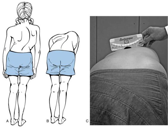
2.Clinical features. Scoliosis is typically asymptomatic. Asymmetry of the shoulder height, scapular position, and the waistline may be present. As the patient bends forward, a hump, representing rib or transverse process rotation of the curved spine, may be seen (forward bending test; Figure 17-1). Curves are right thoracic 90% of the time. Pain or left-sided curves are signs of underlying disorders, and should prompt investigation for nonidiopathic causes.
3.Diagnosis is on the basis of clinical findings and radiologic confirmation. Standing posterior–anterior (PA) films of the spine confirm the curvature and are used to calculate the Cobb angle (Figure 17-2). The Cobb angle, which measures the degree of scoliosis, is measured by drawing a line along the superior aspect of the most angulated vertebrae at the top of the curvature, and drawing another line along the inferior aspect of the lowest most angulated vertebrae of the curvature. The angle at the intersection of these lines is the Cobb angle. A lateral radiograph is helpful to rule out kyphosis, spondylolisthesis, and congenital anomalies of the spine.
4.Management. Treatment options include observation, bracing, and surgery, and management decisions are based on the magnitude of the curve and the remaining growth potential. Bracing prevents progression of the curvature; therefore, bracing is not likely helpful after skeletal growth is completed. Progression of scoliosis generally occurs only during growth. It is important to remember that almost all growth in females ceases within 6 months of menarche. Two years postmenarche is considered skeletally mature. Surgery is required for very severe curves or to stop curves that progress in spite of bracing. If a thoracic curve reaches 50°, there is always progression, even after skeletal maturity. Table 17-1 summarizes management approaches based on skeletal maturity and the degree of curvature.
5.Complications. Severe thoracic curves (>70°) can be associated with restrictive or obstructive lung disease.
C.Kyphosis (roundback) is defined as anterior–posterior (AP) curvature of the thoracic spine.
Fixed kyphosis beyond 50° is considered pathologic.
1.Flexibility in the rounded area should be assessed by having the patient extend his or her
613
back while prone. Most adolescents with kyphosis have flexible kyphosis, in which they can voluntarily correct the rounded area. This is called “postural roundback,” and it is managed with physical therapy and postural exercises.
2.Scheuermann kyphosis is a rigid kyphosis greater than 50°, in which at least three consecutive vertebrae are wedged. End plate changes and disk space narrowing are noted on radiographs. It develops in otherwise healthy adolescents and is managed with referral to physical therapy and orthopedics.
D.Back pain. Any complaint of back pain that interferes with play should be further evaluated. The differential diagnosis is extensive and includes congenital or traumatic musculoskeletal problems such as fractures; infections such as osteomyelitis or pyelonephritis; neoplasms; and rheumatologic conditions. See Table 17-2. Some common causes include the following:
1.Back strain is muscular soreness from overuse or poor body mechanics. Back strain is the most common cause of back pain in children.
a.Clinical features. Diffuse muscular pain is present without neurologic deficits. Physical examination is normal.
b.Management. Treatment includes rest, stretching, and analgesics.
2.Spondylolysis is a stress fracture in the pars interarticularis (i.e., the bone that connects the superior and inferior articular facets of a vertebral body) usually secondary to repetitive hyperextension of the spine as occurs in gymnastics, tennis, and diving. It typically involves the lumbar region, particularly L5. Pain is localized and increases with hyperextension.
a.Diagnosis is on the basis of AP, lateral, and oblique views of the lumbar spine. However, because spondylolysis is a stress fracture, plain films may not detect the fracture. Magnetic resonance imaging (MRI), bone scan, or a single photon-emission computed tomographic (SPECT) scan may be used for diagnosis if the fracture is acute.
b.Management. Treatment includes rest and analgesics for pain. Bracing for immobilization may be necessary if pain persists. Surgery is rarely required.
c.Complications include spondylolisthesis, which is discussed below.
3.Spondylolisthesis occurs when the vertebral body involved in the spondylolysis slips anteriorly (i.e., subluxation of the vertebra). The subluxated vertebra can impinge on nerve roots. Diagnosis and treatment is the same as for spondylolysis. Indications for surgery include nerve impingement, persistent pain, or progression of the subluxation beyond 50% of the vertebral width.
4.Discitis is defined as infection (Staphylococcus aureus is the most commonly identified organism) or inflammation (idiopathic, traumatic, or rheumatologic) of the intervertebral disk.
a.Clinical features. Discitis typically begins with signs and symptoms of an upper respiratory illness or minor trauma, followed by back pain with tenderness over the involved disk. Fever is sometimes present. Children refuse to bend forward, and young children may refuse to walk.
b.Diagnosis. Erythrocyte sedimentation rate (ESR) is typically elevated. MRI or bone scan confirms the diagnosis.
c.Management. Antistaphylococcal antibiotics are given to patients who are suspected to have infection. Treatment also includes bed rest and analgesics.
5.Herniated intervertebral disk is much more common in adults, but may occur in adolescents. The lumbar region is most commonly affected. Unlike in adults, herniation in adolescents is caused by repetitive activity and rarely by trauma. Initial treatment includes nonsteroidal anti-inflammatory drugs (NSAIDs) and rest. Epidural steroids may be considered. Surgery to remove the disk is necessary only if symptoms persist or if the
614
neurologic examination is abnormal.
FIGURE 17.1 Scoliosis. A. Curve is visible when standing. B. Rib hump is visible when the child performs forward bending test. C. Scoliometer is used to quantify the degree of curvature on the forward bend test.
(C) Reprinted with permission from Weinstein SL, Flynn JM. Lovell and Winter’s Pediatric Orthopaedics. 7th Ed. Philadelphia: Lippincott Williams & Wilkins, 2013.
615
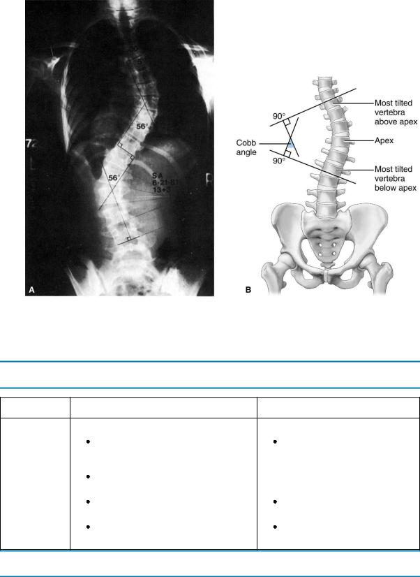
FIGURE 17.2 Cobb angle measurement noted on radiograph (A) and figure (B).
Reprinted with permission from Tecklin JS. Pediatric Physical Therapy. 5th Ed. Philadelphia: Lippincott Williams & Wilkins, 2014.
Table 17-1
Management of Adolescent Idiopathic Scoliosis
Scoliosis
Skeletally Immature Skeletally Mature
Curvature
Before or During Growth |
Growth Completed |
10°–25° |
No treatment required |
Follow-up scoliosis films 6 months after |
|
diagnosis |
|
|
|
≥7° progression—refer to orthopedic surgeon |
|
|
|
25°–40° |
No treatment required |
Bracing indicated |
|
|
|
>40° |
Surgery may be indicated if |
Surgery may be indicated |
|
|
scoliosis > 50° |
Table 17-2
Common Causes of Back Pain in Children and Distinguishing Features
|
Back Strain |
Spondylolysis |
Spondylolisthesis |
Discitis |
Key features |
Most common cause |
Repetitive hyperextension |
Complication of |
Staphyloccocus |
|
of back pain |
of the spine |
spondylolysis |
aureus is most |
|
|
|
|
common |
Etiology |
Overuse or poor |
Stress fracture in pars |
Anterior subluxation of |
Infection or |
|
body mechanics |
interarticularis |
the vertebral body after |
inflammation of |
|
|
|
|
|
616
|
|
|
spondylolysis |
intervertebral disk |
|
Diagnosis |
History and |
Localized painWorse with |
Same as spondylolysis |
Tenderness over |
|
|
physicalNormal |
hyperextensionAP, lateral, |
|
involved disc |
|
|
examination with |
oblique radiographs of |
|
|
|
|
possible tenderness |
lumbar spine confirm |
|
|
|
|
of paraspinal muscles |
diagnosis |
|
|
|
|
|
|
|
|
Refusal |
|
|
|
|
|
to bend |
|
|
|
|
|
forward |
|
|
|
|
|
Elevated |
|
|
|
|
|
ESRBone |
|
|
|
|
|
scan/MRI |
|
|
|
|
|
confirm |
|
|
|
|
|
diagnosis |
Management |
Rest, stretching, and |
Rest, analgesicsBracing to |
Same as |
Antistaphylococcal |
|
|
analgesics |
immobilize if pain is |
spondylolysisSurgery if |
antibiotics |
|
|
|
severe |
signs of nerve |
|
|
|
|
|
impingement or |
|
|
|
|
|
subluxation >50% of |
|
|
|
|
|
vertebral width |
|
|
|
|
|
|
Rest, analgesics |
|
AP = anteroposterior; ESR = erythrocyte sedimentation rate; MRI = magnetic resonance imaging.
617
III.Hip
A.Developmental dysplasia of the hip (DDH) occurs when the acetabulum is shallow or poorly formed, leading to the subluxation or dislocation of the head of the femur. The spectrum of disease severity ranges from hip joint laxity to frankly dislocated hips. DDH was formerly termed congenital hip dysplasia; however, this name was abandoned to reflect the spectrum of findings possible, including a normal examination at birth with progression to dislocation later in life.
1.Epidemiology
a.DDH is more common in girls than in boys.
b.Risk factors include female sex, firstborn, breech presentation, and family history of DDH.
2.Clinical features. DDH is asymptomatic in infants. Physical examination may show the following findings:
a.Positive Barlow maneuver. (The Barlow maneuver attempts to dislocate a dislocatable hip.) With the hips at 90° flexion, the thumb is placed on the medial side of the thigh and the middle finger on the greater trochanter, and gentle pressure is applied posteriorly and laterally to dislocate the hip. The hip is then gently abducted, and a “clunk” is felt as the hip relocates.
b.Positive Ortolani maneuver. (The Ortolani maneuver attempts to reduce a dislocated hip.) In the same position described above, the hip is abducted with pressure gently applied upward with the middle finger to slide the head of the femur back into the acetabulum. In a positive Ortolani maneuver, the hip can be felt slipping into the acetabulum as it is being reduced. An Ortolani-positive hip is one in which the hip is dislocated but can be reduced.
c.Galeazzi sign, which assesses asymmetry of the apparent femoral length. The examination is performed by placing the hips in flexion at 90° with the feet flat on the table, heels apposed to the buttocks, while the infant is supine. If the hip is dislocated, the affected femur, and therefore knee, appears shorter compared with the normal limb. If both hips are dislocated, Galeazzi sign will not be positive as the knees will be at the same height.
d.Asymmetric abduction of the hips and asymmetric thigh or buttock folds. Ortolani and Barlow maneuvers are not useful after about 4 months of age because the hip stabilizes in either the dislocated or reduced position. After this age, decreased ability to abduct the affected hip, asymmetric thigh or buttock folds, or Galeazzi sign is more useful in the diagnosis of DDH.
3.Diagnosis. Physical examination alone may be the basis of diagnosis. However, imaging is helpful if the examination is equivocal or the patient is at high risk, such as an infant who was breech in utero or children with positive family histories.
a.Ultrasound is used to assess for DDH in young infants because the femoral head does not ossify until 4–6 months of age.
b.AP radiographs of the pelvis may be used to assess for DDH if the infant is older than 6 months.
4.Management. Treatment depends on the age of the patient at the time of diagnosis. The earlier the diagnosis, the less likely surgical intervention will be necessary.
a.The Pavlik harness holds the head of the femur against the acetabulum to stimulate formation of the normal cup shape of the acetabulum. It is typically used for 2–
3 months if the diagnosis is made by 6 weeks of age. It is successful in 90–95% of cases.
618
b.Surgery may be required if the diagnosis is made beyond 6 weeks of age, if the hips are bilaterally dislocated, if the hips are not reducible on physical examination, or if the Pavlik harness fails to stabilize the hip. Surgery may involve closed or open reduction of the hip. After surgery, the infant is in a cast for 2–3 months.
5.Complications
a.Avascular necrosis of the femoral head
b.Limb length discrepancy, limp, and osteoarthritis if DDH is not treated
B.Limp is a common symptom during childhood. The differential diagnosis is extensive and is listed in Table 17-3. The more common causes include the following:
1.Septic arthritis is a bacterial infection of the joint, which can be caused by hematogenous spread (most common), contiguous spread, or direct inoculation. Septic arthritis of the hip is an orthopedic emergency.
a.Epidemiology
1.The peak age is 1–3 years.
2.The hip is most commonly affected in younger children, whereas the knee is more commonly affected in older children.
b.S. aureus, Streptococcus pneumoniae, and Streptococcus pyogenes are the most common organisms. Relevant Gram-negative organisms include Neisseria gonorrhoeae in neonates and adolescents and Kingella kingae in children younger than 3 years.
c.Clinical features
1.Fever and irritability are common.
2.Limp, refusal to walk, and pain with movement of the joint may occur. Attempts to move the joint result in significant pain. If the hip is affected, it is usually held in flexion, abduction, and external rotation to relieve the pressure within the joint capsule.
3.Erythema, swelling, and asymmetry of soft tissue folds may be present.
d.Diagnosis
1.Laboratory studies include an elevated white blood cell (WBC) count, ESR, and C-reactive protein (CRP).
a.Blood culture is positive in 30–50% of cases.
b.Analysis of the synovial fluid for cell count, Gram stain, and culture is important. Synovial fluid with a WBC count > 50,000–100,000 cells/mm3 with neutrophilic predominance is suggestive of septic arthritis. Culture of synovial fluid is positive in only 60% of cases. Unless the child is toxicappearing, cultures should be obtained before administering antibiotics.
2.Imaging complements physical examination and laboratory findings.
a.Ultrasound is the best imaging screen and demonstrates fluid in the joint capsule.
b.Plain radiographs may reveal a widened joint space and may exclude other diagnoses in the differential (e.g., fracture) but are often normal.
e.Management
1.Surgical decompression is necessary to avoid avascular necrosis and to obtain synovial fluid for analysis.
2.Empiric intravenous antibiotics to cover Gram-positive organisms, including consideration for methicillin-resistant S. aureus, should be started after blood and joint cultures are obtained. Duration of antibiotic treatment is 4–6 weeks.
f.Complications. The femoral head is vulnerable to ischemic injury. Ischemia may lead to avascular necrosis and cartilaginous damage.
2.Transient synovitis (also known as toxic synovitis) is a common self-limited, irritation of
619
the hip joint. It may be a postinfectious inflammatory reaction. A viral prodrome (upper respiratory infection or diarrheal illness) often precedes transient synovitis. Transient synovitis is a diagnosis of exclusion, and it is extremely important to rule out septic arthritis, especially if a patient is ill-appearing, given the potentially disabling consequences of missed septic arthritis (Table 17-4).
a.Epidemiology
1.Transient synovitis is the most common cause of a painful limp in toddlers.
2.Its peak age of presentation is 2–7 years.
3.It is more common in males.
b.Clinical features
1.Low-grade fever, limp, and mild irritability may be present. Patients usually appear relatively well.
2.Hip pain may be acute or insidious in onset. Although patients may be more willing to move their legs than patients with septic arthritis, they may also hold their hip in a flexed, abducted, and externally rotated position, similar to the position seen in septic arthritis.
c.Diagnosis is often on the basis of the history and physical examination.
1.The WBC count and ESR are normal or only slightly elevated.
2.There may be an effusion in the hip, and if fluid is present with clinical concern for the possibility of septic arthritis, it should be aspirated and analyzed to rule out septic arthritis. Synovial fluid WBC counts may be elevated above the normal reference range, but are less than that seen in septic arthritis (see Table 17-4).
d.Management. Treatment includes NSAIDs, rest, and observation.
e.Prognosis is excellent. Pain usually improves within 3 days with analgesics and rest. Complete resolution of symptoms usually occurs by 3 weeks. Recurrence is unusual unless vigorous activities are resumed too soon.
3.Legg–Calvé–Perthes (Perthes) disease is idiopathic avascular necrosis of the femoral head.
a.Epidemiology
1.The disease is more common in Caucasians and Asians.
2.The disease is more common in boys and has a male-to-female ratio of 4:1.
3.Patients are typically active, thin boys who are small for their age.
4.The disease most commonly affects children between 4 and 8 years of age.
b.Clinical features. Children have a slightly painful limp with decreased internal rotation and abduction of the hip. This can be assessed on examination by logrolling the leg internally. Pain may be referred to the knee and to the groin.
c.Diagnosis is on the basis of AP and frog-leg lateral radiographs of the pelvis, which show increased density in the affected femoral head, or a crescentic subchondral fracture in the femoral head, termed the “crescent sign.”
d.Management. Treatment involves containment or ensuring that the femoral head is positioned within the acetabulum to facilitate its remolding and reossifying.
1.Physical therapy and restriction of vigorous exercise are effective. If pain persists, analgesia, traction, casting, or crutches may be used.
2.Surgery is indicated if there is more than 50% involvement of the femoral head, or if there is subluxation of the femoral head out of the acetabulum.
e.Prognosis is best in younger patients. If children develop the disease when they are younger than 9 years, complete resolution within 3 years is common. The majority of patients who develop the disease at age 9 years or older go on to develop osteoarthritis in the affected hip as adults.
620
4.Slipped capital femoral epiphysis (SCFE) is slipping of the femoral head off the femoral neck.
a.Epidemiology
1.Age of onset is usually during peak linear growth in adolescence.
2.The male-to-female ratio is 2–3:1.
3.The typical patient is an obese adolescent male.
b.Clinical features. Patients have a painful limp with pain in the groin, hip, or knee. The degree of pain varies from severe (acute slip) to mild (chronic slip).
Internal rotation, flexion, and abduction are usually reduced in the affected hip.
SCFE is bilateral in 30% of cases, and patients with hypothyroidism are especially more likely to develop bilateral disease. Diagnosis of SCFE outside the typical age range, or in patients who are thin or have short stature, should prompt consideration of endocrine, renal, or genetic disorders.
c.Diagnosis. AP and frog-leg lateral radiographs of the pelvis confirm the slipped epiphysis. A line drawn flanking the superior edge of the femoral neck (Klein line) crosses 10–20% of the epiphysis in a normal hip on AP pelvis films. However, in SCFE, the Klein line may not cross the epiphysis at all.
d.Management. Treatment involves pinning the epiphysis to prevent further slippage. The femoral head is not placed back into a normal position with the femoral neck because the force required may cause avascular necrosis.
e.Complications
1.Avascular necrosis and collapse or deformity of the femoral head
2.Chondrolysis (degeneration of articular cartilage of the hip)
3.Limb length discrepancy, which may cause limp and pain
4.Osteoarthritis
5.Osteomyelitis is an infection of the bone.
a.Epidemiology
1.The peak ages of onset are less than 1 year and between 9 and 11 years.
2.The male-to-female ratio is 2:1.
b.Etiology
1.S. aureus and S. pyogenes are the most common organisms. Salmonella should be considered in patients with sickle cell anemia. Pseudomonas aeruginosa infection of a bone in the foot can occur if a child has a puncture wound passing through his or her sneaker into the foot.
2.Although contiguous spread and direct inoculation can cause osteomyelitis, children most commonly acquire infection by hematogenous seeding.
3.The causative organism is not isolated in up to 30–50% of cases.
c.Clinical features
1.Fever and irritability
2.Bone pain, erythema, swelling, and induration, which may develop in the area over the osteomyelitis
3.Painful limp
d.Diagnosis
1.Laboratory studies include an elevated WBC count, ESR, and CRP.
a.Blood culture is positive in 50% of cases.
b.Blood culture in combination with a culture of the aspirated bone may identify the organism in 70% of cases. Unless the child is toxic-appearing, cultures should be obtained before administering antibiotics.
2.Imaging. Although a plain radiograph may not show bone changes early, soft tissue swelling (displacement of fat planes) can often be detected. Bone scan
621

or MRI can detect osteomyelitis a few days after the onset of symptoms. After 10–14 days of infection, a plain radiograph begins to reveal elevation of the periosteum, suggesting osteomyelitis.
e.Management
1.Antibiotics should be given for 4–6 weeks, although the length of treatment varies depending on the organism and patient response. Unless the patient is septic, antibiotics should be held until blood cultures are obtained and the bone has been aspirated for culture.
2.Improving symptoms and decreasing ESR/CRP generally indicate response to intravenous antibiotics, at which time oral antibiotics can be used to complete the antibiotic course.
3.Surgery is necessary to drain and debride a subperiosteal abscess if fever and swelling persist despite 48 hours of intravenous antibiotics.
f.Complications
1.Spread of infection may occur, either by contiguous spread infecting a joint or distant seeding of other organs.
2.Chronic osteomyelitis results from a nidus of residual infection, such as a sequestrum (i.e., focus of necrotic bone) or an involucrum (i.e., formation of new bone or fibrosis surrounding the necrotic, infected bone). Symptoms of chronic osteomyelitis are usually insidious, so the diagnosis is often delayed and complications are common. Both surgery and antibiotics are needed.
3.Pathologic fracture at the site of the infection may occur as a result of healing and remodeling of bone, bone necrosis, or a sequestrum. Activity should be restricted during antibiotic therapy to prevent a fracture, especially if the diagnosis of osteomyelitis was initially delayed.
4.Angular deformity or limb length discrepancy may occur if the growth plate is involved in the infection.
Table 17-3
Differential Diagnosis of a Painful Limp
Mnemonic: “The Joint STARTSSSS HOTT” |
Other Causes of Limp |
|
S Septic arthritis |
D Developmental hip dysplasia |
|
T Transient synovitis |
D Discitis |
|
A Acute rheumatic fever |
L Legg–Calvé–Perthes |
|
R Rheumatoid arthritis |
L Limb length discrepancy |
|
T Trauma |
L Lyme disease |
|
Fracture |
|
|
Strain or sprain |
|
|
|
|
|
S Sickle cell disease |
|
|
Pain crisis (vaso-occlusive crisis) |
|
|
Osteomyelitis |
|
|
|
|
|
S Systemic lupus erythematosus |
|
|
S Steroid use (chronic) |
|
|
S Slipped capital femoral epiphysis (SCFE) |
|
|
H Henoch–Schönlein purpura |
|
|
O Osteomyelitis |
|
|
T Tuberculosis |
|
|
T Tumor |
|
|
Osteosarcoma |
|
|
Leukemia |
|
|
|
|
|
|
|
|
622

Table 17-4
Septic Arthritis Versus Transient Synovitis
|
Septic Arthritis |
Transient Synovitis |
Organisms |
Staphylococcus aureus |
Typically sterile inflammationPossibly |
|
|
postviral |
|
Streptococcus pyogenes |
|
|
Streptococcus pneumoniae |
|
Clinical features |
Peak 1–3 years |
Peak 2–7 years |
Acute |
Acute or subacute |
|
Ill-appearing |
Well-appearing |
|
Fever |
Mild viral prodrome |
|
Limp |
Limp |
|
Refusal to bear weight |
Refusal to bear weight |
|
Flexion, abduction, external rotation of |
Flexion, abduction, external rotation of |
|
the hip |
the hip |
|
Typical joints involved |
Hip, knee, ankle |
Hip |
Laboratory findings |
Elevated ESR (>40 mm/hour) |
Mildly elevated ESR |
Elevated CRP (>2 mg/dL) |
Mildly elevated CRP |
|
Elevated WBC |
Mildly elevated WBC |
|
Blood culture positive 30–50% |
Blood culture negative |
|
All laboratory results may be normal |
|
|
Joint aspirate |
>50,000–100,000 cells/mm3 |
<50,000 cell/mm3 |
Gram stain may have organisms |
Gram stain without organisms |
|
Treatment |
Surgical decompression |
NSAIDs |
Intravenous antibiotics |
Rest |
|
ESR = erythrocyte sedimentation rate; CRP = C-reactive protein; WBC = white blood cell; NSAIDs = nonsteroidal anti-inflammatory drugs.
623
IV. Lower Extremity
A.Torsional abnormalities
1.In-toeing (“pigeon-toe”), a common parental concern, is typically normal and corrects with growth (see Table 17-5). The following are the most common causes of in-toeing:
a.Metatarsus adductus is medial curvature of the mid-foot (metatarsals).
1.Epidemiology and etiology
a.Metatarsus adductus occurs in children younger than 1 year.
b.It is usually caused by intrauterine “packaging” or “molding.”
2.Clinical features include a C-shaped or kidney bean-shaped foot that can be straightened to varying degrees by gentle manipulation. Dorsiflexion is normal, but if the ankle cannot dorsiflex, clubfoot should be considered.
3.Diagnosis is by physical examination. Radiographs are not needed.
4.Management
a.Flexible foot that can overcorrect with passive motion needs observation only.
b.Flexible foot that can correct with passive motion, but not overcorrect, may benefit from gentle stretching of the foot.
c.Stiff foot that cannot be straightened warrants evaluation by a pediatric orthopedic specialist. Casting of the foot for 3–6 weeks may be necessary.
5.Prognosis is excellent. Resolution occurs in almost all patients by 1 year of age.
b.Talipes equinovarus (clubfoot) is a fixed inversion and equinus position of the foot with minimal flexibility. Bilateral involvement occurs in 50% of cases.
1.Epidemiology
a.Incidence is 1 in 1000; it is increased to 6 in 1000 in Pacific Islanders.
b.The male-to-female ratio is 2:1.
2.Etiology is multifactorial, but genetics is thought to play a strong role because of the increased incidence of clubfoot within families. Talipes equinovarus is usually isolated but can have associations with DDH, myelomeningocele, myotonic dystrophy, trisomy 18, and some skeletal dysplasias. It is not an in utero packaging problem.
3.Clinical features. The ankle is held in plantar flexion and inversion. The forefoot is curved medially (metatarsus adductus). The deformity is rigid with very little range of motion in the ankle.
4.Diagnosis is generally on the basis of physical examination.
5.Management. Treatment involves casting within the first few weeks of life. Achilles tendon release is often required. If the deformity is severe or if there is no improvement after 3 months of casting, surgical correction may be necessary.
6.Prognosis is good, with 50–80% of patients improving with casting alone.
c.Internal tibial torsion is a medial rotation of the tibia, causing the foot to point inward.
1.Epidemiology and etiology
a.Internal tibial torsion is the most common cause of in-toeing in children younger than 3 years.
b.It is caused by in utero positioning.
2.Clinical features include a foot that points medially when the knee is flexed to 90°. The patella faces forward. Bilateral torsion is more common, but
624
unilateral torsion also occurs. Torsion is present at birth but is often noted between 1 and 3 years of age when the child starts to stand. The thigh–foot angle is determined laying the patient prone, flexing the knee to 90° with the plantar surface of the foot parallel to the ceiling (Figure 17-3). The angle between the line along the length of the femur and the line from the heel through the third web space of the foot is assessed. A normal angle is 10°–15° of external rotation.
3.Management involves observation only.
4.Prognosis is excellent with improvement by 3 years of age and resolution by 5 years of age.
d.Femoral anteversion (or medial femoral torsion) is an inward rotation of the femur.
1.Epidemiology. Femoral anteversion is the most common cause of in-toeing in children older than 3 years. The female-to-male ratio is 2:1.
2.Clinical features
a.Feet and patella point medially.
b.Hips are able to internally rotate more than normal. External rotation is more limited than normal.
c.Child prefers to sit in a “W” position (opposite of sitting crossed-legged on the floor).
3.Management is observation only.
4.Prognosis is excellent with resolution by 8 years of age.
2.Angulation of the knee. Children go through developmentally normal periods of genu varum and genu valgum. Identifying a deviation from the expected trajectory is most important.
a.Physiologic bowed legs (genu varum) is symmetric bowing of the legs in children younger than 2 years. Bowed legs are developmentally normal until about 2 years of age, when the legs begin to naturally straighten.
1.Clinical features
a.“Cowboy” stance. When the child stands erect with the feet together, the knees bow laterally and the patella point forward.
b.Normal gait, with the body weight held in midline throughout the gait cycle. If the weight seems to shift from side to side, then Blount disease may be considered [see section IV.A.2.b].
2.Diagnosis is on the basis of physical examination. Genu varum is present at birth but is usually not noticed until the child begins to walk. A standing AP radiograph of the lower extremities is indicated only if bowing is unilateral, severe, painful, or persists after 2 years of age to assess for pathologic bowing as may occur with rickets, growth plate injury, Blount disease, or skeletal dysplasias.
3.Management is observation. Progression of bowing may be monitored by measuring the intercondylar distance when the patient stands erect with the feet together. Bracing or corrective shoes are not necessary.
4.Prognosis is excellent with resolution by 2 years of age.
b.Blount disease (progressive tibia vara) is a progressive angulation at the proximal tibia.
1.Epidemiology and etiology
a.Risk factors may include obesity and early walking.
b.Incidence of the disease is increased with African American race/ethnicity.
c.The disease is thought to be a result of overload injury to the medial
625
tibial growth plate, causing inhibited growth only on the medial side.
2.Clinical features
a.Angulation just below the knee
b.Lateral thrust with gait (i.e., the shifting of weight away from midline while walking)
3.Diagnosis. Blount disease should be suspected in any child with progressive bowing, unilateral bowing, or persistent bowing after 2 years of age.
Diagnosis is on the basis of a standing AP radiograph of the lower extremities.
4.Management
a.Bracing may be attempted if the bowing is mild and if the patient is 2– 3 years of age.
b.Osteotomy may be performed if there is no improvement when the patient is approaching 4 years of age, if there is recurrence of angulation, or if the deformity is very severe.
5.Prognosis
a.Osteoarthritis is common if angulation is not corrected.
b.Recurrence of angulation is common in obese children if treatment is started after 4 years of age, or if the epiphysis is fragmented from injury.
c.Knock-knees (genu valgum) is idiopathic angulation of the knees toward the midline.
1.Epidemiology and etiology
a.Developmentally normal between 3–5 years of age
b.Most cases are physiologic as a result of overcorrection of normal genu varum.
2.Clinical features
a.Separation of the ankles when standing erect with knees together
b.Swinging of legs laterally with walking or running
3.Diagnosis is on the basis of physical examination.
4.Management is observation for the majority of patients. Surgical intervention is indicated only if genu valgum persists beyond 10 years of age or causes knee pain.
5.Prognosis is excellent as the condition spontaneously resolves in the majority of patients.
B.Osgood–Schlatter disease is an inflammation or microfracture of the tibial tuberosity caused by overuse injury.
1.Epidemiology. Osgood–Schlatter disease is the most common apophysitis. Apophysitis is inflammation at the site of tendon insertion onto bone.
a.Age of onset is most commonly 10–17 years.
b.Osgood–Schlatter disease usually occurs in children who participate in sports involving repetitive jumping or cutting, such as basketball or soccer. The disease is more common in boys.
2.Clinical features include swelling of the tibial tuberosity and knee pain with point tenderness over the tibial tubercle. Pain occurs with extension of the knee against resistance. The pain worsens with running, jumping, and kneeling.
3.Diagnosis is on the basis of history and physical examination. Radiographs are not necessary.
4.Management includes ice, NSAIDs, stretching, and strengthening of the quadriceps and hamstrings.
C.Sever disease (calcaneal apophysitis), caused by overuse, is inflammation at the site of Achilles tendon insertion on the posterior calcaneus.
626

1.Epidemiology. Sever disease is one of the most common causes of heel pain. Similar to Osgood–Schlatter, Sever disease usually occurs in children participating in sports with repetitive jumping or cutting and repetitive heel striking, such as soccer, gymnastics, basketball, and track.
2.Clinical features include heel pain with an insidious onset. Physical examination demonstrates pain with calcaneal compression. Symptoms are exacerbated by wearing footwear without adequate heel support.
3.Diagnosis is on the basis of history and physical examination. Radiographs are not necessary.
4.Management includes ice, NSAIDs, stretching, use of heel cups and strengthening of the Achilles tendon. Rarely, a walking cast may be required in severe cases.
D.Patellofemoral pain syndrome (formerly patellar chondromalacia) is increased contact pressure between the patella and the femur, causing knee pain.
1.Epidemiology. Patellofemoral pain syndrome is common in adolescent girls.
2.Clinical features
a.Anterior knee pain is directly under or around the patella.
b.Pain is worse with activity or with walking up and down stairs. It is relieved with rest, although pain may be noted after prolonged periods of knee flexion (e.g., during movies or air travel).
3.Diagnosis is on the basis of history and physical examination.
4.Management includes stretching and strengthening of the medial quadriceps.
E.Growing pains are idiopathic bilateral leg pains that occur in the late afternoon or evening but do not interfere with play during the day. Pain is not due to growth but rather is an overuse syndrome.
1.Epidemiology. Growing pains are very common and occur in children 4–12 years of age, although this should be considered a diagnosis of exclusion.
2.Clinical features. Children may awaken at night crying in pain; however, the physical examination is normal.
3.Management. Treatment includes analgesics and reassurance.
Table 17-5
Typical Causes of In-toeing in Children
|
Metatarsus Adductus |
Tibial Torsion |
Femoral Anteversion |
Typical age |
<1 year |
1–3 years |
>3 years |
Etiology |
Medial curvature of the |
Internal rotation of the tibia |
Internal rotation of the |
|
foot |
|
femur |
Physical examination |
C-shaped foot |
Patella faces forward |
Patella points medially |
Flexible (passively straightened) or |
Thigh–foot angle |
Increased internal hip |
|
stiff |
abnormal |
rotation |
|
|
|
“W” sitting position |
|
627
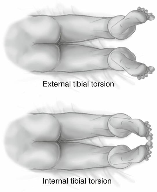
FIGURE 17.3 Determining the thigh–foot angle.
Reprinted with permission from Bowden V, Greenberg CS. Children and Their Families. 3rd Ed. Philadelphia: Lippincott Williams & Wilkins, 2013.
628
V.Common Fractures
A.Describing fractures is critical because it may dictate the management of a fracture.
1.Open or closed fracture describes whether a break in the skin overlies a fracture. In a closed fracture, the skin is intact. In an open fracture, the skin is broken and antibiotics are required because of the risks of local infection and osteomyelitis.
2.Spatial relationship of the fractured ends describes the orientation of the fractured ends.
a.Nondisplaced or nonangulated describes fracture ends that are well approximated and in normal position.
b.Displaced describes fractured ends that are shifted.
c.Angulated describes fractured ends that form an angle.
d.Overriding describes a fracture whose ends override without cortical contact.
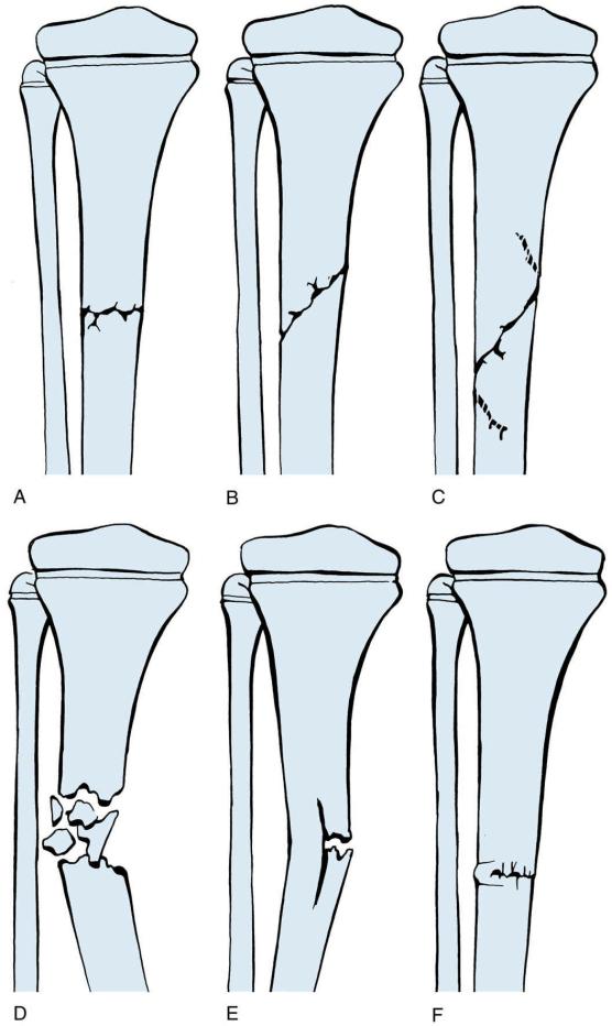
3.Types of fractures (Figure 17-4)
a.Compression fracture (torus or buckle fracture) occurs if the soft bony cortex buckles under a compressive force. This type of fracture commonly occurs in the metaphysis and requires only splinting for 3–4 weeks.
b.Incomplete fracture (greenstick fracture) occurs if only one side of the cortex is fractured with the other side intact. The intact side is the site of compression injury and may be bent, whereas the fractured side receives the tension and fractures. Because angulation can increase even within a cast, reduction may include fracturing the other side of the cortex.
c.Complete fractures
1.Transverse describes a fracture that is horizontal across the bone.
2.Oblique describes a diagonal fracture across the bone.
3.Spiral describes an oblique fracture encircling the bone. Spiral fractures may occur with twisting injury.
4.Comminuted describes a fracture that is composed of multiple fracture fragments.
d.Any fracture can be a sign of child abuse—there is no fracture that is pathognomonic for abuse.
4.Location of the fracture
a.Epiphyseal fracture involves the end of the bone.
b.Physeal fracture involves the growth plate. The Salter–Harris classification describes fractures involving the physis. Table 17-6 contains a mnemonic to help remember the classification, and Figure 17-5 illustrates this classification. The Salter–Harris classification may be an important prognostic factor in determining subsequent growth of the limb. For example, subsequent bone growth is affected in some type II–III Salter–Harris fractures and all type IV–V Salter–Harris fractures.
c.Metaphyseal fracture involves the ends of the central shaft (i.e., between the epiphysis and diaphysis).
d.Diaphyseal fracture involves the central shaft of the bone.
B.Clavicular fractures are common fractures in childhood and are usually caused by falling onto the shoulder. Birth injury is the major cause of clavicular fractures in neonates.
1.Clinical features
a.Infants may be asymptomatic or may present with an asymmetric Moro reflex or pseudoparalysis (refusal to move extremity because of pain). Crepitus may be felt over the fracture.
b.Children typically hold the affected limb with the opposite hand, and the child’s head is often tilted toward the affected side. Point tenderness and deformity may be
629
noted over the fracture.
2.Diagnosis is on the basis of plain radiographs showing the fracture. Most fractures caused by trauma involve the middle and lateral aspects of the clavicle.
3.Management. Treatment includes placement in a sling for 4–6 weeks to assist in immobilizing the limb. Neonates with clavicle fractures often do not require any treatment.
4.Complications. Injury to nerves and vessels is rare. However, brachial plexus injuries can coexist with clavicular fractures in neonates as a result of birth injury.
C.Supracondylar fracture of the humerus occurs when a child falls onto an outstretched arm or elbow. Children younger than 10 years are mostly at risk for this type of fracture because the mechanical force of the impact is transmitted to the supracondylar area. A supracondylar fracture is an orthopedic emergency if the fracture is displaced and angulated, because of the risk of neurovascular injury and compartment syndrome.
1.Clinical features
a.Point tenderness, swelling, and deformity of the elbow may be seen.
b.It is important to assess the pulse, sensation, and movement of the fingers to detect neurovascular injury. If the fractured fragment is angulated or displaced, it can cause injury by stretching the radial or median nerves or the brachial artery.
c.Pain with passive extension of the fingers is suggestive of compartment syndrome.
2.Diagnosis is on the basis of AP and lateral radiographs. A triangular fat pad shadow posterior to the humerus may be observed if an occult fracture is present (i.e., “posterior fat pad sign”).
3.Management. Passive movement of the elbow (i.e., when the examiner moves the arm) may increase the risk of further neurovascular injury. Therefore, if a supracondylar fracture is suspected, never passively move the arm.
a.Nondisplaced and nonangulated fractures require casting.
b.Displaced or angulated fractures require surgical reduction and pinning.
4.Complications
a.Compartment syndrome occurs when the pressure within the anterior fascial compartment is greater than 30–45 mm Hg, leading to ischemic injury and Volkmann contracture (flexion deformity of the fingers and the wrist). The “5 P’s” of compartment syndrome (pallor, pulselessness, paralysis, pain, and paresthesias) are late signs. A more sensitive indication of impending compartment syndrome is pain with passive extension of the fingers.
b.Injury to the radial, median, or ulnar nerve may cause temporary palsy but usually resolves.
c.Cubitus varus is a decreased or absent carrying angle as a result of poor positioning of the distal fragment. This is a permanent complication.
D.Forearm fractures
1.Common types of forearm fractures
a.Distal radius fracture is the most common upper extremity fracture in children.
b.Monteggia fracture is a fracture of the proximal ulna with dislocation of the radial head.
c.Galeazzi fracture is a fracture of the radius with distal radioulnar joint dislocation.
2.Diagnosis is on the basis of AP and lateral radiographs of the forearm.
3.Management. Treatment includes open or closed reduction and splinting. A cast replaces the splint in 4–7 days after the swelling has resolved. Forearm fractures heal within 6–
8 weeks.
E.Femur fractures require a great deal of mechanical force. Therefore, it is important to assess
630
for other injuries in the joint above and below the fracture.
1.Clinical features include erythema, swelling, deformities, and point tenderness.
2.Diagnosis is on the basis of AP and lateral radiographs that include the joint above and below the area of injury.
3.Management. Treatment depends on the age of the patient as well as other factors. It may include using a Pavlik harness in neonates, spica casting in young children, traction, or surgical stabilization.
F.Toddler’s fracture is a spiral fracture of the tibia. This type of fracture may occur after very mild or no identified trauma. The fibula remains intact.
1.Epidemiology. Toddler’s fractures usually occur between 9 months and 3 years of age. Typically, they occur when a child trips and falls while running or playing.
2.Clinical features. The child refuses to bear weight but is willing to crawl. Erythema, swelling, and mild point tenderness may be found over the distal tibia on examination.
3.Diagnosis. Oblique views of the tibia may show the fracture line, although sometimes plain radiographs do not visualize the fracture.
4.Management. Treatment involves a long leg cast for 3–4 weeks.
G.Fractures typical of child abuse. Any fracture before walking age should raise the concern for abuse. (See also Chapter 20, section VI.B.2.d.) A “skeletal survey” (radiographic studies of all bones) is the screening tool used to assess for current and previous bony injuries. A “babygram” is not an adequate skeletal survey. The following fractures should make the clinician suspect abuse:
1.Metaphyseal fractures (corner or bucket handle fractures)
2.Posterior or first rib fractures
3.Multiple fractures at various ages of healing
4.Complex skull fractures
5.Scapular, sternal, and vertebral spinous process fractures
6.Any fracture whose mechanism does not fit the history provided or the child’s developmental abilities (e.g., fracture of the lower extremities in a nonambulatory child)
631
632

FIGURE 17.4 Common types of fractures. A. Transverse. B. Oblique. C. Spiral. D. Comminuted. E.
Greenstick. F. Buckle.
Table 17-6
Salter–Harris Classification
Type |
Acronym |
Description of Fracture |
I |
S |
Same: Fracture is within the physis |
II |
A |
Above: Fracture is in the physis and above into the metaphysis |
III |
L |
Low: Fracture is in the physis and below into the epiphysis |
IV |
T |
Through and through: Fracture is in the physis through both the metaphysis and the epiphysis |
V |
R |
Crush: Crushing of the physis |
FIGURE 17.5 Salter–Harris classification of fractures. Type I is a fracture within the physis. Type II is a fracture within the physis and into the metaphysis. Type III is a fracture within the physis and into the epiphysis. Type IV is a fracture through the metaphysis, physis, and epiphysis. Type V is a crush fracture of the physis.
Reprinted with permission from Flynn JM, Skaggs DL, Waters PM. Rockwood and Wilkins’ Fractures in Children. 8th Ed. Philadelphia: Lippincott Williams & Wilkins, 2014.
633