

- •Copyright
- •Contents
- •Dedication
- •Preface
- •Acknowledgments
- •Contributors
- •Contributors to the Previous Edition
- •Review Test
- •Answers and Explanations
- •Review Test
- •Answers and Explanations
- •Review Test
- •Answers and Explanations
- •Review Test
- •Answers and Explanations
- •Review Test
- •Answers and Explanations
- •Review Test
- •Answers and Explanations
- •Review Test
- •Answers and Explanations
- •Review Test
- •Answers and Explanations
- •Review Test
- •Answers and Explanations
- •Review Test
- •Answers and Explanations
- •IV. Hypertension
- •VI. Nephrotic Syndrome (NS)
- •VII. Hemolytic Uremic Syndrome (HUS)
- •VIII. Hereditary Renal Diseases
- •IX. Renal Tubular Acidosis (RTA)
- •XI. Chronic Kidney Disease (CKD) and End-Stage Renal Disease (ESRD)
- •XII. Structural and Urologic Abnormalities
- •XIII. Urolithiasis
- •XIV. Urinary Tract Infection (UTI)
- •Review Test
- •Answers and Explanations
- •Review Test
- •Answers and Explanations
- •Review Test
- •Answers and Explanations
- •Review Test
- •Answers and Explanations
- •IV. Food Allergy
- •VI. Urticaria (Hives)
- •VII. Drug Allergy
- •VIII. Asthma
- •IX. Immunology Overview
- •X. Disorders of Lymphocytes (Figure 15-2)
- •XI. Disorders of Granulocytes (Figure 15-3)
- •XII. Disorders of the Complement System
- •Review Test
- •Answers and Explanations
- •Review Test
- •Answers and Explanations
- •Review Test
- •Answers and Explanations
- •Review Test
- •Answers and Explanations
- •Review Test
- •Answers and Explanations
- •Review Test
- •Answers and Explanations
- •Comprehensive Examination
- •Index
Review Test
1.A 1-week-old female infant has several vesicles on her scalp. She also has a 1-day history of fever (temperature up to 38.2°C [100.8°F]) and irritability. She is breastfed and is eating slightly less vigorously than usual. The mother’s pregnancy was uncomplicated. Which of the following statements regarding the presumptive diagnosis is correct?
A.Herpes simplex virus type 1 is the most likely causative pathogen.
B.The infection was likely acquired after birth.
C.A Tzanck smear of the base of one of the vesicles may demonstrate epidermal giant cells.
D.Oral acyclovir should be started immediately.
E.Oral antibiotics to cover staphylococcal and streptococcal infection should be started promptly.
2.A 9-year-old girl is brought to the office by her parents. She has a 1-month history of two large well-demarcated areas of hair loss in the parietal scalp. On examination, no inflammation or scaling of the skin is apparent, and the underlying skin is smooth and soft. Which of the following statements regarding the likely diagnosis is correct?
A.The fingernails are likely to be normal.
B.Autoimmune destruction of the hair follicle is likely the cause of this disorder.
C.Oral griseofulvin should be prescribed after cultures.
D.The child and her parents should be counseled to loosen her braids and ponytails when styling her hair.
E.The hair loss is likely to be permanent.
3.A 6-year-old boy presents with a 1-month history of patchy alopecia in the occipital region of the scalp. Examination reveals a well-circumscribed area of hair loss in which all the hairs appear to be broken off at the scalp surface. Occipital lymph nodes are prominent. Which of the following statements regarding the likely diagnosis is correct?
A.This infection is contagious, and brushes, combs, and hats should not be shared.
B.Topical management with clotrimazole is the initial appropriate treatment.
C.Exposure to dogs or cats is the most likely cause of infection.
D.Hairs are likely to fluoresce under Woods light examination.
E.Oral antifungal therapy should be administered for 2 weeks.
4.A 5-year-old boy presents with a 6-week history of scaling, nongreasy erythematous plaques in the occipital region of the scalp and in the inguinal region. The lesions have a “silvery scale” appearance. Which of the following statements regarding the likely disorder is correct?
A.Low-potency corticosteroids are likely to be effective in treating this condition.
B.It would be unusual for other family members to also have this disorder.
C.New lesions may develop at any site of skin trauma.
D.Nail changes would not be expected with this condition.
E.Arthritis is likely to occur in this patient.
5.A 10-year-old girl presents with a history of malaise and a headache that was followed 4 days later with a body rash. Examination reveals a 3-cm scaly erythematous plaque on the upper arm and oval, red macules and papules on the back that follow skin lines. Which of the following management steps is the least appropriate?
A.Therapy with topical corticosteroids for pruritus
B.Natural ultraviolet exposure
C.Topical clotrimazole
D.Oral antibiotics
E.Reassurance only
6.A 4-month-old male infant is brought to the office by his parents for a routine health
683
maintenance examination. On examination, you note significant hypopigmentation in the inguinal area. Medical history is remarkable for suspected diaper dermatitis treated with zinc oxide and high-potency corticosteroids for 3 weeks. Which of the following statements regarding this patient and his clinical findings is most correct?
A.Topical antifungal therapy should now be prescribed to treat suspected fungal superinfection.
B.Zinc oxide should be immediately discontinued.
C.Atrophy of skin in the inguinal area may also be present.
D.The child should be referred to genetics to consider the diagnosis of tuberous sclerosis.
E.Ultraviolet light therapy should be prescribed.
7.A 4-year-old boy with a history of atopic dermatitis presents for evaluation of his skin. Examination shows that the skin on his upper and lower extremities is very dry and irritated. You would like to suggest a moisturizer in addition to topical corticosteroids. Which of the following types of moisturizers is most appropriate?
A.Lotion
B.Cream
C.Ointment
D.Solution
E.Gel
8.A 13-year-old boy is brought to the office by his parents because of concerns regarding his acne. Examination reveals scattered open and closed comedones on the forehead and nose. Which of the following statements regarding this condition is correct?
A.Topical benzoyl peroxide is an appropriate first-line treatment.
B.Oral isotretinoin is an appropriate first-line treatment.
C.Oral clindamycin is an appropriate first-line treatment.
D.Sebum plays little role in the pathophysiology at this stage of acne.
E.Reassurance only should be given because the patient’s skin findings are currently of little consequence.
For statements 9–13, the response options are the same. You will be required to select one answer for each statement in the following set.
A.Papule
B.Macule
C.Pustule
D.Nodule
E.Vesicle
F.Cyst
G.Wheal
H.Target lesion
I.Plaque
Match the disorder with its most characteristic skin lesion.
1.A 3-year-old boy with herpes simplex virus infection involving the upper and lower lips.
2.A 7-year-old boy with suspected vitiligo involving both hands.
3.A 15-month-old girl with molluscum contagiosum on the cheeks and forehead.
4.A 6-year-old boy with erythema multiforme major.
5.A 10-year-old girl with suspected Gianotti–Crosti syndrome.
For statements 14 and 15, the response options are the same. You will be required to select one
684
answer for each statement in the following set.
A.Neurofibromatosis type 1
B.Neurofibromatosis type 2
C.Tuberous sclerosis
For each patient, select the most likely associated neurocutaneous syndrome.
1.A 3-year-old boy with seizures and Lisch nodules on ophthalmologic examination.
2.A 12-month-old infant with infantile spasms.
685
Answers and Explanations
1.The answer is C [V.C.7.c–d]. This patient’s clinical presentation is most consistent with neonatal herpes simplex virus (HSV) infection. Diagnosis is by identification of (1) the virus by culture or (2) the viral antigen by rapid testing techniques. Infection may also be diagnosed by identification of epidermal giant cells on a Tzanck preparation. Two-thirds of HSV infections acquired during the neonatal period are caused by HSV-2, and this infection is most often acquired during passage through the birth canal of a mother infected with the virus. Neonatal HSV infection is a medical emergency that requires prompt admission and management with intravenous acyclovir. Oral antibiotics are not useful.
2.The answer is B [VII.A]. This patient’s presentation with well-demarcated hair loss with-out scalp inflammation is likely caused by alopecia areata. The cause of alopecia areata is thought to be an autoimmune lymphocyte-mediated injury to the hair follicle. Associated findings include nail pitting in 40% of patients. The clinical presentation is not consistent with tinea capitis, which would present with localized areas of hair loss associated with scalp inflammation, scaling, and broken hairs. Thus, griseofulvin is not indicated in this case. This presentation is also not consistent with traction alopecia due to hair care or styling practices, which is characterized by patchy and jagged, irregularly shaped areas of alopecia, especially along the hair line, containing small, thin, and broken hairs. In the case of alopecia areata, regrowth of hair within months to years occurs in the majority of patients.
3.The answer is A [V.A.1]. Clinical features of alopecia with hairs broken off at the scalp and occipital lymphadenopathy suggest tinea capitis, a fungal infection of the hair. The most common causal pathogen is Trichophyton tonsurans, acquired from human-to-human contact, including by sharing hats, brushes, and combs. Topical antifungal therapy is not effective for tinea capitis. Dogs and cats are a source of infection with Microsporum canis, which currently causes only 5% of tinea capitis infections. Infection with M. canis is characterized by thickened, white broken hairs, and only hairs infected with M. canis fluoresce under Woods light, whereas hairs infected with Trichophyton tonsurans do not fluoresce. Treatment of tinea capitis involves 6–8 weeks of oral antifungal therapy.
4.The answer is C [III.E.3]. Skin lesions with a silvery scale appearance suggest psoriasis. Lesions of psoriasis may also demonstrate the Koebner phenomenon, in which new lesions develop at sites of skin injury. Corticosteroids of moderate to high potency are the most effective treatment agents. Psoriasis is inherited in an autosomal dominant fashion, and therefore, other family members may also have the disorder. Nail involvement is often seen in individuals with psoriasis. Psoriatic arthritis is uncommon during childhood.
5.The answer is C [III.D.3–4]. This patient’s clinical presentation and examination findings are consistent with pityriasis rosea. Pityriasis rosea may be an immune response to a viral infection, although its true cause is unknown. Pityriasis rosea resolves without medications, although antihistamines and topical corticosteroids may relieve any associated itching. Exposure to ultraviolet light and short courses of oral antibiotics (for anti-inflammatory properties) may shorten the disease course. Clotrimazole is not an appropriate therapy for pityriasis rosea.
6.The answer is C [I.D.6.a]. The hypopigmentation in the inguinal region is most likely a side effect of the high-potency corticosteroid therapy. Only low-potency corticosteroids should be
applied to the groin because the epidermis of the groin is very thin and absorption of corticosteroids is higher in this area. Local side effects of high-potency topical steroids
include pigmentation changes, skin atrophy, acne, folliculitis, and telangiectasias. Neither antifungal medications nor ultraviolet light is indicated. Discontinuation of zinc oxide will not affect this patient’s skin findings. Although tuberous sclerosis is associated with
686
hypopigmented skin lesions, the history in this case is more suggestive of localized topical steroid use as the cause of the hypopigmentation. In the case of suspected tuberous sclerosis, the individual should have more than one hypopigmented skin lesion, a history of seizures or infantile spasms, and/or a family history of the disorder.
7.The answer is C [I.D.2]. Adequate hydration of the skin is crucial in many dermatologic conditions, especially atopic dermatitis. Ointments are especially useful for very dry skin because they have maximal water-retaining properties and would be the best choice for this patient. Lotions are helpful for minimal dryness, and creams are useful for average dryness. Both solutions and gels are most useful for hair-bearing surfaces.
8.The answer is A [VIII.C]. This patient’s skin findings are consistent with acne. The open and closed comedones are characteristic of noninflammatory acne, and a topical agent is most appropriate. Benzoyl peroxide is very effective for both noninflammatory and inflammatory acne and is an appropriate initial treatment for this patient. Neither oral antibiotics nor isotretinoin is a first-line treatment for noninflammatory acne. Oral antibiotics are most useful in inflammatory acne. Systemic isotretinoin is helpful in all types of acne; however, it is indicated for severe cystic or nodular acne. Obstruction to sebum outflow from the follicle leads to comedone development and therefore is important in the pathophysiology. Acne should never be dismissed as of no consequence, even in the early stages, because it may be associated with future scarring and may have effects on a child’s psychosocial development.
9.The answers are E, B, A, H, and A, respectively [V.C.7.b, VI.A.3, V.C.10.b.(1), IV.C.2, V.C.5, and Table 19-1]. Herpes simplex virus is characterized by grouped vesicles on an erythematous base. Vitiligo is a depigmentation disorder characterized by hypopigmented macules. Molluscum contagiosum is characterized by flesh-colored papules with central
umbilication. Erythema multiforme major is characterized by target lesions. Erythematous or flesh-colored papules are characteristic of Gianotti–Crosti syndrome.
10.The answers are A and C, respectively [Table 19-3]. The 3-year-old boy has neurofibromatosis type 1, which is characterized by café-au-lait spots, axillary freckling, and neurofibromas in the skin and other organs. Seizures may occur because of central nervous system involvement that may include intracranial calcifications, neurofibromas, and optic glioma. Characteristic eye findings include Lisch nodules or iris hamartomas. The 12-month-old infant has tuberous sclerosis, which is associated with characteristic skin findings including ash-leaf spots, angiofibromas, and shagreen patch. Almost all children with tuberous sclerosis have seizures because of central nervous system involvement, and the condition is frequently associated with infantile spasms.
687

C H A P T E R 2 0
688

Emergency Medicine
Calvin G. Lowe
689
I.Infant and Child Cardiopulmonary Resuscitation (CPR)
A.General concepts
1.The most common cause of cardiac arrest in a child is a lack of oxygen supply to the heart secondary to a pulmonary problem (e.g., choking, suffocation, airway or lung disease, drowning), respiratory arrest, or shock. Cardiac arrest can often be prevented if assisted breathing is initiated promptly.
2.Heart disease is an uncommon cause of cardiac arrest in infants and children.
3.Chances for survival increase dramatically if CPR and advanced life support are begun quickly.
4.If a patient is found unresponsive and not breathing, then CPR should be initiated.
B.Sequence of CPR is C–A–B: Circulation–Airway–Breathing. In 2010 (and subsequently - reaffirmed), the American Heart Association recommended that CPR for infants and children be initiated with chest compressions rather than rescue breaths (C–A–B rather than A–B–C). When approaching an unresponsive victim, the rescuer should shout for nearby help and activate the emergency response system. The rescuer should look for breathing or gasping and simultaneously check for a pulse.
1.Circulation
a.Chest compressions are administered for asystole or bradycardia.
b.Rescuers should compress the chest at least one-third of the anterior–posterior diameter of the chest.
1.1½ inches in most infants
2.2 inches (5 cm) in most children
c.Chest compressions are delivered at a rate of 100/minute.
2.Airway
a.After the first set of compressions are given, the vicitim’s airway is opened.
b.The victim’s tongue is the most common cause of airway obstruction.
c.The airway may be opened by the head tilt method, which lifts the tongue from the back of the throat, or by the jaw-thrust method if the child has suspected neck or cervical spine injury. (Cervical spine injury should be suspected in face, head, or neck trauma.)
3.Breathing
a.After the airway is opened, assessment for breathing is performed by the look, listen, and feel method, in which the rescuer looks for a rise and fall in the chest, listens for exhaled air, and feels for exhaled airflow.
b.Rescue breathing must be performed if spontaneous breathing is absent.
C.CPR cycles
1.Ratio of chest compressions for one-rescuer CPR is 30 compressions:2 breaths.
2.Ratio of compressions of two-rescuer CPR is 15 compressions:2 breaths.
3.After 2 minutes of compressions/breaths or five cycles, a reassessment of the patient for a pulse or spontaneous respirations should be performed.
690
II.Shock
A.General concepts
1.Definition. Shock is a clinical state characterized by inadequate delivery of oxygen and metabolic substrates to meet the metabolic demands of tissues.
2.Shock may be present with normal or decreased blood pressure.
B.Classification. Shock may be classified by the degree of compensation and by the cause.
1.Shock classification by the degree of compensation includes compensated, decompensated, or irreversible.
a.Compensated shock is characterized by normal blood pressure and cardiac output with adequate tissue perfusion but maldistributed blood flow to essential organs.
b.Decompensated shock is characterized by hypotension, low cardiac output, and inadequate tissue perfusion.
c.Irreversible shock is characterized by cell death and is refractory to medical treatment.
2.Shock may also be classified on the basis of the cause.
a.Hypovolemic shock is the most common cause of shock in children and is caused by any condition that results in decreased circulating blood volume, such as hemorrhage or dehydration (e.g., from acute gastroenteritis). The amount of volume loss determines the success of compensatory mechanisms, such as endogenous catecholamines, in maintaining blood pressure and cardiac output. Volume losses greater than 25% result in decompensated shock.
b.Septic shock occurs secondary to an inflammatory response to invading microorganisms and their toxins, and it results in abnormal blood distribution. There are two clinical stages:
1.Hyperdynamic stage is characterized by normal or high cardiac output with bounding pulses, warm extremities, and a wide pulse pressure.
2.Decompensated stage follows the hyperdynamic stage if aggressive treatment has not been initiated. It is characterized clinically by impaired mental status, cool extremities, and diminished pulses.
c.Distributive shock is associated with distal pooling of blood or fluid extravasation, and it is typically caused by anaphylactic or neurogenic shock or as a result of medications or toxins.
1.Anaphylactic shock is characterized by acute angioedema of the upper airway, bronchospasm, pulmonary edema, urticaria, and hypotension because of extravasation of intravascular fluid from permeable capillaries (see Chapter 15, section I).
2.Neurogenic shock, typically secondary to spinal cord transection or injury, is characterized by a total loss of distal sympathetic cardiovascular tone, with hypotension resulting from pooling of blood within the vascular bed.
d.Cardiogenic shock occurs when cardiac output is limited because of primary cardiac dysfunction. Causes include dysrhythmias (e.g., supraventricular tachycardia), congenital heart disease (e.g., any lesion that impairs left ventricular outflow), and cardiac dysfunction after cardiac surgery. Clinical features are the signs and symptoms of congestive heart failure (CHF; see Chapter 8, section I.D).
C.Diagnosis
1.Recognition of shock may be difficult because of the presence of compensatory mechanisms that prevent hypotension until 25% of intravascular volume is lost. Therefore, the index of suspicion for shock must be high.
691
2.Historic features that may suggest the presence of shock include the following:
a.Severe vomiting and diarrhea
b.Trauma with hemorrhage
c.Febrile illness, especially in an immunocompromised patient
d.Exposure to a known allergic antigen
e.Spinal cord injury
3.Physical examination
a.Blood pressure may be normal in the initial stages of hypovolemic and septic shock.
b.Tachycardia almost always accompanies shock and occurs before blood pressure changes in children.
c.Tachypnea may be present as a compensatory mechanism for severe metabolic acidosis.
d.Mental status changes may indicate poor cerebral perfusion.
e.Capillary refill may be prolonged with cool and mottled extremities.
f.Peripheral pulses may be bounding in early septic shock.
4.Laboratory studies should include the following:
a.Complete blood count (CBC) to assess for blood loss and infection
b.Electrolytes to assess for metabolic acidosis and electrolyte abnormalities
c.Blood urea nitrogen and creatinine to evaluate renal perfusion and function
d.Lactate to assess adequacy of tissue perfusion
e.Calcium and glucose to assess for frequently encountered metabolic derangements
f.Coagulation factors to evaluate for disseminated intravascular coagulation (DIC), which may accompany shock
g.Toxicology screens to evaluate for a poisoning, which could cause shock
h.Blood and urine cultures to evaluate for infections that could cause shock
D.Management
1.Initial resuscitation includes:
a.Supplemental oxygen
b.Early endotracheal intubation to secure the airway and decrease the patient’s energy expenditure
c.Vascular access with appropriate fluid resuscitation. Fluids should initially include a 20 mL/kg bolus of normal saline or lactated Ringer solution.
2.To restore intravascular volume, intravenous crystalloid or colloid solutions should generally be used before administration of inotropic and vasopressor agents.
3.Inotropic and vasopressor medications (e.g., dobutamine, dopamine, epinephrine) are indicated if the blood pressure increase in response to fluids is inadequate.
4.Metabolic derangements, such as metabolic acidosis, hypocalcemia, or hypoglycemia, should be treated.
5.Other considerations include administration of broad-spectrum antibiotics for septic shock, blood products for hemorrhage, and fresh-frozen plasma for DIC.
692

III.Trauma
A.General concepts
1.Trauma is the leading cause of death in the United States in children older than 1 year.
2.Motor vehicle accidents are the leading cause of trauma.
3.Anatomic and physiologic differences between a child and an adult account for the child’s unique response to trauma.
a.Head injuries are common because a child’s head comprises a larger percentage of total body mass.
b.The neck of a child is shorter and supports a relatively greater weight.
c.The rib cage of a child is more pliable, leading to greater energy transmitted to internal organs, such as the spleen and liver.
d.The growth plates in the bones of a growing child result in a relatively weak epiphyseal–metaphyseal junction. Ligaments are stronger than the growth plate; therefore, with injury, the growth plate is at the highest risk for fracture.
B.Primary survey. This rapid initial assessment of the patient should be performed within 5– 10 minutes of arrival in the emergency department. The primary survey can be recalled using the mnemonic ABCDE: Airway maintenance, Breathing and ventilation with 100% oxygen, Circulation and control of hemorrhage, Disability assessment using the Glasgow coma score (GCS, which is used to assess the extent of neurologic impairment based on physical examination (Table 20-1), and Exposure/Environmental control, in which the patient is undressed completely to facilitate examination and then warmed to prevent hypothermia.
C.Adjuncts to the primary survey
1.Electrocardiographic (ECG) monitoring is mandatory for all patients.
a.Dysrhythmias may indicate cardiac injury.
b.Pulseless electrical activity may indicate cardiac tamponade, tension pneumothorax, or profound hypovolemia.
2.A urinary catheter should be placed to monitor urine output, and a nasogastric tube to reduce abdominal distension.
3.Diagnostic studies typically include radiographs of the cervical spine, chest, and pelvis, and computed tomographic (CT) scans of the head and abdomen.
D.Secondary survey. This head-to-toe evaluation includes a thorough physical examination.
E.Specific injuries in the pediatric trauma patient
1.Head trauma
a.Seizures are common after head trauma but are self-limited.
b.Infants are at risk for bleeding in the subgaleal and epidural spaces because of open fontanelles and cranial sutures. However, these open structures may also allow infants to be more tolerant of expanding intracranial masses.
c.Intracranial bleeding may occur in the epidural space, subdural space, or within the brain parenchyma itself after even mild head trauma without skull fracture or loss of consciousness.
1.Epidural hematoma is bleeding between the inner table of the skull and the dura. It is associated with tearing of the middle meningeal artery typically following a high-risk injury (direct blow to the side or back of the head, or fall from a significant height).
a.Clinical features are the signs and symptoms of increased intracranial pressure (ICP; see Table 20-2 and section III.E.1.d). A classic history could include an initial loss of consciousness, a lucid interval for several hours, followed by abrupt decompensation and coma.
693
b.Diagnosis is by head CT, which shows a lenticular density representing blood within the epidural space.
c.Management is immediate surgical drainage.
d.Prognosis is generally good if surgery can be performed rapidly.
2.Subdural hematoma is blood beneath the dura. It is associated with tearing of the bridging meningeal veins by direct trauma or shaking. It is more common than epidural hematoma and is seen most commonly in infancy.
a.Clinical features include seizures and signs and symptoms of increased ICP (see Table 20-2 and section III.E.1.d). Subdural hematomas are bilateral in 75% of cases, and symptoms develop more slowly than with an epidural bleed.
b.Diagnosis is by head CT, which shows a crescentic density representing blood in the subdural space.
c.Management includes neurosurgical consultation and usually surgical drainage.
d.Prognosis is variable and may be poor if the underlying brain is also injured.
3.Intracerebral hematoma is bleeding within the brain parenchyma. Frontal and temporal lobes are most often affected, usually on the opposite side of the impact injury (contrecoup injury). Management is surgical drainage if the hematoma is accessible.
d.Increased ICP
1.Clinical features
a.Headache is the first symptom.
b.Pupillary changes and altered mental status are the first signs.
c.Table 20-2 lists the clinical features of increased ICP.
2.Complications. Increased ICP may lead to cerebral herniation, most commonly transtentorial or uncal herniation, in which the temporal lobe or uncus is displaced into the infratentorial compartment. Clinical features of herniation include the following:
a.Bradycardia, which is an early sign of herniation in children younger than 4 years
b.Fixed and dilated ipsilateral pupil
c.Contralateral hemiparesis
d.Pupils will eventually become bilaterally fixed and dilated.
e.Bilateral hemiparesis will also eventually occur.
f.Cushing triad, a late sign, is characterized by bradycardia, hypertension, and irregular breathing.
3.Management. The goal of treatment is to prevent secondary brain injury and includes the following:
a.Mild hyperventilation with 100% oxygen to lower PaCO2 to 30– 35 mm Hg, which in turn mildly vasoconstricts cerebral vessels.
Aggressive hyperventilation can lead to worsening cerebral ischemia.
b.Elevation of the head to 30°–45°, which encourages venous drainage
c.Diuretics (e.g., mannitol, 3% hypertonic normal saline)
d.Neurosurgical consultation
2.Spinal cord injury. Injury to the spinal cord may occur in children. Even in the presence of serious injury, radiographs of the cord may be normal. Note that spinal cord injury without radiographic abnormality (“SCIWORA”) occurs more commonly in children than in adults.
694

3.Chest trauma
a.A child’s soft and pliable chest wall allows transmission of forces to the lung parenchyma.
b.Tension pneumothorax may occur and is life-threatening.
1.Clinical features include distended neck veins, decreased breath sounds, hyperresonance to percussion, displaced trachea, pulseless electrical activity, and shock.
2.Management is emergent chest decompression by needle thoracotomy.
Waiting for radiographic confirmation of the diagnosis can lead to a patient’s death.
4.Abdominal trauma is common because of underdeveloped abdominal musculature.
a.Duodenal hematoma often occurs secondary to injury to the right upper quadrant, commonly from a bicycle handle bar. Clinical features include abdominal pain and vomiting. Bowel obstruction is found on radiographic evaluation.
b.Lap belt injuries from a motor vehicle accident include a chance fracture (flexion disruption of the lumbar spine), liver and spleen lacerations, and bowel perforation.
c.Spleen, liver, and kidneys are often injured by blunt trauma.
TABLE 20-1
Glasgow Coma Scale (GCS)*
Verbal Patient |
GCS Score |
Nonverbal Patient (Child) |
|
Eye opening |
|
Spontaneously |
4 |
Spontaneously |
Response to voice |
3 |
Response to voice |
Response to pain |
2 |
Response to pain |
No response |
1 |
No response |
Best motor response |
|
|
Obeys commands |
6 |
Normal movements |
Localizes pain |
5 |
Localizes pain |
Flexion withdrawal |
4 |
Flexion withdrawal |
Decorticate posturing |
3 |
Flexion abnormal |
Decerebrate posturing |
2 |
Extension abnormal |
No response |
1 |
No response |
Best verbal response |
|
|
Oriented/appropriate |
5 |
Cries normally, smiles, and coos |
Disoriented conversation |
4 |
Cries |
Inappropriate words |
3 |
Inappropriate crying and screaming |
Incomprehensible words |
2 |
Grunts |
No response |
1 |
No response |
*This scale is used to assess the level of neurologic impairment on the basis of a patient’s physical examination (eye opening, motor response, and verbal response). A GCS of 13–15 indicates mild head injury; a GCS of 9–12 indicates moderate head injury; and a GCS < 8 indicates severe head injury.
Table 20-2
Symptoms and Signs of Elevated Intracranial Pressure
Symptom |
Sign |
Headache |
Papilledema |
Vomiting |
Cranial nerve palsies |
Stiff neck |
Stiff neck |
Double vision |
Head tilt |
Transient loss of vision |
Retinal hemorrhage |
Episodic severe headache |
Percussion of skull—“cracked pot sound” |
Gait disturbance |
Obtundation |
Dulled intellect |
Unconsciousness |
Irritability |
Progressive hemiparesis |
695
696
IV. Burns
A.Epidemiology. More than 40% of deaths from burns occur in children younger than 4 years.
1.Scalding injuries from hot liquids are the most common type of burn.
2.Not all burns are accidental, and child abuse should be considered (see section VI.B.2.c).
B.Classification of burns is on the basis of degree (depth of skin injured) and body surface area (BSA).
1.First-degree burns involve only the epidermis and are characterized by red, blanching, painful skin that heals without scarring (e.g., sunburn).
2.Second-degree burns (partial-thickness burns) involve the entire epidermis and part of the dermis.
a.Superficial partial-thickness burns involve the entire epidermis and outer portion of the dermis. Burns are moist, painful, and red. They blister but usually do not scar.
b.Deep partial-thickness burns involve destruction of the entire epidermis and lower portion of the dermis. Burns are pale white. They may blister and they heal with scarring.
3.Third-degree burns or full-thickness burns involve the complete destruction of the epidermis, dermis, and part of the subcutaneous tissue. Burns are dry, white, and
leathery to the touch, and skin grafts are needed. Because nerve endings are burned, the victim is usually insensitive to pain.
4.BSA burned is expressed in percentage. The Lund–Browder classification may be used to help measure the burned area. Although the “rule of 9’s” is used to measure percent BSA burned in adolescents and adults (each arm = 9%, each leg = 18%, anterior trunk = 18%, posterior trunk = 18%, head and neck = 9%), this rule overestimates burns in a child because of a child’s relatively larger head and smaller legs. Another estimate of percent BSA burned used in children uses the size of the patient’s palm to measure the burned area; the palm is approximately equivalent to 1% BSA. First-degree burns are not included in the calculation of the BSA burned.
C.Management
1.Initial resuscitation should include the ABCs.
a.Endotracheal intubation should be performed in any victim suspected of inhaling hot gases, which may burn the upper airway and lead to progressive edema and airway obstruction.
b.Assess oxygenation by pulse oximetry. Administer 100% oxygen and assess for carbon monoxide inhalation (see section VIII.D.6).
c.Intravenous access should be obtained through nonburned skin.
2.Fluid resuscitation is critical, because large volumes of fluid may be lost from burned skin and leaky capillaries.
a.Lactated Ringer solution is the isotonic crystalloid fluid of choice in burn resuscitation.
b.The Parkland formula calculates the volume of fluid replacement within the first 24 hours.
1.The replacement volume is approximately 4 mL/kg body weight per percent BSA burn.
2.Half of the replacement volume is given over the first 8 hours postburn, and the remaining half over the next 16 hours.
3.Skin care depends on the degree of burn.
697
a.First-degree burns require only moisturizers and analgesics.
b.Second-degree burns require appropriate analgesics (e.g., opiates) and debridement of dead skin to prevent infection. Bullae (large blisters), if intact, are generally not removed because the skin of the bullae forms a barrier to infection and prevents fluid loss. Bullae that have ruptured should be removed.
c.Third-degree burns require skin grafting and hydrotherapy. Escharotomy (i.e., surgical removal of a constricting scar) may be needed if the burn restricts blood flow or chest expansion.
d.Antibiotics, usually topical 1% silver sulfadiazine, are applied to secondand thirddegree burns to decrease the risk of infection.
4.Hospitalization is required for partial-thickness burns >10% BSA, full-thickness burns >2% BSA, burns to specific areas of the body (e.g., face, perineum, hands, feet, burns overlying a joint, or circumferential burns), suspected inhalation injury, and suspected nonaccidental trauma (i.e., inflicted burn).
698
V.Drowning
A.Definition. Drowning is the process of experiencing respiratory impairment from submersion/immersion of liquid.
1.Nonfatal drowning refers to a person who is rescued at any time, and the process of drowning is interrupted.
2.Fatal drowning refers to a person who dies any time as a result of drowning.
3.Terms such as “near drowning,” “dry or wet drowning,” “secondary drowning,” or “active and passive drowning” are no longer used.
B.Epidemiology. Submersion-related injuries are the leading cause of death among children ages 1– 4 years in the United States. There is a bimodal age distribution in childhood.
1.Older infants and toddlers, who may wander into unfenced pools or tip over into water containers (e.g., toilets and buckets)
2.Adolescents, most commonly males, whose submersion injury is typically associated with alcohol or drug ingestion
C.Pathophysiology
1.Victims may suffer asphyxia from aspirating liquid or from laryngospasm.
2.Both fresh and salt water drowning result in denaturing of surfactant, alveolar instability and collapse, and pulmonary edema.
3.The end result is decreased pulmonary compliance, increased airway resistance, increased pulmonary artery pressures, and impaired gas exchange.
D.Clinical features
1.Respirations may be absent or irregular, and the victim may cough up pink, frothy material.
a.Physical examination may reveal rales, rhonchi, and wheezes.
b.Pneumonia from aspiration of fluid containing mouth flora may develop after 24 hours.
c.Slow deterioration of pulmonary function (e.g., hypoxemia and hypercarbia) may occur during the first 12–24 hours.
2.Neurologic insult (hypoxic central nervous system [CNS] injury) is directly related to the length and severity of the hypoxia. The victim may appear alert initially or may be agitated, combative, or comatose.
3.Cardiovascular abnormalities include dysrhythmias and myocardial ischemia.
4.Hematologic abnormalities include hemolysis and DIC.
5.Renal failure may occur.
E.Management. Treatment is the same regardless of whether the drowning occurred in salt or fresh water.
1.Initial resuscitation includes the ABCs, cervical spine immobilization (because of the possibility of coexistent head trauma), and removal of wet clothing to reduce heat loss.
2.Intubation and mechanical ventilation with high positive end-expiratory pressures (PEEPs) are indicated for patients with respiratory failure.
3.Rewarming of body core with warm saline gastric lavage, bladder washings, or peritoneal lavage should be performed if needed. In severely hypothermic patients, resuscitation should continue until the patient is rewarmed to 32°C (89.6°F).
4.Attention should be paid to fluid and electrolyte imbalance.
F.Prognosis. In general, children have a better outcome from drowning because their primitive dive reflex shunts blood to vital organs, such as the heart, brain, and liver. However, prognosis is poor for the following victims:
1.Children younger than 3 years
699
2.Submersion time > 5 minutes
3.Resuscitation delay > 10 minutes
4.CPR required
5.Abnormal neurologic examination or seizures
6.Arterial blood pH < 7
700
VI. Child Abuse
A.General concepts
1.In most states, health care personnel have a legal obligation to report suspected child abuse or neglect to appropriate protective service or law enforcement agencies.
2.The index for suspicion of abuse should be high, especially in situations in which injuries found on examination are unaccounted for or are inconsistent with the caregiver’s history or the child’s developmental abilities.
3.Child abuse includes physical abuse, psychological abuse, neglect, and sexual assault.
B.Physical abuse
1.Epidemiology
a.Any child is at risk for abuse. The risk of abuse is greatest, however, in children with the following characteristics:
1.Age younger than 4 years, especially younger than 1 year
2.Mental retardation, developmental delay, severe handicaps, hyperactivity, or challenging temperament (including colic or frequent tantrums)
3.History of premature birth, low birth weight, neonatal separation from parents, or multiple births
4.Chronic illness
b.Child abusers come from all socioeconomic, cultural, and ethnic groups. Risk factors for an abusive caregiver include the following:
1.Low self-esteem, social isolation, depression, or history of substance abuse
2.History of abuse as a child
3.History of mental illness
4.History of violent temperament
5.Family dynamics that include single parenthood, unemployment, poverty, marital conflicts, domestic violence, poor parent–child relationships, and unrealistic expectations of the child
2.Clinical features
a.Bruises
1.Bruises on fleshy or protected areas, such as the face, neck, back, chest, abdomen, buttocks, and genitalia, are often consistent with inflicted injury. In contrast, bruises on exposed areas, such as the shins, knees, elbows, and forehead, are typically from noninflicted trauma.
2.Patterns of bruising may help determine the type of object used to inflict the trauma (e.g., distinctive marks are left by belt loops, buckles, hangers, and hands).
b.Human bites may be found anywhere on the body, including the genitalia and buttocks of infants.
c.Burns often have distinguishable patterns.
1.Accidental burns have an irregular, splashlike configuration. In contrast, nonaccidental burns typically have a clear line of demarcation (e.g., “stocking” or “glovelike” pattern, suggesting submersion injury).
2.Objects used to burn may be branded to the skin (e.g., irons and cigarettes).
d.Fractures that are inconsistent with the history or with the child’s developmental ability may be secondary to abuse (see Chapter 17, section V.G). The following fractures are considered highly suggestive of abuse:
1.Metaphyseal fractures (“bucket handle” or corner fractures), which are caused by torsional force on the limb (i.e., pulling and twisting) or by violent
701
shaking
2.Fractures of the posterior or first ribs, sternum, scapula, and vertebral spinous process
3.Multiple fractures in different stages of healing
e.Head injuries, caused by trauma, asphyxiation, or shaking, are the leading cause of death and morbidity from child abuse. Shaken baby syndrome may occur in a child younger than 2 years of age who is violently shaken. (This syndrome is termed “shaken impact syndrome” if the child is thrown after the shaking.) Retinal hemorrhages, subdural hematomas, metaphyseal fractures, and significant brain injury are characteristic.
f.Visceral injuries are the second leading cause of death from child abuse and include rupture and injury of the intestinal tract, liver, and spleen.
3.Diagnosis
a.History is critical in differentiating inflicted from noninflicted trauma. Child development should correlate with the nature of the injury. Delays in seeking medical attention, implausible histories, and histories that change or are inconsistent among caregivers are suspicious for abuse.
b.Physical examination should focus both on acute injuries and on identifying old lesions that may be secondary to abuse. If shaken baby syndrome is suspected, a dilated ophthalmoscopic evaluation for retinal hemorrhages should be performed (see Chapter 18, section V.A).
c.Accessory tests should include a skeletal survey to evaluate for old or healing fractures and head and abdominal CT scans to evaluate for acute injuries.
4.Management. Child protective services or law enforcement agencies must be notified if there is a suspicion of abuse. Hospitalization may be required if medically indicated or until a safe location for the child has been identified.
C. Sexual abuse
1.General concepts
a.Unlike physical abuse, there are typically no overt physical signs of trauma.
b.Perpetrators are often known to the child before the abuse.
2.Epidemiology. Eighty percent of sexual abuse occurs in females.
3.Diagnosis
a.History is critical to confirm abuse.
1.Obtaining a history of abuse from a young child is difficult. Ideally, the history should be obtained with open-ended questions from an interviewer trained in sexual abuse evaluation.
2.Sexually abused children typically present with multiple nonspecific complaints, including abdominal and urogenital symptoms.
3.Sexual behavior in young children raises red flags for abuse.
b.Physical examination should be performed after rapport with the patient has been established.
1.Signs of trauma should be noted.
2.Genital and perianal examination should be performed last and should include inspection of the hymen, vagina, and perianal areas (penis, scrotum, and perianal areas in males), with notation of any discharge, injury, or bleeding.
3.Physical examination is normal in most victims.
c.Laboratory studies to collect forensic evidence should be performed if the abuse occurred within 72 hours of presentation, and should include cultures or serologic testing for sexually transmitted infections (STIs), including human
702
immunodeficiency virus (HIV). If appropriate, testing for pregnancy and assessment of vaginal fluid for spermatozoa should also be performed.
d.Management
1.Safety of the child should be the highest priority in determining placement.
2.Child protective services or social services must be notified and should arrange follow-up and support.
3.Pregnancy may be prevented with high-dose oral contraceptives (morningafter pills).
4.Antibiotics are often prescribed to empirically treat STIs.
703
VII. Sudden Infant Death Syndrome (SIDS)
A.Definition. SIDS is the death of an infant younger than 1 year whose death remains unexplained after a thorough case investigation that includes autopsy, death scene evaluation, and review of the clinical history.
B.Epidemiology
1.SIDS is the most common cause of death in children younger than 1 year.
2.Incidence is approximately 1 in 2500 live births. Peak incidence is at 2–4 months of age.
3.The typical victim is found dead in the morning in bed after being put to sleep at night.
4.Risk factors associated with SIDS may be found in Chapter 9, section IV.E.2.b.
C.Management
1.Resuscitation should be attempted on all patients because of the difficulty in ascertaining the period of time the infant has been apneic and pulseless.
2.If resuscitation is unsuccessful, the child’s body is referred to the medical examiner for autopsy. Postmortem examination may demonstrate intrathoracic petechiae (the most common autopsy finding in 80% of cases, but one whose cause is unknown), pulmonary congestion or edema, small airway inflammation, and evidence of hypoxia.
704
VIII. Poisonings
A.General concepts
1.Epidemiology
a.Sixty percent of all poisonings occur in children younger than 6 years.
b.Ninety percent of poisonings are accidental. The majority of poisonings occur at home when the child’s caregiver is distracted.
c.Most poisons are ingested, although poisons may also be inhaled, spilled on the skin or into the eyes, or injected intravenously.
d.Mortality is < 1%.
2.Etiology. The most common toxic exposures involve commonly used household products.
a.Cosmetics and personal care products (most common toxic exposure)
b.Cleaning agents
c.Cough and cold preparations
d.Vitamins, including iron
e.Analgesics (e.g., acetaminophen, nonsteroidal anti-inflammatory drugs, aspirin)
f.Plants (6–7% of all ingestions)
g.Alcohols (e.g., ethanol) and hydrocarbons (e.g., gasoline, paint thinner, furniture polish)
h.Carbon monoxide (see section VIII.D.6)
i.Prescription medications
B.Evaluation
1.Consider poisoning in patients presenting with nonspecific signs and symptoms, such as seizures, severe vomiting and diarrhea, dysrhythmias, altered mental status or abnormal behaviors, shock, trauma, or unexplained metabolic acidosis.
2.History obtained from caregivers typically identifies the poison.
a.Information about the toxin should include the type or name of toxin, toxin concentration (if known), and the route of exposure.
1.Potential poison dose is calculated for the worst-case scenario. Toxicity is typically on the basis of the amount ingested per kilogram of body weight.
2.Consider multiple agents in adolescents.
b.Information about the environment should include location of victim when discovered, and medications, plants, vitamins, herbs, and chemicals in the home. Time of occurrence, if known, is very important.
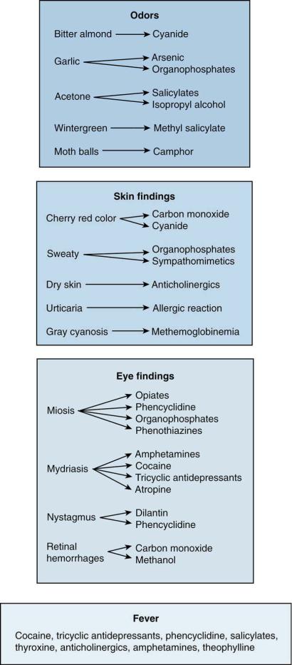
3.Physical examination should be comprehensive and may provide additional clues to the identity of the toxin. Figure 20-1 shows the link between some typical physical findings and associated toxins.
4.Laboratory studies
a.Screening laboratory tests include serum glucose, serum and urine toxicology screens, and electrolytes.
1.Anion gap [Na+ − (Cl− + HCO3−)] should be calculated.
2.Causes of an increased anion gap (>16) may be recalled using the mnemonic
AMUDPILES (alcohol, methanol, uremia, diabetic ketoacidosis, paraldehyde, iron and isoniazid, lactic acidosis, ethylene glycol, salicylates).
b.Radiographic imaging of the abdomen may reveal radiopaque substances. These may be recalled using the mnemonic CHIPE (chloral hydrate and calcium, heavy metals, iodine and iron, phenothiazines, enteric-coated tablets).
C.General management principles
705
1.The ABCs are the initial management priority.
2.If the patient has altered mental status, administer dextrose for hypoglycemia and naloxone for possible opiate overdose.
3.A poison control center may be consulted to assist with management.
4.Gastric decontamination
a.Activated charcoal has a very large adsorptive surface area that binds toxins and minimizes their absorption.
1.Activated charcoal should be considered for all poisonings. However, it is ineffective for some poisons such as iron, lithium, alcohols, ethylene glycol, iodine, potassium, and arsenic. In addition, activated charcoal interferes with visualization during endoscopy and therefore should not be used for caustic ingestions.
2.Evidence suggests that activated charcoal improves clinical outcomes, especially if given within 1 hour after an ingestion.
b.Whole-bowel irrigation (WBI) is rapid, complete emptying of the intestinal tract accomplished using polyethylene glycol (an osmotic agent) and an electrolyte solution (to prevent electrolyte imbalance). Preliminary studies show that WBI may be effective for ingestions of iron and other heavy metals and sustained-release medications.
c.Antidotes exist for a relatively small number of compounds (Table 20-3).
D.Specific poisonings
1.Acetaminophen. This drug is one of the most common medications ingested by children and adolescents. Doses >150 mg/kg are associated with toxicity.
a.Pathophysiology
1.Hepatic damage, the major sequelae of toxicity, is directly related to the depletion of glutathione, a cofactor used during the metabolism of acetaminophen by the cytochrome P-450 system.
2.Toxic intermediates produced when glutathione is depleted bind directly to hepatocytes, causing hepatocellular necrosis.
b.Clinical features. There are four stages of acetaminophen poisoning (Table 20-4).
c.Management
1.Activated charcoal
2.Obtain serum acetaminophen level 2–4 hours after ingestion. The level should be plotted on the Matthew–Rumack nomogram to determine the potential for hepatitis.
3.If the nomogram predicts hepatitis, the antidote, N-acetylcysteine (NAC), a glutathione precursor, is indicated.
a.NAC is given orally as a 140 mg/kg loading dose and is followed with 70 mg/kg every 4 hours for 17 doses.
b.NAC may be given intravenously as an infusion for more than 21 hours.
c.NAC is hepatoprotective if given within 8 hours of ingestion. It may still be helpful up to 72 hours after ingestion.
2.Salicylates. Salicylate poisoning has decreased as acetaminophen’s usage has increased; however, salicylates remain as an ingredient in many over-the-counter compounds, such as Pepto-Bismol, Ben-Gay, and oil of wintergreen. Doses >150 mg/kg are associated with toxicity.
a.Pathophysiology
1.Salicylates directly stimulate respiratory centers. This causes hyperventilation that may overcompensate for metabolic acidosis produced by
706
the salicylate (it is a weak acid), resulting in a respiratory alkalosis.
2.Salicylates uncouple oxidative phosphorylation, producing lactic acidosis and enhancing ketosis.
b.Clinical features. Common signs and symptoms include fever, diaphoresis, and flushed appearance; tinnitus; vomiting; headache; lethargy, restlessness, coma, and seizures; hyperpnea; and dehydration.
c.Laboratory findings
1.Respiratory alkalosis with an anion-gap metabolic acidosis is the most common acid–base disturbance.
2.Hyperglycemia, followed later by hypoglycemia
3.Hypokalemia
d.Management
1.Activated charcoal is effective and may be readministered every 4 hours in severe poisonings.
2.Obtain serum salicylate level at least 6 hours after ingestion. The level should then be plotted on the Done nomogram to assess for potential toxicity.
3.Alkalinization of urine with sodium bicarbonate to a urine pH > 7 and largevolume intravenous fluids enhance renal excretion of salicylates.
4.Dialysis may be required for life-threatening ingestions.
3.Iron
a.Epidemiology
1.Iron is one of the most common and potentially fatal childhood poisonings. As little as 20 mg/kg of iron is toxic.
2.Adult-strength ferrous sulfate tablets and iron in prenatal vitamins are the most common sources of accidental iron ingestion.
b.Pathophysiology
1.Direct damage to the gastrointestinal tract leading to hemorrhage
2.Hepatic injury and necrosis
3.Third spacing and pooling of blood in the vasculature leading to hypotension
4.Interference with oxidative phosphorylation
c.Clinical features. There are four stages of iron toxicity (Table 20-5).
d.Management
1.Activated charcoal does not bind to iron. However, if a polyingestion is suspected, activated charcoal should be given.
2.Hypovolemia, blood loss, and shock should be anticipated and treated.
3.WBI should be considered for life-threatening ingestion.
4.Serum iron level should be obtained 2–6 hours after ingestion.
5.Intravenous deferoxamine, an iron-binding ligand, should be given if any of the following condition exists:
a.If serum iron levels > 500 µg/dL, or if > 300 µg/dL and acidosis, hyperglycemia, or leukocytosis are present
b.If severe gastrointestinal symptoms are present
c.If more than 100 mg/kg of iron is ingested
6.Before the serum iron level is known, a test dose of deferoxamine may be administered. If the patient’s urine then turns red or pink (vin rose) (the color of chelated iron), the challenge is considered positive, indicating a clinically significant iron ingestion. Intravenous deferoxamine should then be continued.
4.Lead
a.Sources of lead include ingestion of lead-based paint chips, water carried by
707
outdated lead pipes, improperly glazed or foreign-made ceramic food or water containers, and pica (compulsive eating of nonnutrient substances such as dirt, paint, and clay). (See Chapter 1, section IV.J.)
b.Clinical features. Lead poisoning is typically a chronic ingestion; however, children may also present with acute lead intoxication.
1.Abdominal complaints include colicky pain, constipation, anorexia, and vomiting.
2.CNS complaints include listlessness, irritability, seizures, and decreased consciousness with encephalopathy.
3.Peripheral blood smear may show microcytic anemia, basophilic stippling, and red blood cell precursors.
4.Radiopacities may be seen on abdominal radiographs, and dense metaphyseal bands may be seen on radiographs of the knees and wrists (lead lines).
c.Diagnosis. An elevated lead level or elevated erythrocyte protoporphyrin is the basis of diagnosis.
d.Management. Treatment for significant toxicity includes dimercaprol, British antilewisite (BAL), or calcium disodium ethylenediaminetetraacetic acid (EDTA).
5.Caustic agents. These are acids or alkalis with corrosive potential.
a.Pathophysiology
1.Acids (e.g., toilet bowl cleaner) cause coagulation necrosis that leads to superficial damage to the mouth, esophagus, and stomach. More severe injury results from compounds that have a pH <2.
2.Alkalis (e.g., oven and drain cleaners, bleach, laundry detergent) cause liquefaction necrosis that produces deep and penetrating damage, most commonly to the mouth and esophagus. More severe injury results from compounds that have a pH >12.
b.Clinical features
1.Immediate burning sensation with intense dysphagia, salivation, retrosternal chest pain, and vomiting
2.Obstructive airway edema (especially with acid ingestion)
3.Gastric perforation and peritonitis may follow acid ingestion.
4.Esophageal perforation with mediastinitis may follow alkali ingestion.
c.Management. Treatment initially includes the ABCs.
1.No attempt should be made to neutralize the caustic agent, because the combination of acid and alkali will generate an exothermic reaction and worsen any burn.
2.Activated charcoal is contraindicated because it interferes with endoscopy.
3.Endoscopy is performed to assess the degree of damage.
4.Household bleach has less corrosive potential and generally does not require treatment.
6.Carbon monoxide poisoning
a.Epidemiology. Carbon monoxide (CO) is a by-product of incomplete combustion of carbon-containing material. Excessive exposure may occur from fires, tobacco, faulty home heaters, car exhaust, and industrial pollution. CO is odorless, tasteless, and colorless.
b.Pathophysiology. CO interferes with oxygen delivery and utilization.
1.Carbon monoxide displaces oxygen from the hemoglobin molecule, forming carboxyhemoglobin (CO-Hb), which can no longer carry oxygen. The bond between CO and hemoglobin is more than 200 times stronger than the bond
708
between oxygen and hemoglobin.
2.The oxygen–hemoglobin dissociation curve is shifted to the left. This leads to tighter binding of the remaining oxygen bound to hemoglobin and impaired release of oxygen to tissues.
3.Carbon monoxide also interferes with cellular oxidative metabolism.
c.Clinical features depend on the CO-Hb level.
1.Low levels are associated with nonspecific symptoms such as headache, flulike illness, dyspnea with exertion, and dizziness.
2.High levels are associated with visual and auditory changes, vomiting, confusion and later syncope, slurred speech, cyanosis, myocardial ischemia, coma, and death.
3.Classic physical examination findings, although uncommon, include cherry red skin (venous blood carries more oxygen than normal as a result of impaired release of oxygen to tissues) and retinal hemorrhages. Tachycardia and tachypnea may be present.
4.Young children (<8 years) have more symptoms at lower CO-Hb levels. Young children are also more likely to have gastrointestinal symptoms (e.g., vomiting and diarrhea) instead of neurologic symptoms.
5.Delayed permanent neuropsychiatric syndrome, consisting of memory loss, personality changes, deafness, and seizures, may occur in some victims up to 4 weeks after CO exposure.
d.Diagnosis is made by measuring the CO-Hb level. It is important to remember that CO-Hb levels are not always indicative of the degree of CO exposure and may even be low in victims with significant intoxication. Other abnormal findings include anion-gap metabolic acidosis, low oxygen saturation (however, PaO2 may be normal), and evidence of myocardial ischemia on ECG or elevated cardiac enzymes.
e.Management
1.One hundred percent oxygen is administered to displace CO from hemoglobin.
2.If available, hyperbaric oxygen more rapidly displaces CO from hemoglobin as compared with oxygen alone and also improves oxygen delivery to tissues.
3.Hospitalization is indicated for CO-Hb levels > 25%, CO-Hb levels > 10% during pregnancy, history or presence of neurologic symptoms, or presence of metabolic acidosis or ECG changes.
709
710
FIGURE 20.1 Selected physical examination findings and their associated toxins.
Table 20-3
Selected Toxins and Their Antidotes
Toxin |
|
|
|
Antidote |
|
Acetaminophen |
|
|
NAC |
||
Anticholinergic agents |
|
|
Physostigmine |
||
Benzodiazepines |
|
|
Flumazenil |
||
Black widow spider envenomation |
Antivenin Latrodectus mactans |
||||
Carbon monoxide |
|
|
Oxygen |
||
Coral snake envenomation (Eastern U.S. or Texas coral |
Antivenin Micrurus fulvius |
||||
snake) |
|
|
|
|
|
Cyanide |
|
|
Cyanide antidote kit (contains amyl nitrite, sodium nitrite, sodium |
||
|
|
|
|
thiosulfate) |
|
Hydroxocobalamin (vitamin B12) |
|
|
|||
Digitalis glycosides |
|
|
Digoxin-specific Fab antibodies |
||
Heavy metals (e.g., mercury, manganese, copper, gold, |
d-Penicillamine (for lead, mercury, arsenic, copper) |
||||
nickel, zinc, lead, arsenic) |
|
|
|
|
|
|
|
|
|
Dimercaprol (British anti-lewisite [BAL] in oil) for all the heavy metals |
|
|
|
|
|
and lewisite (chemical weapon) |
|
|
|
|
|
DMSA (for lead and possibly mercury, arsenic, other metals) |
|
|
|
|
|
EDTA, calcium (for lead, nickel, zinc, manganese) |
|
Inducers of dystonia |
|
|
Diphenhydramine |
||
Benztropine |
|
|
|
|
|
Inducers of methemoglobinemia |
Methylene blue |
||||
Iron |
|
|
|
Deferoxamine |
|
Isoniazid |
|
|
Pyridoxine (vitamin B6) |
||
Methanol; ethylene glycol |
|
|
Ethanol, fomepizole |
||
Narcotics |
|
|
Naloxone |
||
Organophosphates; carbamate pesticides |
Atropine |
||||
Pralidoxime (for organophosphates) |
|
|
|||
Pit viper snake bite (rattlesnake, water moccasin, |
Crotalidae polyvalent, antivenin, Crotalidae polyvalent Fab antibodies |
||||
copperhead envenomation) |
|
|
|
|
|
β-Blockers; calcium-channel blockers |
Glucagon |
||||
Sulfonylurea oral hypoglycemic agents |
Octreotide |
||||
|
|
|
|
Glucagon |
|
EDTA = ethylenediaminetetraacetic acid; DMSA = dimercaptosuccinic acid; NAC = N-acetylcysteine. |
|||||
Table 20-4 |
|
|
|
|
|
Stages of Acetaminophen Toxicity |
|
|
|||
|
|
|
|
|
|
|
|
|
|
|
|
Stage |
Time After |
Signs and Symptoms |
|
|
|
Ingestion |
|
|
|||
|
|
|
|
|
|
1 |
30 minutes– |
Asymptomatic, or vomiting and diarrhea |
|||
|
24 hours |
|
|
|
|
2 |
24–72 hours |
Gastrointestinal symptoms resolve; at 36 hours, hepatic transaminases begin to increase |
|||
3 |
72–96 hours |
Hepatic necrosis, jaundice, hypoglycemia, lactic acidosis, hepatic encephalopathy, coagulopathy, |
|||
|
|
and renal failure |
|
|
|
4 |
4 days–2 weeks |
Resolution of symptoms, progressive liver damage requiring liver transplantation, or death |
|||
Table 20-5 |
|
|
|
|
|
Stages of Iron Toxicity |
|
|
|
|
|
|
|
|
|
|
|
|
|
|
|
|
|
Stage |
Time After Ingestion |
Signs and Symptoms |
|
|
|
1 |
1–6 hours |
|
Abdominal pain, vomiting, diarrhea, GI bleeding |
|
|
|
|
|
Shock from bleeding and vasodilation |
|
|
|
|
|
Fever and leukocytosis |
|
|
2 |
6–12 hours |
|
Resolution of stage 1 symptoms |
|
|
3 |
12–36 hours |
|
Metabolic acidosis |
|
|
|
|
|
|
|
|
711
|
|
Circulatory collapse |
|
|
Hepatic and renal failure |
|
|
DIC |
|
|
Neurologic deterioration |
4 |
2–6 weeks |
Late sequelae includes pyloric or intestinal scarring with stenosis |
DIC = disseminated intravascular coagulation; GI = gastrointestinal.
712
IX. Mammalian Bites
A.Epidemiology
1.Dogs (80% of bites), cats, rodents, other wild or domesticated animals, and humans (2– 3% of bites) may all cause a bite injury.
2.Most bites occur in boys during the spring and summer months.
B.Dog bites
1.Clinical features
a.Bites range in severity from scratches, punctures, and lacerations to severe soft tissue injury. Note that the jaw pressure of a dog may exceed 200–450 pounds per square inch.
b.Young children are typically bitten on the head and neck, whereas older children are bitten predominantly on the extremities.
c.Secondary infections may result from anaerobic and aerobic organisms, such as
Staphylococcus aureus, Pasteurella multocida, and Streptococcus species.
2.Management. Treatment includes meticulous and prompt local wound care.
a.Copious wound irrigation
b.Wounds on the face or large wounds should be sutured if less than 12 hours old.
Facial wounds <24 hours old can be sutured because the face has increased vascularity, which assists with healing and minimizes the risk of infection.
c.Wounds at high risk for infection include those on the hand, wrist, and foot, and small puncture wounds.
d.Antibiotic prophylaxis, such as amoxicillin–clavulanic acid, should be administered.
e.Tetanus prophylaxis should be given if needed.
1.Tetanus toxoid vaccine should be given if the most recent tetanus vaccine was given greater than 5 years ago.
2.Tetanus immune globulin may also be recommended if the date of the last tetanus vaccine is unknown.
C.Cat bites
1.Clinical features
a.Puncture wounds to the upper extremity are most common.
b.Victims have a high risk of infection due to P. multocida.
c.Cat scratch disease (regional lymphadenitis) may also develop (see Chapter 7, section XVII.B).
2.Management. Treatment is similar to the management of dog bites. Because the injury is typically a puncture wound, adequate irrigation is often difficult.
D.Human bites
1.Clinical features
a.Wound is typically located on the trunk or face in young children. If the wound occurred during a fistfight, it is typically located at the metacarpophalangeal (MP) joint. Wounds to the MP joint are extremely serious, because infection may penetrate the avascular fascial layers, resulting in deep infection and tendonitis.
b.Infection rate is high.
1.Mixed bacterial infection is often present. Pathogens include Streptococcus viridans, S. aureus, and anaerobic bacteria such as Bacteroides, Peptostreptococcus, and Eikenella corrodens.
2.Other systemic infections, such as hepatitis B, HIV, and syphilis, may also be transmitted.
713
c.Management. Treatment includes copious wound irrigation, closure of large lacerations, and antibiotics (e.g., amoxicillin–clavulanic acid).
714
X.Biologic Poisonings (Venoms)
A.Black widow spider (Latrodectus species)
1.General concepts
a.The black widow spider is characterized by a red or orange hourglass marking on the ventral surface.
b.Only the female spider is dangerous, and the female spider only bites if provoked.
c.The web is located in dark recesses, such as closets, woodpiles, and attics.
d.Black widows are found in temperate regions throughout the world, which include the United States, southern Europe and Asia, Australia, Africa, and much of South America.
2.Clinical features result from the venom, a potent neurotoxin.
a.The bite causes few local symptoms, except for burning or a sharp pinprick sensation.
b.Pathognomonic signs and symptoms include severe hypertension and muscle cramps.
c.Nonspecific symptoms, such as headache, dizziness, nausea, vomiting, anxiety, and sweating, may also occur.
3.Management
a.Local wound care, including wound irrigation and tetanus prophylaxis (if needed), is important.
b.Benzodiazepines and narcotics may relieve muscle cramps.
c.Latrodectus antivenin is given for signs and symptoms suggesting severe envenomation.
B.Brown recluse spider (Loxosceles species)
1.General concepts
a.The brown recluse spider (also called the fiddleback spider) is characterized by a brown violin-shaped marking on the dorsum of the thorax.
b.The spider only bites if provoked.
c.The web is located in dark recesses.
d.The known geographic range of the brown recluse spider extends from Nebraska to Ohio and across the south from Texas to Florida.
2.Clinical features result from the venom, a cytotoxic compound containing tissuedestructive enzymes.
a.The bite results in little initial pain. However, 1–8 hours later, a painful itchy papule that increases in size and discolors during the course of 3–4 days develops at the site of the bite.
b.Some patients develop a necrotic and ulcerated deep lesion at the bite site.
c.Systemic reactions may occur 24–48 hours after the bite with fever, chills, weakness, vomiting, joint pain, DIC, hemolysis, and renal failure from myoglobinuria.
3.Management. Treatment includes local wound care and tetanus prophylaxis, if needed. Treatment of a necrotic ulcer is controversial but may include steroids, skin grafting, dapsone, and hyperbaric oxygen. There is no antivenin.
C.Pit viper snakes (family Crotalidae) account for more than 95% of all snakebites. Rattlesnake, cottonmouth, and copperhead snakes are members of the Crotalidae family.
1.Pathophysiology
a.Bite location and amount of venom injected determine the severity of envenomation. Head and trunk bites are most severe.
715
b.Venom is a complex mixture of proteolytic enzymes.
2.Clinical features
a.Local findings include puncture marks and progressive severe swelling and ecchymosis.
b.Systemic effects include paresthesias of the scalp, periorbital fasciculations, weakness, diaphoresis, dizziness, nausea, and a metallic taste in the mouth.
c.Coagulopathy, thrombocytopenia, hypotension, and shock may also develop.
3.Management. Treatment involves local wound care, tetanus prophylaxis if needed, immobilization of the bitten extremity, and immediate transport to the nearest emergency department.
a.Incision and suction are not recommended.
b.Tourniquets, ice, and direct pressure on the wound may cause more injury.
c.Crotalidae polyvalent antivenin should be considered for all bites.
1.Children require more antivenin because they receive proportionally more venom per kilogram body weight.
2.Antivenin is most effective if given within 4–6 hours of the bite.
3.Complications of antivenin are common and include serum sickness and anaphylaxis.
d.Crotalidae polyvalent immune Fab is also available for envenomation and is safe, more potent, and very effective.
D.Coral snakes (family Elapidae) account for 1–2% of all snakebites. Coral snakes may be identified by their stripe pattern using the mnemonic “red next to yellow, kill a fellow; red next to black, venom lack.”
1.Clinical features result from the neurotoxic venom and include mild local swelling and tenderness and severe systemic symptoms, such as paresthesias, vomiting, weakness, diplopia, fasciculations, confusion, and respiratory depression.
2.Management should be aggressive and includes antivenin (available only for the Eastern U.S. and Texas coral snake), local wound care, and supportive care.
716