


Dysmorphic erythrocytes on electronic microscopy (A – normal erythrocyte)

Leucocyturia - more then 2000 cells in 1 ml
N 80% neutrophiles, 20% lymphocytes
Lymphocytes > 20% |
Active SLE nephritis |
|
Subacute GN |
|
Exacerbation of chronic GN |
|
Nephrotic syndrome |
|
Interstitial nephritis |
|
Transplant regection |
Neutrophiles 90-100% |
UTI |
|
|
Macrophages |
Amiloidosis |
|
|
Eosinophiles 5-60% |
Drug-induced nephritis |
|
Interstitial nephritis |
|
Rapidly progressing GN |
|
Ig A- nephropathy |


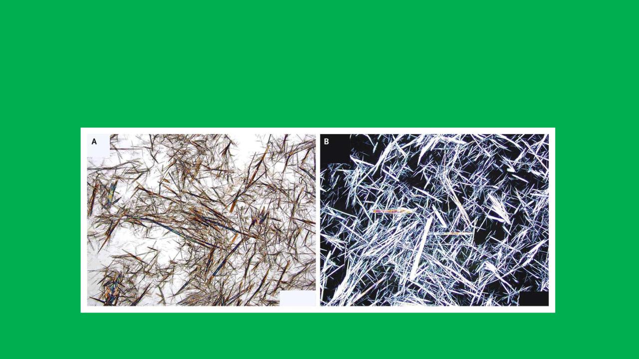
Crystalluria


Measurement of the glomerular filtration rate
Method
Urea
Plasma creatinine
Creatinine clearance
51Cr-EDTA practice
Inulin clearance
Comments
Poor surrogate
variable production ratevariable excretion rate
Better than urea
Poor discrimination at near-normal GFR
Reasonable surrogate but depends on accurate timed urine collection (uaually 24 hours)
The best surrogate in clinical Expensive
Near perfect measurement of GFR but
needs continuous infusiondifficult urine and plasma assays
research studies only – not suited to clinical practice
31