
- •Contents
- •Methods of Otoscopy
- •The Normal Tympanic Membrane
- •Secretory Otitis Media (Otitis Media with Effusion
- •Cholesterol Granuloma
- •Atelectasis, Adhesive Otitis Media
- •Non-Cholesteatomatous Chronic Otitis Media
- •Chronic Suppurative Otitis Media with Cholesteatoma
- •Congenital Cholesteatoma of the Middle Ear
- •Petrous Bone Cholesteatoma
- •Glomus Tumors (Chemodectomas)
- •Meningoencephalic Herniation
- •Postsurgical Conditions
- •References
- •Index

83
11 Glomus Tumors (Chemodectomas)
The glomus body was first described by Guild in 1941 as a small highly vascular mass of epithelioid cells located in the region of the adventitia of the jugular bulb. In 1953, Guild described glomus formations along the tympanic branches of the glossopharyngeal and vagus nerves (Jacobson's and Arnold's nerves, respectively).
Glomus bodies are mainly found in the tympanic region, jugular bulb, at the carotid bifurcation, and related to the vagus nerve. They are classified as paraganglia that are derived from the neural crest. While the carotid and vagal bodies function as chemoreceptors stimulated by the changes in the oxygen tension, tympanic and jugular bulb paraganglia do not exhibit this function.
The term glomus tympanicum is reserved for tumors that originate from the mesotympanum while the term glomus jugulare is attributed to those cases that arise from the jugular bulb or the hypotympanum with secondary invasion of the bulb. These tumors are highly vascular and they derive the blood supply mainly from the ascending pharyngeal artery. It is claimed that they have a hereditary transmission as autosomal dominant traits with penetrance that increases with age.
In the majority of cases, the initial symptoms are hearing loss (conductive, sensorineural, or mixed) and pulsatile tinnitus synchronous with pulse. The tumor can extend into the labyrinth, causing vertigo of peripheral origin; towards the jugular foramen, leading to deficits of one or more of the lower cranial nerves (IX-XI); or towards the occipital condyle, leading to hypoglossal nerve paralysis. Patients suffering from preoperative affection of the lower cranial nerves run a better postoperative course as compensation of the contralateral side would have already started. The contralateral vocal cord compensates by crossing the midline to meet the paralyzed cord, thereby markedly reducing the risk of aspiration pneumonia. On the other hand, patients with preoperative intact lower cranial nerves in whom the nerves are sacrificed during the operation suffer from deglutition problems in the postoperative course. Nasogastric feeding is used in such cases and oral feeding is resumed only when compensation from the contralateral side occurs. A useful alternative is vocal cord medialization either by Teflon injection or by medialization thyroplasty using cartilage or silicon.
The tumor can also extend into the petrous apex, leading to paralysis of the abducent nerve and trigeminal neuralgia, or invade the mastoid, resulting in facial nerve paralysis. Further extension can also occur in the external auditory canal. Tumors occupying the exter-
nal auditory canal can lead to serous or purulent otorrhea due to irritation of the skin and retention of squamae and epithelial debris. Hemorrhagic discharge rarely occurs.
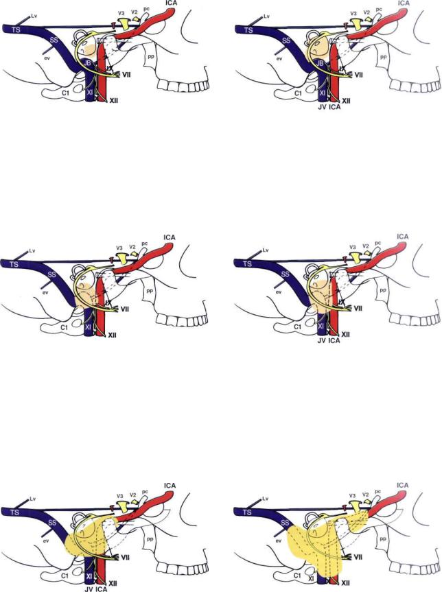
Fisch classified glomus tumors into four classes based on location and extension seen on high-resolu- tion CT scans (see Table 11.1 and Figs. 11.1-11.6).
On otoscopy, a retrotympanic pulsatile mass is usually seen in the inferior quadrants. The mass is red or bordeaux red. In some cases the mass may have a red- dish-blue color due to the presence of middle ear effusion secondary to eustachian tube blockage. The tumor may be seen as a polyp in the external auditory canal either due to erosion of the floor of the canal or to the tumor breaking through the tympanic membrane.
The diagnosis can be made clinically (history and otoscopic findings). Computed tomography (CT) with contrast and magnetic resonance imaging (MRI) with gadolinium allow exact definition of the tumor extension. Radiology also helps to differentiate between glomus tumors and other lesions such as aberrant carotid artery, high jugular bulb, cholesterol granuloma, or meningioma extending into the middle ear. Carotid and vertebral angiography allows identification of the arteries supplying the tumor; and they should be embolized before surgery to avoid excessive intraoperative bleeding.
In cases in which the horizontal carotid artery is engulfed by the tumor, the balloon occlusion test is indispensable for studying the perfusion by the contralateral carotid artery as well as for the safety of the closure of the involved carotid.
Table 11.1 Classification of glomus tumors according to Fisch (1978)
Class A: Glomus tympanicum
Class B: Tympanomastoid
Class C: Glomus jugulare
CI: Carotid foramen
C2: Vertical ICA until genu
C3: Horizontal ICA
C4: ICA + FL
Class D: Intracranial extension
De (1-2): Intracranial extradural
Di (1-2): Intracranial intradural
ICA = internal carotid artery; FL = anterior foramen lacerum
84 11 Glomus Tumors (Chemodectomas)
JVICA
Figure 11.1 The class A tumor originates from glomus formations along the course of Jacobson's nerve. They are localized to the middle ear. Abbreviations are given in Figure 10.1.
JVICA
Figure 11.3 The class C tumor originates in the dome of the jugular bulb and destroys the infralabyrinthine compartment. The tumor may spread in the following directions: interiorly, along the internal jugular vein and cranial nerves IX—XII; superiorly, towards the otic capsule and the internal auditory canal; posteriorly, into the sigmoid sinus; anteriorly, to the internal carotid artery; more medially, to the petrous apex and the cavernous sinus; or laterally, to the hypotympanum and middle ear. Class C tumors are further subdivided according to the degree of erosion of the carotid canal. The C1 tumor erodes the carotid foramen without involvement of the carotid artery.
Figure 11.5 The class C3 tumor involves the horizontal segment of the carotid.
Figure 11.2 The class B tumor originates at the level of the promontory and invades the hypotympanum without affecting the jugular bulb. The tumor also can extend into the mastoid and the retrofacial air cells.
Figure 11.4 The class C2 tumor erodes the vertical carotid canal up to the carotid genu.
JV ICA
Figure 11.6 The class C4 tumor grows to the anterior foramen lacerum and extends to the cavernous sinus. Class D indicates intracranial extension of the tumor. This might be extradural (De) or intradural (Di).
Glomus Tumors (Chemodectomas) |
85 |
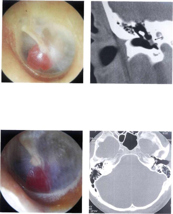
Figure 11.7 Left ear. Glomus tympanicum or class A tumor. The small red mass behind the anteroinferior quadrant is localized on the promontory and does not extend towards the hypotympanum (see Fig. 11.7).
Figure 11.8 CT scan of the case presented in Figure 11.7. The lesion is limited to the region of the promontory. There are no visible signs of bone erosion.
Figure 11.9 Left ear. Class A glomus tumor. The tumor is |
Figure 11.10 CT scan of the case described in Figure 11.9. |
|
again limited to the promontory (see Figs. 11.10 and 11.11). |
||
|
86 11 Glomus Tumors (Chemodectomas)
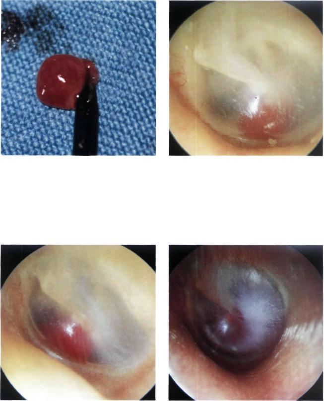
Figure 11.11 The tumor was removed using a transcanal |
Figure 11.12 Left ear. Another example of a small class A |
approach after having bipolarly coagulated the tympanic |
glomus tumor. |
arteries that supply the tumor. |
|
Figure 11.13 Left ear. This small glomus tympanicum tumor is situated in the anteroinferior quadrant of the middle ear near the tubal orifice. Further growth of the tumor can block the tubal orifice, leading to middle ear effusion.
Figure 11.14 Left ear. Class B glomus tumor or hypotympanic tumor. The reddish mass is visible through the inferior quadrants of the tympanic membrane.
Glomus Tumors (Chemodectomas) |
87 |
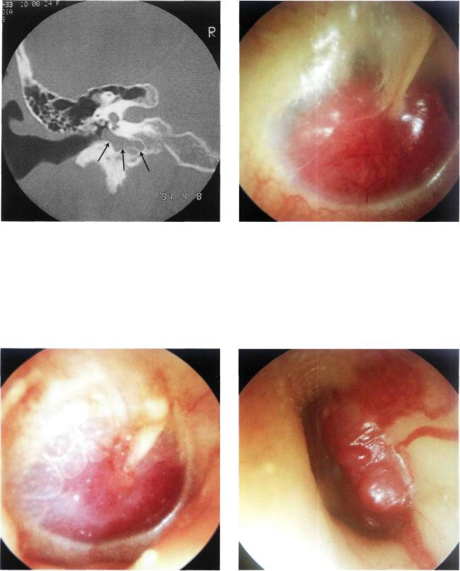
Figure 11.15 CT of the case presented in Figure 11.14. Tumor extension towards the hypotympanum is observed. There is no erosion of the bony plate covering the jugular bulb.
Figure 11.16 Right ear. Class B glomus tumor. The highly vascular red tumor mass pushes the tympanic membrane laterally. A middle ear effusion is present.
Figure 11.17 Right ear. Class B glomus tumor. An air-fluid level due to middle ear effusion is seen together with the tumor. A tympanoplasty removed all of the tumor while conserving the excellent preoperative hearing.
Figure 11.18 Left ear. Type B glomus tumor. The tumor causes bulging of the posterior quadrants of the tympanic membrane (see CT scan, Fig. 11.19).
88 11 Glomus Tumors (Chemodectomas)
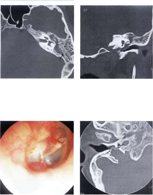
Figure 11.19 CT scan of the case in Figure 11.18. An axial section demonstrates the presence of effusion in the mastoid due to retention.
Figure 11.20 CT scan of the case in Figure 11.18. The tumor extends to the hypotympanum but does not erode the bone overlying the dome of the jugular bulb.
Figure 11.21 Right ear. Reddish mass protruding from the inferior wall of the external auditory canal.
Figure 11.22 CT scan of the previous case. Axial view demonstrating the erosion caused by the tumor of the bone overlying the jugular bulb. This tumor can be considered an intermediate class between B and C. The tumor is localized in the hypotympanum and extends to the jugular bulb but does not invade it.
Glomus Tumors (Chemodectomas) |
89 |
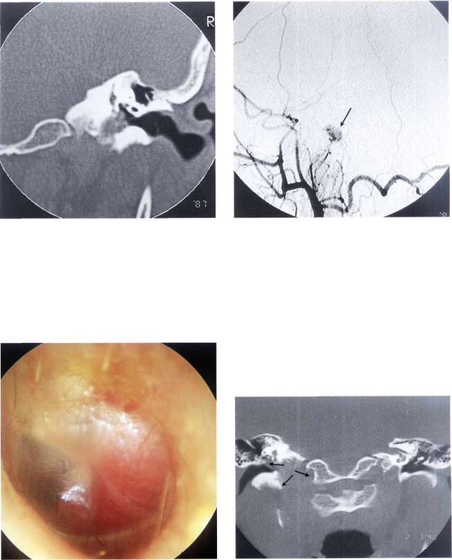
Figure 11.23 Coronal section giving a better view of the tumor extension towards the jugular bulb. Intraoperatively, no invasion of the bulb was noted and the integrity of the bulb was thus conserved.
Figure 11.24 Angiography of the same case. The blood supply of the tumor (arrow) is derived from the ascending pharyngeal artery that is a branch of the external carotid artery.
Figure 11.25 Left ear. Class C1 glomus tumor. The only complaint of the patient was ipsilateral pulsatile tinnitus of 4 years' duration (see following figures).
Figure 11.26 CT scan, coronal view showing enlargement of the jugular foramen with extension of the tumor into the middle ear.
90 11 Glomus Tumors (Chemodectomas)
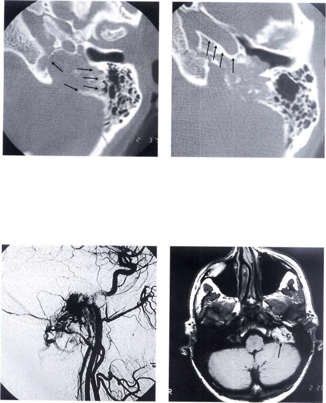
Figure 11.27 CT scan, axial view. The jugular foramen is enlarged. Irregular erosion of the borders of the jugular foramen can be observed (differential diagnosis with lower cranial nerves' schwannoma).
Figure 11.28 Axial view demonstrates that the horizontal segment of the internal carotid artery is free of tumor.
Figure 11.29 Angiography demonstrating that the blood supply of the tumor comes from the ascending pharyngeal, the occipital, and the posterior auricular arteries.
Figure 11.30 MRI with gadolinium. The tumor is enhancing except for some flow-void zones corresponding to large vascular spaces. This picture is pathognomonic of glomus tumors.
Glomus Tumors (Chemodectomas) |
91 |
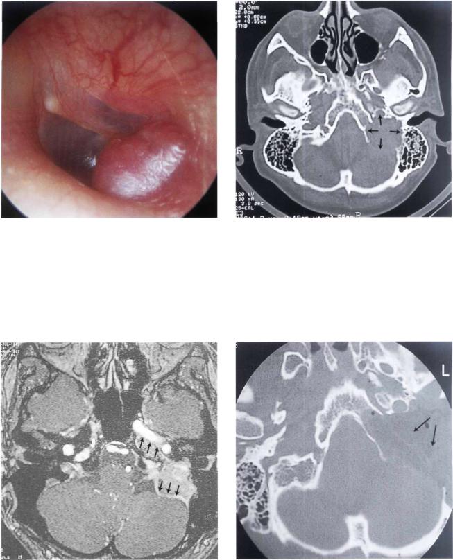
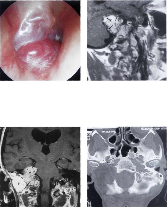
Figure 11.31 Class C2 De2 glomus jugulare tumor of the left ear. The patient complained of pulsatile tinnitus, hearing loss, and 2 months before presentation started to suffer from dysphonia, dysphagia, and hypoglossal paresis. The affection of the lower cranial nerves was progressive in nature. It resulted from compression by the slowly growing tumor.
Figure 11.32 CT scan of the case presented in Figure 11.31. The marked erosion of the jugular foramen and the vertical portion of the carotid canal can be appreciated.
Figure 11.33 MRI demonstrating tumor in contact with the medial aspect of the horizontal carotid artery and the posterior fossa dura without infiltrating it.
Figure 11.34 Postoperative CT scan demonstrating tumor removal using an infratemporal fossa approach type A.
92 11 Glomus Tumors (Chemodectomas)
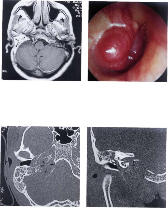
Figure 11.35 Right ear. Class C3 Di2 glomus jugulare tumor. The patient complained of pulsatile tinnitus and mixed hearing loss of 12 months' duration.
Figure 11.36 MRI, sagittal view demonstrating intradural extension of the tumor.
Figure 11.37 MRI, coronal view after first-stage removal of the extradural component of the tumor using an infratemporal fossa approach type A. The fat (F) obliterating the operative cavity can be seen. The intradural tumor residue (T) is also observed. Staging is necessary in such cases to avoid communication between the subarachnoid space and the wide open neck spaces.
Figure 11.38 Postoperative CT scan after the second-stage removal of the tumor through a petro-occipital trans-sigmoid approach (POTS).
Glomus Tumors (Chemodectomas) |
93 |
Figure 11.39 MR I demonstrating obliteration of the operative cavity with abdominal fat.
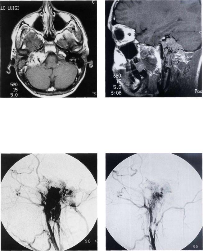
Figure 11.40 Right ear. Class C3 Di2 glomus jugulare tumor. The patient complained of ipsilateral total hearing loss, diplopia, grade IV facial paralysis, and dysphonia (see following figures).
Figure 11.41 CT scan axial section demonstrating the involvement of the jugular foramen and the horizontal segment of the internal carotid artery. The artery was closed preoperative^ with a balloon.
Figure 11.42 CT scan, coronal section. The tumor involves the internal auditory canal.
94 11 Glomus Tumors (Chemodectomas)
Figure 11.43 MRI, axial view giving a global idea of the |
Figure 11.44 MRI, sagittal view, |
extraand intradural extension of the tumor. |
|
Figure 11.45 Angiography before embolization. |
Figure 11.46 Angiography showing marked reduction of |
|
the tumor vascularity following embolization. |
Glomus Tumors (Chemodectomas) |
95 |
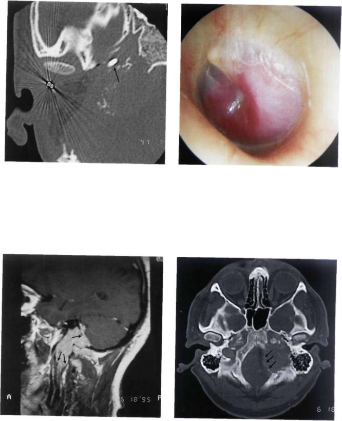
Figure 11.47 CT scan performed after first-stage removal of the extradural part of the tumor using an infratemporal fossa approach type A. Staging is necessary to avoid communication between the subarachnoid spaces and the neck spaces. The balloon used for the closure of the internal carotid artery can be seen (arrow).
Figure 11.48 Left ear. Class C2 Di2 glomus jugulare tumor. The patient complained of hearing loss and pulsatile tinnitus of 2 years' duration. He also complained of dysphonia, dysphagia, paralysis of the left half of the tongue, and paresis of the lower face.
Figure 11.49 MRI, sagittal view demonstrating the intradural extension of the tumor as well as the inferior extension towards C1 and C2.
Figure 11.50 Preoperative CT scan. The jugular foramen is enlarged, with involvement of the foramen magnum.
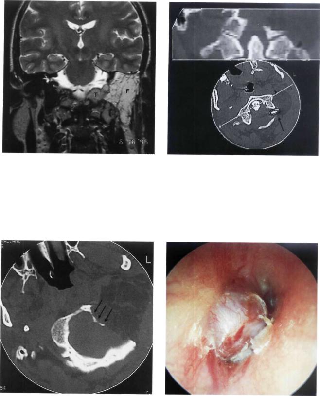
96 11 Glomus Tumors (Chemodectomas)
Figure 11.51 MRI with gadolinium after removal of the extradural part using an infratemporal fossa approach type A. Fat is seen obliterating the operative cavity (F). The intradural tumor residue at the level of the foramen magnum is noted
(T).
Figure 11.52 CT scan following the second-stage removal of the intradural portion of the tumor using an extreme lateral approach. The balloon used to close the vertebral artery is visible.
Figure 11.53 CT scan following the second-stage removal |
Figure 11.54 Another example of a large class C3 Di2 glo- |
|
of the intradural portion of the tumor. The removal of a large |
||
mus tumor. |
||
part of the left occipital condyle is also shown. |
|

Figure 11.55 MRI of the case in Figure 11.54 (T= tumor).
Summary
Because of the complex anatomy of the temporal bone and the structures at the base of the skull, as well as the invasiveness, rich vascularity, and aggressive behavior of glomus tumors, surgery for these difficult lesions is problematic.
Glomus tumors generally present with hearing loss and pulsatile tinnitus. When the lower cranial nerves are invaded, a jugular foramen syndrome becomes manifest.
Otoscopy usually reveals a reddish retrotympanic mass. A definitive diagnosis is obtained after neuroradiological studies are performed. These include a high-resolution CT scan with bony window, MRI with and without gadolinium, and digital subtraction angiography. Radiological studies are essential not only to confirm the diagnosis and define the exact tumor class, but also to properly evaluate these tumors. The neuroradiologist should be able to inform the surgeon about the following:
•Details of the osseous lesion
•Involvement of the jugular bulb and foramen
•Exact involvement of the temporal bone
•The presence of inner ear invasion
•The relationship between the fallopian canal and the tumor
•Carotid canal erosion and exact involvement of the internal carotid artery
Glomus Tumors (Chemodectomas) |
97 |
•Invasion of the petrous apex and clivus
•Details regarding the relationship between the tumor and surrounding soft tissues, e.g.:
-degree of neck extension
-infratemporal fossa involvement
-intracranial and intradural extension
Radiology also helps to determine the superior and inferior extension of the tumor, the possibility of other associated lesions (e.g., contralateral glomus or carotid body tumor), as well as the patency of the contralateral sigmoid sinus and internal jugular vein. In class C and D tumors, selective digital subtraction angiography is essential. Arteriography is performed for both ipsilateral and contralateral internal and external carotids and for the vertebrobasilar system. A study of the venous phase is also of great importance.
Arteriography of the external carotid artery defines the exact feeding vessels for further embolization. In all tumors of class C and D, embolization is fundamental.
Arteriography of the internal carotid artery shows vascularization from the caroticotympanic artery and from the cavernous branches of the artery as well as the exact status of arterial invasion by the tumor.
Study of the vertebrobasilar system demonstrates the vascularization of intracranial extension of the tumor from muscular, meningeal, and parenchymal (PICA, AICA) branches. Arterial supply from these latter branches indicates a definite intradural extension of the tumor. This study also provides indications for the possibility of embolizing muscular or meningeal branches.
When arteriography shows clear involvement of the internal carotid artery in its horizontal segment (C3 and C4 tumors), a balloon occlusion test to evaluate the collateral circulation and the possibility of sacrificing the artery is necessary. In some selected cases, when the temporary balloon occlusion test is negative, it might be necessary to perform a permanent closure of the artery 30 to 40 days before operation. In 1978, Fisch classified these lesions into four types: A, B, C, and D. He introduced the type A infratemporal fossa approach for the management of tumors localized in the jugular foramen that were considered inoperable at that time due to the presence of the facial nerve in the middle of the operative field and the inaccessibility of the internal carotid artery and petrous apex. To overcome these obstacles, Fisch proposed anterior rerouting of the facial nerve, giving direct access to the whole intratemporal course of the internal carotid artery as well as an excellent control of the large venous sinuses. Hearing loss is the only permanent postoperative deficit in this approach and is the result of obliteration of the middle ear.
The type A infratemporal fossa approach is generally used for the removal of class C and D glomus tumors of the temporal bone according to the Fisch classification. In cases with intradural extension
98 11 Glomus Tumors (Chemodectomas)
exceeding 2 cm in diameter, staging is indicated where the intradural part is removed in a second stage 6 to 8 months after the first operation. This surgical strategy avoids the high risk of having postoperative CSF leak should a single-stage removal be attempted. The reason for such a risk is the need to resect a wide area of the dura infiltrated by the tumor, and hence the subarachnoid space becomes widely connected to the open neck spaces. Using the staging strategy, we never experienced any CSF leak in our cases.
To sum up, the infratemporal fossa approach offers a wide access to the lateral skull base. The adequate exposure and systematic management of the important arteries and venous sinuses greatly reduces the intraoperative hemorrhage. An accurate preoperative study of the tumor extension, the preoperative tumor embolization, and the eventual closure of an invaded internal carotid artery (when feasible) by the neuroradiologist are prerequisites for successful surgery. Therefore, the collaboration between the neuroradiologist and the skull-base surgeon is of paramount importance. Lesions of the skull base are rare and very difficult to treat. Management of such cases should be restricted to specialized centers to avoid any serious problems.
•Meningioma
•Differential Diagnosis with Other Retrotympanic Masses
A variety of diseases can present as a mass behind an intact tympanic membrane. A detailed history of the patient, audiological assessment, and proper radiological evaluation are essential to reach a proper diagnosis. Table 11.2 summarizes the most common conditions causing a retrotympanic mass. For details of each condition, the reader is referred to the relevant chapters.
Table 112 Conditions that may present as a retrotympanic mass
Anomalous anatomy High jugular bulb Aberrant carotid artery
Tumors and tumor-like conditions Congenital cholesteatoma Iatrogenic cholesteatoma Glomus tumor
Facial nerve tumor (neuroma, hemangioma) Carcinoid tumor
Adenoma, adenocarcinoma
Meningioma (Primary or secondary to temporal bone invasion)
Rhabdomyosarcoma of the tensor tympani Miscellaneous
Meningoencephalic herniation
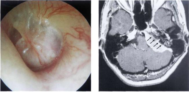
Figure 11.56 Left ear. This patient presented with dysphagia as her only symptom. A nonpulsating retrotympanic mass was noticed. The mass was whitish rather than the reddish color characteristic of glomus tumor. CT scan and MRI demonstrated an en-plaque meningioma invading the posterior surface of the temporal bone.
Figure 11.57 MRI of the case presented in Figure 11.56. Large posterior fossa meningioma located along the posterior surface of the petrous bone.
Glomus Tumors (Differential Diagnosis) |
99 |
• Facial Nerve Neurinoma
Figure 11.58 Postoperative CT scan of the case described in Figure 11.56. The tumor was removed using a modified transcochlear approach. The surgical cavity was obliterated using abdominal fat.
Figure 11.59 Left ear. Otoscopic view similar to the previous case. A whitish retrotympanic mass is seen causing bulging of the posterior quadrants of the tympanic membrane. A small reddish mass is visible in the posterior inferior regions of the external auditory canal (i.e., lateral to the annulus). The patient complained of left hearing loss and nonpulsating tinnitus of 2 years' duration. In the last 3 months before presentation, left facial nerve paresis started to appear (see following figures).
Figure 11.60 CT scan, axial view, of the case presented in Figure 11.59. The tumor is centered on the left iuqular foramen.
Figure 11.61 CT scan, coronal view. The mass eroded the bony plate over the jugular bulb extending into the hypotympanum.
100 11 Glomus Tumors (Chemodectomas)
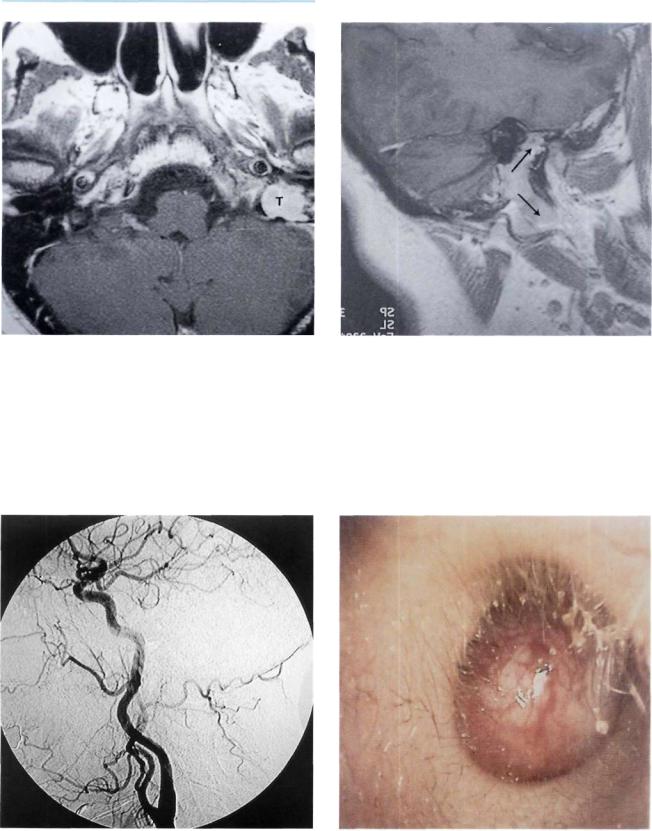
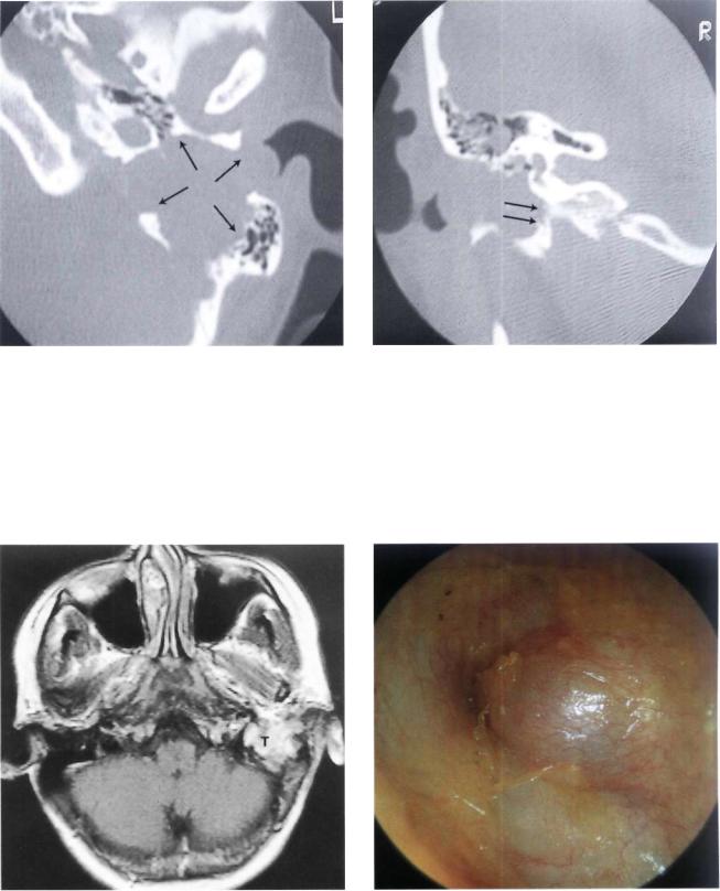
Figure 11.62 MRI, axial view, shows a mass centered on the |
Figure 11.63 MRI, sagittal view, of the case in Figure 11.59. |
jugular foramen (T= tumor). |
|
Figure 11.64 Angiography did not show the characteristic tumor blush of glomus tumors. During surgery, the tumor proved to be a facial nerve neurinoma, as confirmed later by histopathological examination. The tumor was arising from the mastoid segment of the nerve and extended to the jugular bulb.
Figure 11.65 Left ear. The patient complained of left facial twitches of 8 months' duration, sensation of ear fullness associated with pulsating tinnitus of 6 months' duration, and progressive conductive hearing loss of 3 months' duration.
Glomus Tumors (Differential Diagnosis) |
101 |
Figure 11.66 CT scan, axial view, showing the presence of a tumor involving the mastoid, middle ear, and hypotympanum without extension to the carotid canal.
Figure 11.67 CT scan, coronal view. The bony plate over the jugular bulb is not eroded.
Figure 11.68 MRI with gadolinium of the previous case. The tumor shows nonhomogeneous enhancement with contrast. Histopathological examination following tumor removal revealed a facial nerve neurinoma (T= tumor).
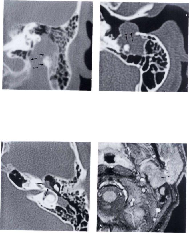
Figure 11.69 Left ear. Mass protruding into the posterior auditory canal. The patient complained of left mild hearing loss and left facial nerve palsy H.B. grade III of 6 months' duration (H.B.: House-Brackmann [see references]).
102 11 Glomus Tumors (Chemodectomas)
Figure 11.70 CT scan demonstrated the presence of tumor involving the vertical portion of the facial nerve.
Figure 11.71 CT scan showed also erosion of the posterior wall of the external canal.
Figure 11.72 CT scan. The tumor extended to the genicu- |
Figure 11.73 MRI showed a mass extending to the parotid |
|
late ganglion. |
||
gland area. |
||
|
Glomus Tumors (Differential Diagnosis) |
103 |
Ectopic Internal Carotid Artery
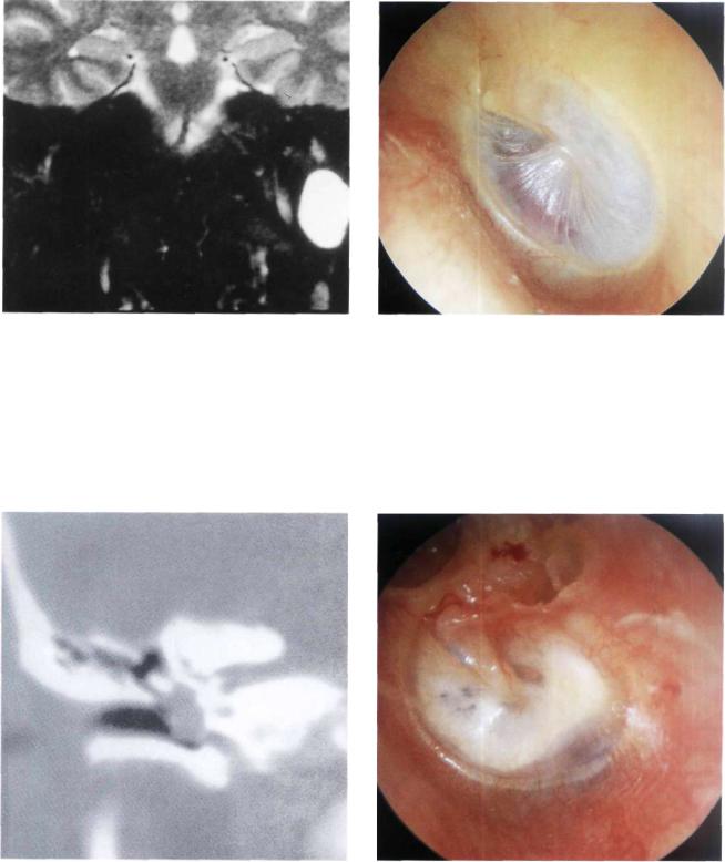
Figure 11.74 Another MR I of the same case. A combined middle fossa-transmastoid approach with parotid extension was performed. During surgery the tumor (T) proved to be a facial nerve neurinoma extended from the parotid to the intralabyrinthine segment of the facial nerve. The nerve was reconstructed with sural graft.
Figure 11.75 Left ear. A small pulsating reddish area in the anteroinferior quadrant of the tympanic membrane. This picture may be confused with a glomus tympanicum tumor.
High Jugular Bulb
Figure 11.76 A high-resolution CT scan established the diagnosis of an ectopic internal carotid artery.
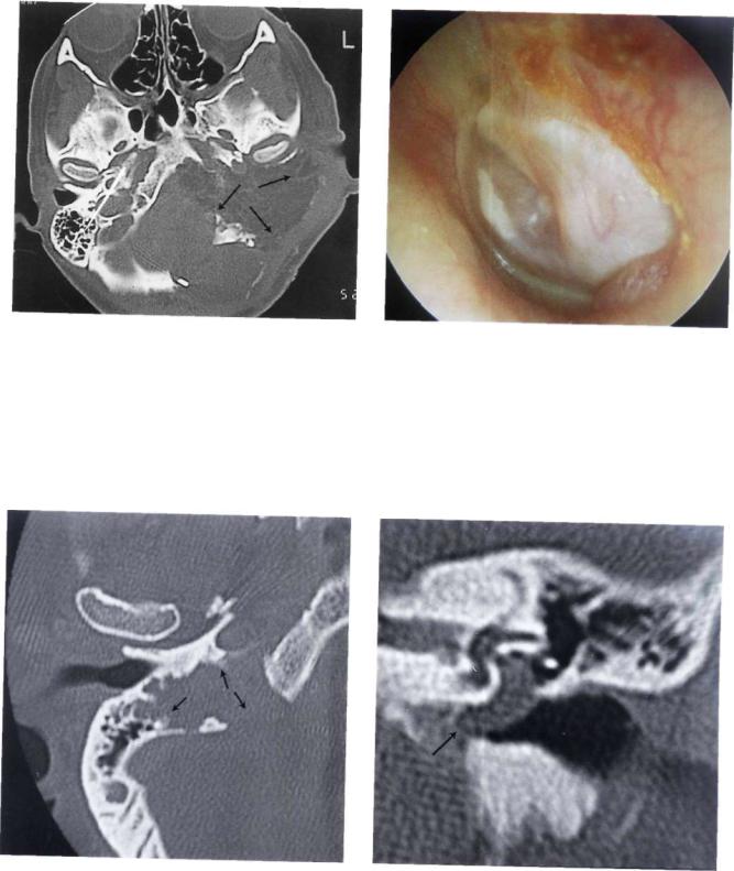
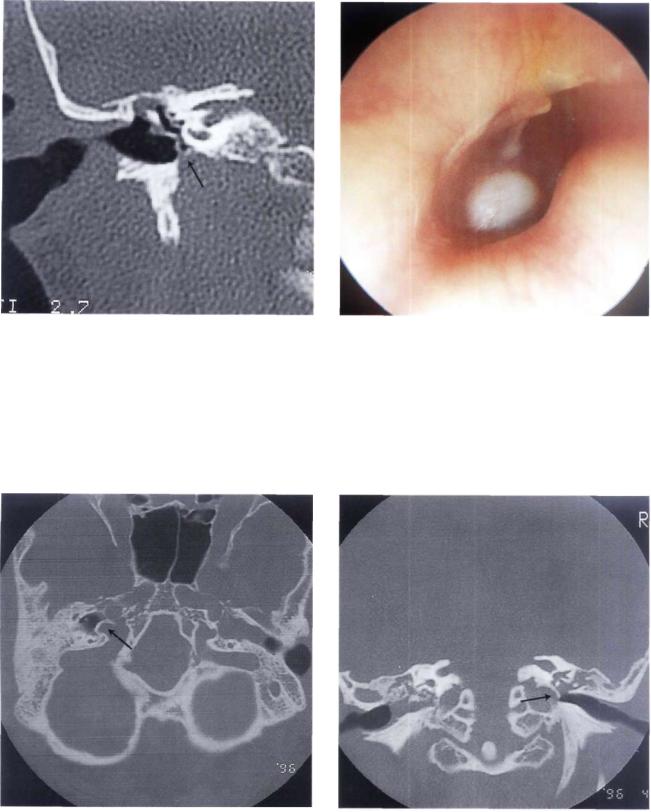
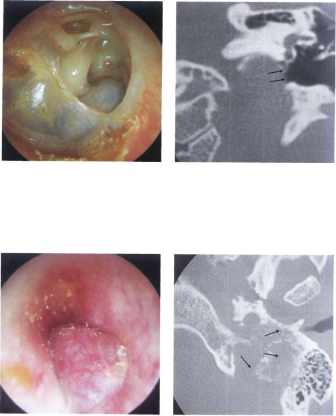
Figure 11.77 Left ear. Tympanosclerosis involving the whole tympanic membrane. An epitympanic erosion with cholesteatoma is also visible. At the level of the posteroinferior quadrant, a bluish mass is observed. A CT scan (see Fig. 11.78) proved this mass to be a high jugular bulb.
104 11 Glomus Tumors (Chemodectomas)
Figure 11.78 CT scan of the previous case. The uncovered jugular bulb is seen protruding into the middle ear.
Figure 11.79 Right ear. Another example of a high jugular bulb covered by a thin bony shell in a young male patient with a skull-base malformation (see following figures).
Figure 11.80 CT scan, axial view. The jugular bulb protrudes into the middle ear.
Figure 11.81 CT scan, coronal view. The high jugular bulb can be observed.
Glomus Tumors (Differential Diagnosis) |
105 |
Figure 11.82 Left ear. A high and uncovered jugular bulb |
Figure 11.83 CT scan of the case in Figure 11.82. |
reaching up to the level of the round window is visible |
|
through a posterior tympanic membrane perforation. |
|
• Polypoidal Pulsating Mass
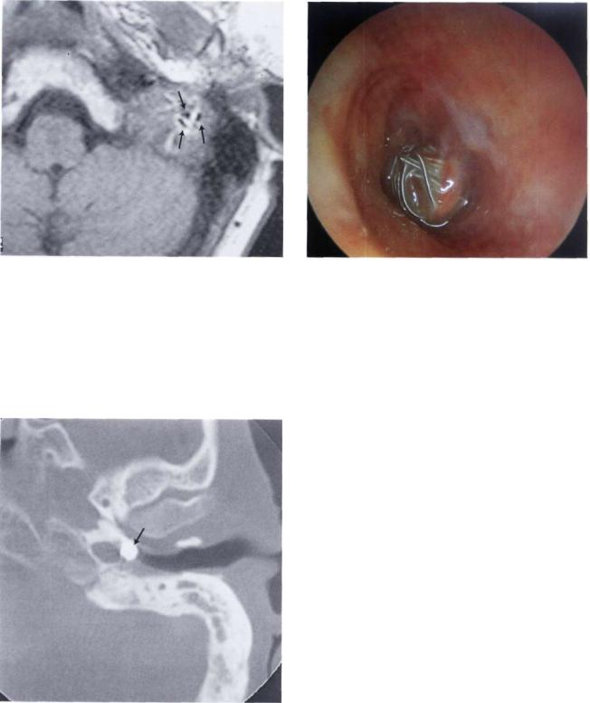
Figure 11.84 Left ear. A polypoidal pulsating red mass is seen in the external auditory canal. This example has been included to emphasize the fact that biopsy of external auditory canal polypi should never be taken without radiological investigations.
Figure 11.85 CT scan, in this case, demonstrated the presence of a glomus tumor eroding the surrounding bone in an irregular way giving a moth-eaten appearance.
106 11 Glomus Tumors (Chemodectomas)
• Internal Carotid Artery Aneurysm
Figure 11.86 MRI demonstrates the presence of fluid voids typical of large intratumoral vessels.
Figure 11.87 Guglielmi coils used to occlude an intrapetrous internal carotid artery aneurysm.
Figure 11.88 CT scan of the case presented in Figure 11.87 demonstrating occlusion of the aneurysm with the coils.
Summary—Meningioma
Posterior fossa meningiomas are the second most common tumor of the cerebellopontine angle. These tumors are characterized by a higher morbidity and mortality than acoustic neurinoma.
Surgical removal of these lesions poses many problems because of the deep location, the involvement of vital neurovascular structures, and the large sizes these tumors usually attain before diagnosis. Moreover, they have an aggressive behavior with frequent involvement of the dura and bone. Total removal is fundamental to avoid recurrence and is better achieved in the first operation. Total removal with minimal morbidity can be obtained utilizing an array of approaches that must be adapted to each individual case.
In general, an ideal approach is that which allows total removal with minimal or no brain retraction. The site of the tumor is the most important factor for the choice of the surgical approach. The size of the tumor, the patient's age and general medical condition, and the preoperative status of the cranial nerves are other factors to consider.
Tumors localized posterior to the internal auditory canal in young patients with good preoperative hearing can be removed using a retrosigmoid approach. In the elderly, however, a translabyrinthine approach is preferred to avoid cerebellar retraction. In cases of involvement of the jugular foramen, a POTS approach is adopted.
In small tumors lying anterior to the internal auditory canal, the middle fossa transpetrous approach is utilized. In large petroclival lesions, which pose more difficulties due to their deep location, the intimate relation with the brain stem, and the involvement of vital neurovascular structures, the modified transcochlear approach should be used, irrespective of the preoperative hearing. This approach permits a wide and direct exposure, and a flat angle of vision with no cerebellar or brain stem retraction. Moreover, it allows the removal of any infiltrated dura or bone.
Though total removal can be obtained in the majority of petroclival meningiomas, it is not always necessary or even safe. Subtotal removal is decided on in the absence of an arachnoid plane of cleavage between the tumor and the brain stem or when the perforating arteries are at risk of interruption during total tumor removal.
Neuroradiologic evaluation is fundamental to plan surgery. A CT scan with contrast to evaluate the bone, MRI with gadolinium, and in some cases, digital subtraction angiography are of paramount importance in each case.
The neuroradiologist should provide the surgeon with information on the following:
•Anatomical relations of the tumor
•Tumor consistency
•Vascularity
•Peritumoral edema
Glomus Tumors (Differential Diagnosis) |
107 |
•Tumor-brain stem interface
•Invasion of the dura and bone
•Relationship between the tumor and the vertebrobasilar and carotid systems
•Necessity of eventual embolization
The main blood supply of these tumors comes from large dural arteries. However, significant contributions may also come from pial arteries or from dural branches of the internal carotid and vertebral arteries. The angiographic data helps the neuroradiologist and the skull-base surgeon to determine the need for embolization. When indicated, it should be performed a few days before surgery. It not only decreases the intraoperative bleeding, but also produces a certain amount of tumor necrosis, rendering some cases easier to remove.
Close cooperation between the neuroradiologist and the skull-base surgeon offers optimal chances for successful management of these challenging tumors.
108 11 Meningioma—Facial Nerve Neurinoma
Summary—Facial Nerve Neurinoma
Tumor involvement of the facial nerve has been estimated to be the cause of facial palsy in 5% of cases. Though uncommon, facial neuromas should be considered in the differential diagnosis of facial nerve dysfunction. Unfortunately, the rarity of facial neuromas and the diversity of their clinical picture, together with the fact that their presentation may mimic other more common pathologies, renders the diagnosis of these tumors difficult.
Facial nerve dysfunction is the most common symptom. It can vary from the classic progressive palsy to sudden or recurrent facial palsy or hemifacial spasm. In limited cases the function of the nerve is normal. Therefore, all patients with progressive facial palsy must be considered to have a tumor until proved otherwise. Moreover, all patients with Bell's palsy persisting for more than 4 weeks and with recurrent facial paralysis should be investigated for the presence of a tumor.
The second most common complaint is hearing loss. Conductive hearing loss is usually associated with tumor involvement of the middle ear with subsequent interference with the ossicular chain. Sensorineural hearing loss is attributable to inner ear erosion or extension of the tumor into the internal auditory canal.
Most diagnosed tumors are of large size. One reason is that the facial nerve can accommodate tumor expansion to some extent before significant pressure, with subsequent dysfunction, can occur. Another reason is the relatively long duration of symptoms before diagnosis is made. Because of the absence of classic symptomatology in such cases, a higher index of suspicion is needed for early diagnosis. Diagnostic work-up includes audiometric testing, vestibular testing, and auditory brain-stem evoked response. Electrophysiologic testing of facial nerve function in such cases is of little or no benefit. The usefulness of these tests in the diagnosis of facial neuromas has been challenged by other authors (Dort and Fish 1991, Neely and Alford 1974).
Advances in radiologic techniques have aided greatly in the diagnosis of these lesions. The characteristic appearance on CT is that of an enhancing soft tissue mass, usually in the perigeniculate region, with sharp bony erosion and enlargement of the fallopian canal. High resolution CT scan is the best method to assess middle and inner ear involvement by tumor. However, MRI with gadolinium is the best available method for the preoperative assessment of tumor extension, especially of those involving the internal auditory canal, cerebellopontine angle, and/or the parotid region. Both methods are believed to be complementary for the preoperative assessment and the choice of the most suitable surgical approach for removal of these tumors. However, because these tumors show intraneural spread, it is still doubtful whether MRI with gadolinium can show the full extent of the lesion. Therefore, the surgeon should be prepared to expose the whole length of the facial nerve.
Differential diagnosis of these lesions includes acoustic neuroma, congenital cholesteatoma, chemodectoma, facial nerve hemangioma, and parotid tumors. Introdural facial nerve neuromas pose a major diagnostic difficulty, usually being mistaken for acoustic neuromas. Apart from the few cases in which tumor extension to the geniculate ganglion could establish the diagnosis, most of these cases were actually diagnosed intraoperatively.
Congenital cholesteatomas of the petrous bone are uncommon lesions that usually present with hearing loss and facial weakness or paralysis and, therefore, can be mistaken for facial neuromas. Moreover, these lesions appear on CT as smoothly marginated expansile lesions, and on MRI as hypo/isointense on Tl and hyperintense on T2 images. Unlike facial neuromas, however, cholesteatomas do not show enhancement following contrast administration, a fact that helps to differentiate between the two lesions.
Treatment generally aims at total removal of the tumor, restoration or preservation of facial nerve function, and conservation of hearing. The surgical approach depends on the extent of the lesion and the preoperative hearing level. There is general agreement that surgical removal is the treatment of choice. There is some controversy, however, regarding facial neuromas and absence of or mild preoperative facial nerve paresis. Some surgeons prefer to delay surgery because the patient is faced with the inevitable postoperative paralysis followed by some degree of recovery that will never be better than H.B. grade III. Patient counseling is important in these cases.
The age at presentation is another factor to be considered. If the patient is young, early surgical resection should be done because these tumors grow inexorably with subsequent intracranial or extra temporal extension, making the approach more difficult and postoperative complications more likely. Moreover, tumor growth causes progressive degeneration and regeneration of facial nerve fibers, leading to collagenization of the distal part of the nerve with consequent poor recovery of facial function following reconstruction. Another reason is that these tumors are potentially invasive: otic capsule erosion may be present in about 20% of the cases. On the other hand, in an elderly patient with an absence of or mild facial nerve paresis, facial nerve decompression may suffice if surgery is to be performed.
When total tumor removal involves resection of a long segment of the nerve, a cable graft is usually needed for reconstruction of the facial nerve. The length of the graft and whether it is from the sural or great auricular nerve has no effect on the eventual recovery of facial function.
In summary, facial nerve neuromas are uncommon tumors requiring a high degree of suspicion for their diagnosis. Recent advances in radiological techniques are the cornerstone for the diagnosis and preoperative assessment of these cases, and early surgical resection gives the best prognosis.
