

59
8 Chronic Suppurative Otitis Media with Cholesteatoma
Cholesteatoma is an epidermal inclusion cyst localized in the middle ear, whose capsule and matrix is formed from stratified squamous epithelium. The desquamating debris includes pearly white lamellae of keratin that accumulate concentrically, forming the cholesteatomatous mass.
The term cholesteatoma is actually a misnomer. It is derived from the Greek "cole" or bile, "steatos" or fat, and "oma" or tumor. There is no relation between cholesteatoma and bile or fat. The suffix "oma" (tumor), however, is more appropriate because cholesteatoma can be considered an epidermal inclusion cyst.
Cholesteatoma can be divided into congenital (middle ear or petrous bone) and acquired (middle ear or petrous bone). Congenital cholesteatoma is derived from entrapped ectodermal cellular debris during embryonic development. When it involves the middle ear, it appears as a whitish retrotympanic mass that may be localized either anterior or posterior to the malleus (see Chapter 9). When it involves the petrous part of the temporal bone, it is termed congenital petrous bone cholesteatoma and in the majority of cases it is localized in the petrous apex (see Chapter 10). In this chapter we will deal exclusively with cholesteatoma involving the middle ear. Petrous bone cholesteatoma is dealt with in a later chapter.
Acquired cholesteatoma of the middle ear can be caused by invasion of the skin of the external auditory canal into the middle ear through a marginal perforation. It can also originate from a epitympanic retraction pocket that becomes so deep that keratin debris can no longer be expelled, leading to their accumulation and subsequent cholesteatoma formation. Such retraction pockets can remain asymptomatic until they become infected, resulting in otorrhea and hearing loss. In other cases, the only symptom might be progressive hearing loss due to erosion of the ossicular chain by the developing cholesteatoma.
Because it is not always easy to establish a clear distinction between epitympanic or posterosuperior retraction pockets and cholesteatoma, we prefer to follow up these patients with otomicroscopy and endoscopy. In cases in which the retraction pocket becomes deep, giving rise to a cholesteatoma, a tympanoplasty is indicated. Because of the early stage of the disease, surgery can be done in a single stage.
Fetid otorrhea and hearing loss are the main complaints in cholesteatoma. In addition, complicated cases can manifest with vertigo and/or facial nerve paralysis. Vertigo occurs as a result of labyrinthine fistula, which is most commonly located in the lateral semicircular canal. Facial paralysis can be caused by pressure of the cholesteatoma sac or neuritis.
In rare cases, the cholesteatoma can invade the labyrinth, cochlea, posterior and middle fossa durae, the internal auditory canal, and the petrous apex, forming a petrous bone cholesteatoma (see Chapter 10).
Treatment of cholesteatoma is exclusively surgical. Early this century, radical mastoidectomy, a destructive procedure for the middle ear, was performed with the only goal being eradication of infection to save the ear.
In the early 1950s, the concept of tympanoplasty was introduced. Tympanoplasty was aimed at eradication of infection as well as reconstruction of the tym- pano-ossicular system. Today, two types of tympanoplasty are employed: closed tympanoplasty in which the posterior canal wall is preserved, and open tympanoplasty in which the posterior canal wall is drilled. Both techniques, when performed appropriately and with the proper indications, can produce excellent results in terms of eradication of cholesteatoma and restoration of hearing. In children, the closed technique is preferred, performed in two stages, in the majority of cases due to their highly cellular mastoids and in an attempt to preserve the anatomy of the ear as much as possible. In adults, particularly in epitympanic cholesteatoma with marked erosion of the scutum, in cases with sclerotic mastoids, or when middle ear atelectasis is present, an open tympanoplasty is performed (see also Chapter 13).

60 8 Chronic Suppurative Otitis Media with Cholesteatoma
Epitympanic Retraction Pocket
Figure 8.1 Right ear. Early epitympanic retraction pocket. The tympanic membrane shows grade I atelectasis. Middle ear effusion with characteristic yellowish coloration of the drum is seen. In the anterosuperior quadrant, the tubal orifice is visible in transparency, whereas the long process of the incus is evident in the posterosuperior quadrant. In the area of the cone of light, an atrophic part of the tympanic membrane due to a previous myringotomy can be appreciated.
Figure 8.2 Right ear. Epitympanic retraction pocket with the onset of tympanosclerosis of the pars tensa of the tympanic membrane.
Figure 8.3 Right ear, similar case. The anterior quadrants of the pars tensa are retracted and thickened.
Figure 8.4 Right ear. A large controllable epitympanic retraction pocket with erosion of the scutum. The head of the malleus is seen. Middle ear effusion gives the tympanic membrane the characteristic yellowish coloration. To prevent progression of the retraction pocket and the formation of adhesions, myringotomy, ventilation tube insertion, and regular fol- low-up are indicated. These cases frequently represent the
transition from a simple retraction pocket to an initial attic cholesteatoma. The distinction between the two is sometimes difficult. In suspected cases, a high-resolution computed tomography (CT) scan (bone window) is beneficial for better evaluation of the extension of the retraction pocket. In cases where the condition remains stable with regular follow-up and where hearing is normal, no surgery is required. If the pocket extends deeper, giving rise to a frank cholesteatoma, surgery is indicated. If hearing is normal, an open tympanoplasty (modified Bondy technique) is performed in a single stage.

• Epitympanic Cholesteatoma
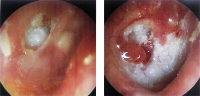
Figure 8.5 Right ear. Epitympanic erosion with cholesteatoma. The patient complained of fetid otorrhea and attacks of bloody ear discharge of several years' duration. Inflammatory tissue is seen surrounding the area of epitympanic erosion. As preoperative hearing was nearly normal (see audiogram, Fig. 8.6), a single-stage open tympanoplasty in the form of a modified
Epitympanic Cholesteatoma |
61 |
0 dBHL
10
20
30
40
50
60
70
80
90
100
110
120
125 |
250 |
500 |
1K |
2K |
4K |
8K |
16KHz |
Bondy technique was performed. This technique allows eradication of the cholesteatoma and also conserves hearing (for the modified Bondy technique, see Chapter 13).
Figure 8.6 Audiometry of the case described in Figure 8.5. Normal preoperative hearing.
Figure 8.7 Right ear. Epitympanic erosion with cholesteatoma. The tympanic membrane is completely tympanosclerotic. The patient did not complain of otorrhea (dry cholesteatoma).
Figure 8.8 Right ear. Epitympanic erosion with cholesteatomatous squamae. The patient did not complain of otorrhea. The pars tensa is intact. Intraoperatively, the cholesteatoma was found to have partially eroded the head of the malleus and the short process of the incus. The ossicular chain, however, maintained its continuity. A modified Bondy technique was performed and the normal preoperative hearing was conserved.

62 |
8 Chronic Suppurative Otitis Media with Cholesteatoma |
Figure 8.9 CT of the previous case, coronal view. The cholesteatoma is located in the epitympanic area. The middle ear is free.
Figure 8.10 Right ear of a 46-year-old patient suffering from bilateral cholesteatoma. An epitympanic erosion with cholesteatoma and middle ear effusion showing an air-fluid level can be seen. CT scan (Fig. 8.12) demonstrates cholesteatoma extension into the mastoid. Intraoperatively, a fistula of the lateral semicircular canal was encountered, as well as erosion of the incus. A single-stage open tympanoplasty was performed with autologous incus interposition between the handle of the malleus and the head of the stapes. In patients with bilateral cholesteatoma, an open technique is preferred.
Figure 8.11 Left ear of the same patient. Cholesteatoma with marked erosion of the scutum and epidermization of the attic and mesotympanum. The cholesteatoma debris was partially cleaned. The residual pars tensa shows tympanosclerosis. Intraoperatively, the ossicular chain was absent. The otoscopic view of the left ear is apparently more advanced than the right ear. This, however, was not the case intraoperatively since the marked epitympanic erosion shown here allowed
self-cleaning of the cholesteatoma debris (see CT scan, Fig. 8.12). Because of the total destruction of the ossicular chain, a second stage was programmed for functional reconstruction.
Figure 8.12 CT of the previous case showing cholesteatoma extension in the mastoid in the right ear and self-cleaning of the cholesteatoma debris in the left ear.

Epitympanic Cholesteatoma |
63 |
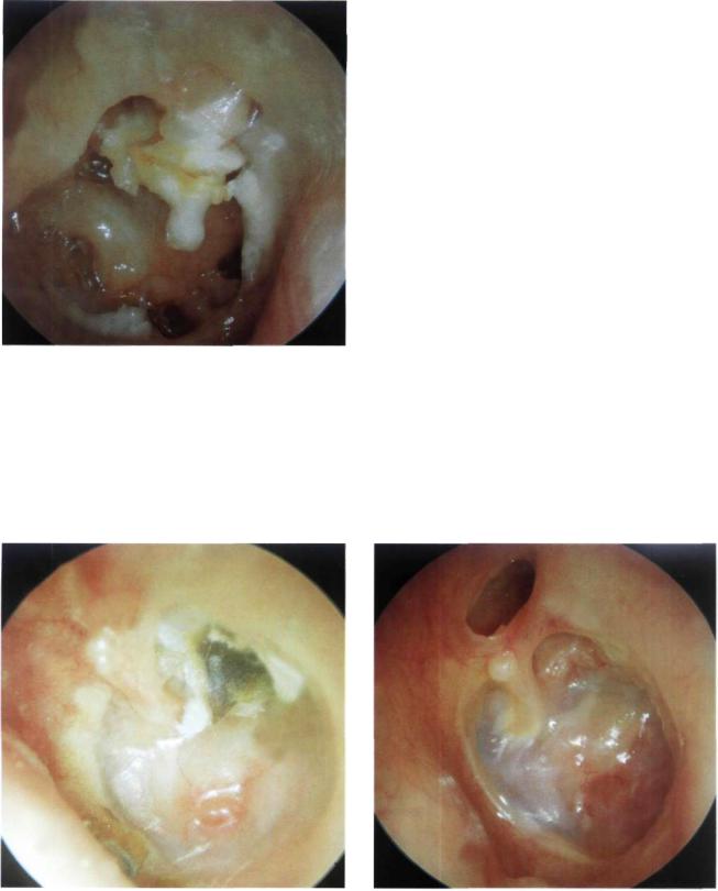
Figure 8.13 Left ear. Small epitympanic erosion with cholesteatoma. The skin surrounding the erosion is hyperemic and everted.
Figure 8.14 Left ear. Large epitympanic erosion with cholesteatoma and fetid otorrhea. The head of the malleus and body of the incus are eroded.
Figure 8.15 Right ear. Large epitympanic erosion with cholesteatoma. This 18-year-old patient did not complain of otorrhea. Ipsilateral hearing was normal, whereas the contralateral side showed severe sensorineural hearing loss secondary to a previous surgery of radical mastoidectomy. Given the intact ossicular chain, an open tympanoplasty (modified Bondy technique) was performed. According to our strategy, cholesteatoma in the only hearing ear is one of the absolute indications for performing an open technique. The reason is
that this technique, if properly performed, ensures complete eradication of the pathology and better long-term follow-up, thus minimizing the risk of recurrence. Further surgical interventions, with their potential risk even in the most experienced hands, are therefore avoided.
Figure 8.16 Right ear. Large epitympanic erosion with cholesteatoma and polypoid tissue that covers the head of the malleus. The pars tensa is intact.
64 |
8 Chronic Suppurative Otitis Media with |
esteatoma |
Figure 8.17 Left ear. Epitympanic cholesteatoma. Extensive erosion of the scutum with excessive cholesteatomatous debris. The pars tensa shows grade I atelectasis with catarrhal middle ear effusion.
Figure 8.18 Left ear. Cystic retrotympanic cholesteatoma situated posterior to the malleus. The tympanic membrane shows bulging at the level of the pars flaccida and slight retraction with tympanosclerosis in the posterior quadrants.
Figure 8.19 Same case as in Figure 8.18 during an acute inflammatory episode. Note the increase in size of the cholesteatomatous cyst.
Figure 8.20 Left ear. Epitympanic erosion occupied by a cholesteatomatous mass that protrudes into the external auditory canal. The mass is visible behind the posterior quadrant of the pars tensa. It engulfs the ossicular chain and extends towards the promontory and the hypotympanum.

Epitympanic Cholesteatoma |
65 |
Figure 8.21 Left ear. A large epitympanic erosion is seen with epidermization of the attic and posterior mesotympanum. The cholesteatoma, visible in transparency, causes bulging of the tympanic membrane in the posterior inferior quadrants. Resorption of the incus and head of the malleus is discernible.
Figure 8.22 Right ear. Epitympanic erosion with cholesteatoma. Extension of the cholesteatoma into the mesotympanum is seen through the bulging posterior quadrants of the tympanic membrane.
Figure 8.23 Left ear. Epitympanic erosion with cholesteatoma. Extension of the cholesteatoma into the mesotympanum (visible through the transparent pars tensa).
Figure 8.24 Left ear. Epitympanic erosion with cholesteatoma. Epidermization of the posterior mesotympanum is seen through a posterior perforation of the tympanic membrane. The tympanic membrane residue has a whitish color. This can be either due to tympanosclerosis or to epidermization of the medial surface of the tympanic membrane. Examination under the microscope can, in many cases, determine the exact cause.
66 8 Chronic Suppurative Otitis Media with Cholesteatoma
Summary
An epitympanic retraction pocket should be regularly checked with otomicroscopy. The 30° rigid endoscope allows visualization of the extent of the retraction pocket that can be difficult with the microscope. When progression of the epithelium into the epitympanum cannot be controlled, the presence of cholesteatoma is considered. In such cases, surgery should be performed. Whenever a minor epitympanic erosion is present, we adopt a closed technique with reconstruction of the attic using cartilage and bone pate. This technique is valid particularly in children where the mastoid is usually very pneumatized. Frequently, surgery is staged in these cases.
When a marked attic erosion is present, especially in adults, we perform an open technique to avoid cholesteatoma recurrence that can occur due to absorption of the material used for reconstruction of the attic defect. When preoperative hearing is normal in the presence of attic cholesteatoma with large bony erosion, we perform an open tympanoplasty in the form of a modified Bondy technique. This technique allows single-stage eradication of the disease with conservation of the normal preoperative hearing.
il Mesotympanic Cholesteatoma
Figure 8.25 Right ear. Mesotympanic cholesteatoma. The epithelial squamae can be seen through the retromalleolar perforation. Anterior to the malleus, the cholesteatomatous mass causes bulging and whitish coloration of the tympanic membrane without perforating it. The entire middle ear is filled with cholesteatoma in this case.
Figure 8.26 Right ear. Posterior mesotympanic cholesteatoma associated with a polyp are seen at the level of the oval window. There is evidence of discharge,

Mesotympanic Cholesteatoma |
67 |
Figure 8.27 Left ear. Small epitympanic erosion and a mesotympanic retraction pocket with wax and cholesteatomatous squamae. Extension of the cholesteatomatous mass into the anteromalleolar region is seen through the retracted tympanic membrane.
Figure 8.28 Right ear. A child with mesotympanic retraction and posterosuperior perforation through which cholesteatomatous debris and inflammatory tissue are visible. Purulent discharge is observed. The patient was operated on using a staged closed tympanoplasty.
Figure 8.29 Right ear. Posterior perforation with cholesteatoma in the posterior mesotympanum. The cholesteatomatous squamae cover the region of the oval window extending towards the attic and progress anterior to and under the handle of the malleus. The promontory and the round window are visible through the perforation.
Figure 8.30 Right ear. Total tympanic membrane perforation. The handle of the malleus is absent. The long process of the incus and part of the stapes are covered by cholesteatoma, which also involves the promontory. The round window, hypotympanic air cells, and tubal orifice are free of pathology. In these cases, a staged closed tympanoplasty can be performed.
68 8 Chronic Suppurative Otitis Media with Cholesteatoma
Figure 8.31 Right ear. Total perforation of the tympanic membrane. A cholesteatoma completely covers the handle of the malleus and the incudostapedial joint.
Summary
The presence of a posterior mesotympanic retraction pocket is usually associated with erosion of the ossicular chain. Surgery is indicated in these cases. The retraction pocket is completely removed after performing canalplasty of the posterior canal wall. In the same stage, the tympanic membrane is grafted, the posterosuperior quadrant of the tympanic membrane is reinforced, and middle ear aeration is restored using Silastic sheeting. One year later, if the tympanic membrane position remains normal (i.e., not retracted), the ossicular chain is reconstructed.
When an extensive erosion of the posterior wall is present, a modified radical mastoidectomy is indicated in the elderly, whereas a staged open tympanoplasty is performed in younger patients. The same strategy is also followed in patients presenting with bilateral cholesteatoma.
•Cholesteatoma Associated with Atelectasis
Figure 8.32 Left ear. Grade IV tympanic membrane atelectasis with posterosuperior mesotympanic retraction pocket. A mixture of wax and cholesteatomatous debris is seen. The middle ear mucosa is visible because of the absence of the epithelial layer.
Figure 8.33 Left ear. Epitympanic erosion through which a cholesteatoma is shown filling the attic and causing erosion of the head of the malleus. A grade IV atelectasis of the tympanic membrane (adhesive otitis) is seen with formation of polypoidal granulation tissue in the middle ear. In the region posterior to the malleus, the cholesteatoma engulfs the ossicular chain.

Cholesteatoma Associated with Atelectasis |
69 |
Figure 8.34 Left ear. Epitympanic erosion with cholesteatoma associated with atelectasis of the tympanic membrane. The incus is absent. A natural myringostapedopexy has been created. The second portion of the facial nerve is seen superior to the stapes; inferiorly the round window is noted. The anterior part of the tympanic membrane is affected with tympanosclerosis. In these cases, as hearing loss is mild (< 30 dB), a modified radical mastoidectomy is indicated to maintain the normal preoperative hearing level obtained as a result of the spontaneous myringostapedopexy.
Figure 8.36 CT scan of the previous case. An epitympanic cholesteatoma is found. Adhesions between the tympanic membrane and the promontory can be observed. This 45- year-old woman underwent a modified radical mastoidectomy with no interference in the middle ear.
Figure 8.35 Right ear. Epitympanic cholesteatoma associated with complete atelectasis of the tympanic membrane (see CTscan, Fig. 8.36).
Summary
In adult patients with extended epitympanic erosion or with bilateral cholesteatoma we prefer to perform an open technique. In all cases in which a spontaneous tympanostapedopexy with normal preoperative hearing or elderly patients with normal contralateral hearing, we prefer to leave the atelectatic tympanic membrane untouched after having verified the absence of any middle ear cholesteatoma. In the presence of mesotympanic cholesteatoma, staging is indicated. In the first operation a closed tympanoplasty is performed with reconstruction of the tympanic membrane, and a Silastic sheet is positioned in the middle ear. Silastic favors regeneration of the middle ear mucosa and prevents the formation of adhesions. In the second stage, performed 6 to 8 months later, the middle ear is checked for the presence of any residual cholesteatoma. The ossicular chain is then reconstructed using, preferably, an autologous incus. In children we always try to perform a staged closed tympanoplasty. If a recurrent cholesteatoma (epitympanic retraction pocket) is encountered in the second stage, we do not hesitate to transform it into an open technique.

70 8 Chronic Suppurative Otitis Media with
•Cholesteatoma Associated with Complications
Figure 8.37 Left ear. Large epitympanic perforation with pars tensa perforation. Cholesteatomatous squamae are present in the attic, whereas the middle ear is completely free. The handle of the malleus is present. The promontory, round window, and hypotympanic air cells are covered with normal mucosa. The tympanic annulus is intact. During surgery, a fistula of the lateral semicircular canal was encountered
(see Fig. 8.38). In such cases, because of the presence of marked epitympanic erosion and of the fistula, an open tympanoplasty is preferred.
Figure 8.38 Intraoperative view of the previous case. A fistula of the lateral semicircular canal is clearly seen.
Figure 8.39 Left ear. Large polyp obstructing the external auditory canal. The patient complained of fetid otorrhea, hearing loss, and vertigo. A high resolution CT scan of the temporal bone was ordered (see Fig. 8.40). A CT scan of the temporal bone should always be ordered in patients with chronic suppurative otitis media suffering from vertigo and/or instability.
Figure 8.40 CT scan of the previous case. A huge cholesteatoma causing a fistula of the lateral semicircular canal and erosion of the tegmen can be seen.

Cholesteatoma Associated with Complications |
71 |
Figure 8.41 Right ear. Epiand mesotympanic cholesteatoma. The cholesteatomatous debris protruded through the epitympanic erosion. In the posterosuperior quadrant, the cholesteatomatous sac can be seen in transparency, causing bulging of the tympanic membrane. The skin surrounding the attic erosion is hyperemic. The pars tensa is intact. The patient complained of frequent episodes of vertigo. A CT scan (see Fig. 8.42) demonstrated the presence of a fistula of the lateral semicircular canal.
Figure 8.42 CT scan of the previous case. The interruption of the lateral semicircular canal caused by the cholesteatoma is apparent.
Figure 8.43 Left ear. Small epitympanic retraction pocket in a patient presenting with hearing loss, tinnitus, and recurrent episodes of otitis media with effusion. The contralateral ear had been operated on elsewhere using an open tympanoplasty that resulted in total hearing loss and facial nerve paralysis. A CT scan of the temporal bone revealed the presence of an epitympanic cholesteatoma that caused a fistula of the superior semicircular canal and erosion of the tegmen (see Fig. 8.44). The patient underwent open tympanoplasty. Being
the only hearing ear, the cholesteatoma matrix was left over the fistula, whereas the tegmental erosion was repaired using cartilage to avoid a meningo-encephalic herniation (see Chapter 12).
Figure 8.44 CT scan of the previous case. Cholesteatoma caused a fistula of the superior semicircular canal and erosion of the tegmen.

72 8 Chronic Suppurative Otitis Media with Cholesteatoma
Figure 8.45 Left ear. This patient had already undergone bilateral radical mastoidectomy elsewhere. He presented with profound bilateral hearing loss and fetid otorrhea from his left ear. During revision surgery, a cholesteatoma causing a cochlear fistula was found. This patient suffered profound hearing loss in the other ear, thus the cholesteatoma matrix was left over the fistula to avoid deaf ear.
Summary
Figure 8.46 Polyp in the external auditory canal with purulent discharge. A cholesteatoma is frequently found behind such a polyp. In such cases, biopsy is not indicated as a CT scan is often used to differentiate cholesteatoma from other pathologies (glomus, carcinoid, or carcinoma). A tympanoplasty revealed the presence of a large cholesteatoma occupying the attic and mesotympanum.
At present, with the diagnostic methods at hand and increased medical care, it is very rare to find a cholesteatoma with intracranial complications (e.g., meningitis, brain abscess, lateral sinus thrombophlebitis, etc.). However, cases of cholesteatoma with massive bone destruction, labyrinthine fistulae, severe sensorineural hearing loss resulting in deaf ear, and facial nerve paralysis are not infrequently encountered. In general, it is not necessary to order a CT scan to diagnose a cholesteatoma. However, in the presence of headache, vertigo, facial nerve paralysis, severe sensorineural hearing loss, or sudden deafness, a high-resolution CT scan of the temporal bone becomes highly important. Axial and coronal cuts without contrast are required. When intracranial complications are suspected, contrast injection is also needed.
A labyrinthine fistula is found in less than 10% of cases. The lateral semicircular canal, being the most superficial, is the most commonly involved. Treatment of a labyrinthine fistula depends on the type (bony or membranous) and size of the fistula. A tegmental erosion can be repaired using cartilage and bone pate.
Facial nerve paralysis is either due to infection of the exposed nerve or secondary to compression by the cholesteatoma. In the majority of cases, removing the
cholesteatoma and clearing the infection are sufficient for the paralysis to resolve. It is very rare to find fibrosis or thinning of the nerve. In these cases, facial nerve reconstruction varies from rerouting and end-to-end anastomosis to nerve grafting, according to the degree of injury and length of the injured segment.