

4
2 The Normal Tympanic Membrane
•Anatomy
The tympanic membrane forms the major part of the lateral wall of the middle ear (see Figs. 2.1-2.3). It is thin, resistant, semitransparent, has a pearly gray color, and is cone-like. The apex of the membrane lies at the umbo, which corresponds to the lowest part of the han-
dle of the malleus. Most of the membrane circumference is thickened to form a fibrocartilaginous ring, the tympanic annulus, which sits in a groove in the tympanic bone called the tympanic sulcus. The fibrocartilaginous ring is deficient superiorly. This deficiency is known as the notch of Rivinus. The anterior and posterior malleolar folds extend from the short process of
Figure 2.1 Right ear. Normal tympanic membrane. 1 = pars flaccida; 2 = short process of the malleus; 3 = handle of the malleus; 4 = umbo; 5 = supratubal recess; 6 = tubal orifice; 7 = hypotympanic air cells; 8 = stapedius tendon; c = chorda tympani; I = incus; P = promontory; o = oval window; R = round window; T = tensor tympani; A = annulus.
Figure 2.2 Right ear. Structures of the middle ear seen after removal of the tympanic membrane. 9 = pyramidal eminence; co = cochleariform process; f = facial nerve; j = incudostapedial joint. See legend to Figure 2.1 for other numbers and abbreviations.

Normal Otoscopy
Normal Otoscopy
Figure 2.3 Right ear. Division of the tympanic membrane into four quadrants: A.S. = anterosuperior; A.I. = anteroinferior; P.S. = posterosuperior; P.I. = posteroinferior. This division facilitates the description of different pathologic affections of the tympanic membrane.
the malleus to the tympanic sulcus, thus forming the inferior limit of the pars flaccida of Sharpnell's membrane. The membrane forms an obtuse angle with the posterior wall of the external auditory canal. It also forms an acute angle with the anterior wall of the canal. It is important to respect this acute angulation in the myringoplasty operation to maintain as much as possible the vibratory mechanism of the tympanic membrane and hence ensure maximum hearing improvement.
The external surface of the tympanic membrane is innervated by the auriculotemporal nerve and the auricular branch of the vagus nerve, whereas the inner surface is supplied by Jacobson's nerve, a branch of the glossopharyngeal nerve.
The blood supply is derived from the deep auricular and anterior tympanic arteries. Both are branches of the maxillary artery.
•Histology
The tympanic membrane consists of three layers: an outer epithelial layer continuous with the skin of the external auditory canal, a middle fibrous layer or lamina propria, and an inner mucosal layer continuous with the lining of the tympanic cavity.
The epidermis or outer layer is divided into the stratum corneum, the stratum granulosum, the stratum spinosum, and the stratum basale, which is the deepest layer that rests on the basement membrane.
The lamina propria is characterized by the presence of collagen fibers. In the pars tensa, these fibers are arranged in two basic layers: an outer radial layer that originates from the inferior part of the handle of the malleus and inserts in the annulus, and an inner circular layer that originates primarily from the short process of the malleus. Such a distinct arrangement, however, is absent in the pars flaccida.
The mucosal layer is formed mainly of a simple cuboidal or columnar epithelium. The free surface of the cells possesses numerous microvilli.
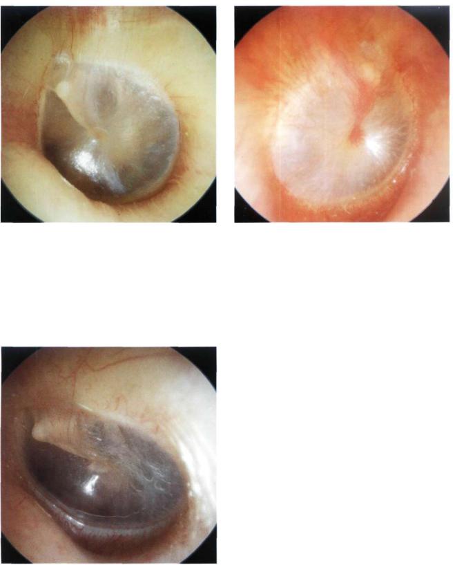
Figure 2.4 Left ear. Normal tympanic membrane. Note the acute angle formed between the tympanic membrane and the anterior wall of the external auditory canal. The pars tensa with the short process of the handle of the malleus, the umbo, the cone of light, the annulus, and the pars flaccida are seen. Note also the presence of early exostosis in the superior wall of the external auditory canal.
Figure 2.5 Right ear. Normal tympanic membrane. In this case, the drum is very thin and transparent. The handle and short process of the malleus as well as the umbo and cone of light are well visualized. Through the transparent tympanic membrane, the region of the oval window, the long process of the incus, the posterior arc of the stapes, the incudostapedial joint, the round window, and the promontory can be distinguished. Anteriorly, at the region of the eustachian tube, the tensor tympani canal and the supratubaric recess can be observed.
6 2 The Normal Tympanic Membrane
Figure 2.6 Left ear. Normal tympanic membrane. The handie of the malleus and cone of light are well visualized through the tympanic membrane; the promontory, the area of the round window, and the air cells in the hypotympanum can be appreciated. The pars flaccida is visualized superior to the short process of the malleus.
Figure 2.7 Right ear. Normal tympanic membrane. The drum, however, is slightly thickened with an accentuated capillary network along the handle of the malleus. The increased thickness of the tympanic membrane obscures all the structures in the middle ear.
Figure 2.8 Left ear. A normal tympanic membrane that is slightly thinned in the anterior quadrant and moderately thickened posteriorly.

7
3 Diseases Affecting the External Auditory
Canal
• Exostosis and Osteoma
Exostoses are defined as new bony growths in the osseous portion of the external auditory canal. They are usually multiple, bilateral, and are commonly sessile. They vary in shape, being either round, ovoid, or oblong. The condition is caused by periostitis secondary to exposure to cold water. This explains the high incidence of exostoses among divers and coldwater bathers. Histologically, they are formed from parallel layers of newly-formed bone. It is postulated that the periosteum stimulates an osteogenic reaction with each exposure to cold water, thus causing this stratification.
When exostoses are small they are asymptomatic. Large lesions, however, can occlude the external auditory canal and lead to conductive hearing loss or reten-
tion of wax and debris with subsequent otitis externa. In such cases, and in cases in which a hearing aid is to be fitted, surgical removal of exostoses is indicated. In some cases, surgery is technically difficult and special care is taken to preserve the skin of the external auditory canal. Other structures at risk are the tympanic membrane and ossicular chain medially, the temporomandibular joint anteriorly, and the third segment of the facial nerve posteroinferiorly. A postauricular incision is preferred because it allows good exposure and proper replacement of the skin of the external auditory canal to prevent postoperative scarring and stenosis.
Osteoma is a true benign neoplasm of the bone of the external auditory canal, usually unilateral and pedunculated. Histologically, it can be differentiated from exostosis by the absence of the laminated growth pattern.
Figure 3.1 Right ear. Small exostosis originating from the superior wall of the external auditory canal. Anterosuperiorly, another exostosis is seen in the early phase of formation.
Figure 3.2 Right ear. A small asymptomatic exostosis of the superior wall of the external auditory canal is observed. A hump of the anterior wall precludes adequate visualization of the entire tympanic membrane.

3 Diseases Affecting the External Auditory Canal
Figure 3.3 Right ear. Osseous neoplasm of the external auditory canal. In this case, given the pedunculated narrow base, an osteoma is a more probable diagnosis. This was confirmed by pathological examination of the removed specimen. Ample bone removal is performed in such cases to avoid recurrence.
Figure 3.4 Exostosis of the superior wall of the left external auditory canal. The lesion prevents complete visualization of the tympanic membrane.
Figure 3.5 Same patient, right ear. Two exostoses are present in the superior wall of the external auditory canal. In addition, the anterosuperior wall shows an additional exostosis. The lesions allow only a limited view of the central part of the tympanic membrane. In this case, a regular follow-up and evaluation is necessary because further growth of the lesion could lead to accumulation of debris and cerumen, necessitating surgical intervention.
Figure 3.6 Right ear. Exostosis of the posterior superior wall of the external auditory canal that precludes visualization of the pars flaccida. A bony hump is also present in the anterior wall of the canal. In such a case, it is useful to photograph the ear for further follow-up within 1-2 years.
Exostosis and Osteoma |
9 |
Figure 3.7a Left ear. Obstructing exostosis that causes subtotal occlusion of the external auditory canal. The patient complains of hearing loss and frequent episodes of otitis externa secondary to retention of water and debris inside the canal. A canalplasty under local anesthesia is indicated to restore the size of the external canal.
Figure 3.8 Obstructing exostosis of the external auditory canal resulting in otitis externa due to accumulation of squamous debris inside the canal. Surgery is essential both to avoid the formation of cholesteatoma and to improve hearing.
Figure 3.7b Computed tomography (CT) of the same case. The bony external canal is particularly narrowed.
Summary
Surgery in cases of exostosis is indicated only in cases with obstructing stenosis with or without hearing loss but with frequent otitis externa due to retention of debris. Surgery can be performed under local anesthesia, preferably using a postauricular incision. This approach allows excellent exposure of the whole meatus, thus minimizing the risk of injury to the tympanic membrane. In addition, it enables the surgeon to preserve the canal skin, thereby avoiding postoperative cicatricial stenosis. After dissecting the posterior limb, the flap is retained by the prongs of the self-retaining retractor. The skin of the anterior wall is incised medial to the tragus and is dissected in a lateral-to-medial direction. While drilling the exostosis, the skin of the canal is protected using an aluminum sheet (the cover of surgical sutures).
Osteoma can be removed by using a curette. In case of recurrence, a wide drilling of the bone around its base is also indicated.

103 Diseases Affecting the External Auditory Canal
•Furunculosis
Furunculosis is pustular folliculitis caused by staphylococcal infection of a hair follicle. Infection occurs as a result of microabrasion or of decreased immunity, as in diabetics. It is characterized by severe pain. A tender swelling is seen in the cartilaginous part of the external auditory canal which may have a central necrotic part.
Figure 3.9 A furuncle almost totally occluding the meatus. Pain is caused by distention of the richly innervated skin. A central necrotic part is seen.
• Myringitis and Meatal Stenosis
Myringitis is an inflammatory process that affects the tympanic membrane. Three forms are recognized: acute myringitis, bullous myringitis, and myringitis granulomatosa.
Acute myringitis is usually seen in association with infection of the external ear (otitis externa) or middle ear (otitis media). It is characterized by hyperemia and thickening of the tympanic membrane, as well as the presence of purulent secretions (Fig. 3.10). Therapy consists of administration of general and/or local antibiotics and local steroids.
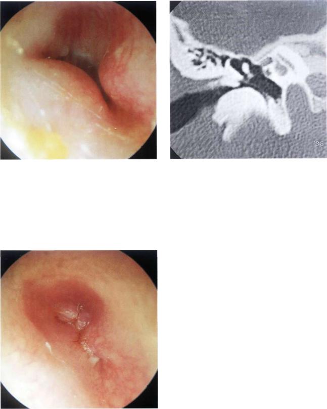
Figure 3.10 Left ear. The tympanic membrane is characterized by thickening and hyperemia. In this case, the skin of the external auditory canal is also hyperemic. The tympanic membrane seems lateralized.

Myringitis and Meatal Stenosis |
11 |
Bullous myringitis is commonly associated with viral upper respiratory tract infection. It is characterized by the presence of bullae filled with serosanguineous fluid. The bullae are located between the outer and middle layers of the tympanic membrane. The patient complains of otalgia and hearing loss. Therapy consists of antibiotics and steroids (Figs. 3.11, 3.12).
In granulomatous myringitis, the outer epidermic layer of the tympanic membrane as well as the adjacent skin of the external auditory canal are replaced by granulation tissue. It is generally seen in patients suffering from frequent episodes of otitis externa. In some cases, it may ultimately lead to stenosis of the most medial part of the external auditory canal. It can usually be cured, however, by removing the granulations in the outpatient clinic using the microscope. This is followed by the administration of local steroid drops for nearly 1 month. In refractory cases, however, surgery in the form of canalplasty with free skin graft is necessary.
Figure 3.11 Left tympanic membrane with a large bulla anterior to the malleus and a smaller one posterior to it.
Figure 3.12 Right tympanic membrane with a large bulla occupying the entire surface of the membrane. The malleus is not visible.
Figure 3.13 Granulomatous myringitis. The granulomatous tissue has replaced the external skin layer of the tympanic membrane and part of the anterior wall of the external canal. This case was treated by removal of the granulation tissue under local anesthesia in the outpatient clinic. Local steroid drops were then administered for 1 month.
12 3 Diseases Affecting the External Auditory Canal
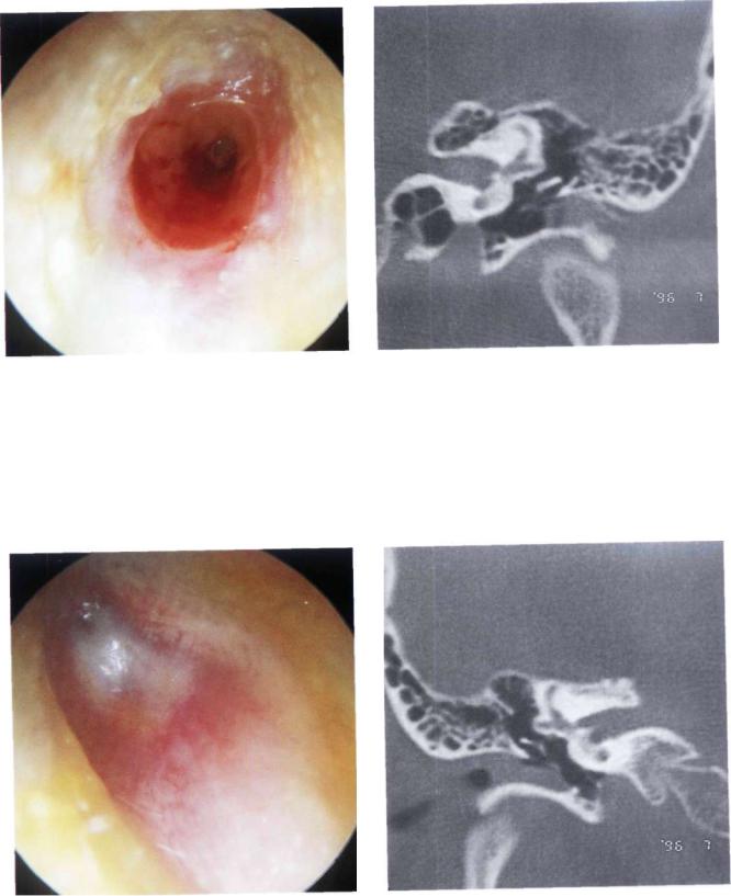
Figure 3.14 Postinflammatory stenosis of the right external auditory canal of a 68-year-old woman. The patient complained of bilateral continuous otorrhea and hearing loss of 3 years' duration. The otorrhea in the left ear stopped 2 months before presentation. The granulations over the tympanic membrane were removed in the outpatient clinic. A cellophane sheet was inserted into the external auditory canal to avoid the reformation of stenosis. Local steroid drops were
administered for 1 month. On follow-up, stenosis was already resolved and the granulation tissue in the external auditory canal was completely replaced by healthy skin.
Figure 3.15 CT of the same case. The bony walls of the external auditory canal are intact. The pathologic skin occupies the lumen of the external auditory canal.
Figure 3.16 Same patient, left ear (see also CT in Fig. 3.18). A canalplasty was performed on this side. After having removed the granulation tissue, myringoplasty and canalplasty were performed. Next, the meatal flaps were repositioned.
Figure 3.17 This CT scan demonstrates a similar lesion on the contralateral side.
Myringitis and Meatal Stenosis |
13 |
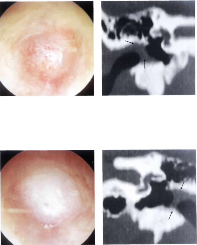
Figure 3.18 Right ear. Case similar to that seen in Figure 3.14. The patient complained of intermittent otorrhea and hearing loss (see CT scan in Fig. 3.19).
Figure 3.19 The CT scan shows thickening of the tympanic membrane and normal bony canal.
Figure 3.20 Same patient, left ear. Two tympanolplasties were previously performed on this ear. Generally, revision surgery is better avoided in patients who have undergone multiple operations and present with canal stenosis associated with lateralization of the tympanic membrane. (For postoperative stenosis of the external auditory canal, see Chapter 13.)
Figure 3.21 CT Scan of the previous case. The tympanic membrane is thickened and lateralized.
14 3 Diseases Affecting the External Auditory Canal
Summary
Postinflammatory stenosis of the external auditory canal is a difficult pathology to treat. In early cases, in which only granulation tissue is present, it is possible to remove the pathologic tissue (under local anesthesia in the outpatient clinic). This is followed by the insertion of a plastic (polyethylene) sheet to be left in place for about 20 days during which regular lavage is performed with 2% boric acid in 70% alcohol and local steroid lotions are applied. Surgery is doubtful in well-established cases with excessive cicatricial tissue leading to marked narrowing of the external auditory canal and lateralization of the tympanic membrane (secondary to thickening of the latter). In the majority of cases, restenosis occurs following operative interference. Therefore, it is preferable not to operate in the case of unilateral postinflammatory stenosis. In bilateral cases with marked hearing loss, a hearing aid is prescribed. By contrast, postoperative stenosis has a better prognosis and the results of treatment are more encouraging.
Figure 3.22 Right ear. Radical mastoid cavity showing cholesteatoma with superimposed fungal infection.
Otomycosis
Otomycosis is more common in tropical and subtropical countries. In the majority of cases, the isolated fungi are of the Aspergillus (niger, fumigatus, flavescens, albus) or the Candida species. Otomycosis is more common in immunocompromised patients and in diabetics. Local factors that favor fungal infections
include chronic otorrhea and the presence of epithelial debris. Clinically, the patient complains of otorrhea, itching, and hearing loss. Therapy consists of cleaning the ear to remove all debris and the instillation of local antimycotic preparations as well as lavage with 2% alcohol boric acid drops.
Figure 3.23 An ear with chronic suppurative otitis media |
Figure 3.24 Another example of otomycosis in a radical |
with cholesteatoma showing a superimposed fungal infec- |
mastoid cavity. |
tion. The blackish fungal masses are easily recognized. They |
|
should be removed before local antifungal solution is instilled. |
|

Cholesteatoma of the External Auditory Canal |
15 |
Eczema
Eczema is a dermo-epidermal process of reactive nature resulting from local or general factors. Local factors include allergy, topical medical preparations, or cosmetics, whereas general factors include hepatic or gastrointestinal dysfunction. It manifests by itching, a bur-ning sensation, vesication, and sometimes serous otorrhea. Treatment consists of discontinuation the suspected causative irritant, correction of the systemic disturbances, as well as lavage with boric acid with alcohol and steroid lotion.
Figure 3.25 Right ear. Chronic eczema of the external auditory canal. Squamous debris covering the skin of the external auditory canal can be noted. Successfully treated by the use of local steroid lotion.
•Cholesteatoma of the External Auditory Canal
Cholesteatoma of the external auditory canal should be differentiated from keratosis obturans. The latter entails accumulation of desquamated squamous epithelium in the external auditory canal forming an occluding cholesteatoma-like mass. The patient complains of pain and hearing loss. Keratosis obturans is generally bilateral and occurs in young patients, whereas cholesteatoma of the external auditory canal is usually unilateral and occurs in the elderly. In about 50% of patients, keratosis obturans is associated with bronchiectasis and chronic sinusitis. Removal of the mass is sufficient in keratosis obturans. However, in cholesteatoma it may also be necessary to remove the underlying bone followed by reconstruction of the external auditory canal and its skin.
Postoperative (iatrogenic) cholesteatoma of the external auditory canal is generally located at the level of the anterior angle of the tympanic membrane. It usually originates from incorrect repositioning of the skin flaps at the end of the procedure.
Figure 3.26 Cholesteatoma of the external auditory canal that occurred as a result of incorrect repositioning of the skin flaps in a previous intact canal wall tympanoplasty. This condition is to be differentiated from exostosis. A probe is used to palpate the mass. If it is tender and of soft consistency, cholesteatoma is diagnosed.

16 3 Diseases Affecting the External Auditory Canal
Figure 3.27 A case similar to that in Figure 3.26. The mass originating from the posterior canal wall inhibits the normal process of epithelial migration towards the outside.
Figure 3.28 Cholesteatoma of the inferior wall of the left external auditory canal being removed in the outpatient clinic. In this case, the squamous debris led to erosion of the underlying bone.
Figure 3.29 Same patient, a few months later. Note the |
Figure 3.30 A case similar to the that in Figure 3.28. The |
bone erosion caused by the cholesteatoma. |
cholesteatoma occupies more than half of the external audi- |
|
tory canal and is in contact with the tympanic membrane. The |
|
CT scan (Fig. 3.31) demonstrates partial erosion of the under- |
|
lying bone. |

Carcinoid Tumors |
17 |
Carcinoid Tumors
A carcinoid tumor is an adenomatous neuroendocrinal tumor of ectodermal origin. It has the same histologic and histochemical characteristics as other carcinoid tumors that involve different parts of the body. A carcinoid tumor is suspected whenever an adenomatous tumor of the middle ear has acinic or trabecular histologic features. The diagnosis is confirmed by electron microscopy and immunohistochemistry to demonstrate the presence of serotonin and argyrophilic granules. Surgical removal is indicated. To avoid recurrence, removal of the whole tumor together with the attached ossicular chain is essential.
Figure 3.31 CT scan of the same case, coronal view. The cholesteatoma is clearly seen in the anteroinferior portion of the external auditory canal with partial erosion of the underlying bone.
Summary
Postoperative (iatrogenic) cholesteatoma can almost always be removed in the outpatient clinic under local anesthesia using an endomeatal approach. The sac is opened and the cholesteatoma is aspirated. It is advisable to insert a plastic sheet in the external auditory canal for about 3 weeks to prevent the formation of adhesions that could lead to reformation of the cholesteatoma pearl.
Cholesteatoma of the external auditory canal should be surgically removed using a postauricular approach. A wide drilling of the floor of the canal is mandatory to avoid recurrences.
Pathologies Extending to the External Auditory Canal
Some middle ear pathologies can extend into the external auditory canal (e.g., cholesteatomas, glomus tumors, meningiomas, carcinoid tumors, and histiocytosis X). These cases are discussed here to underline the importance of their inclusion in the differential diagnosis of "polypi" in the external auditory canal. Moreover, taking a biopsy of these polypi in the outpatient clinic without proper radiological study is sometimes hazardous. For a detailed discussion of these pathologies, the reader is referred to the relevant chapters.
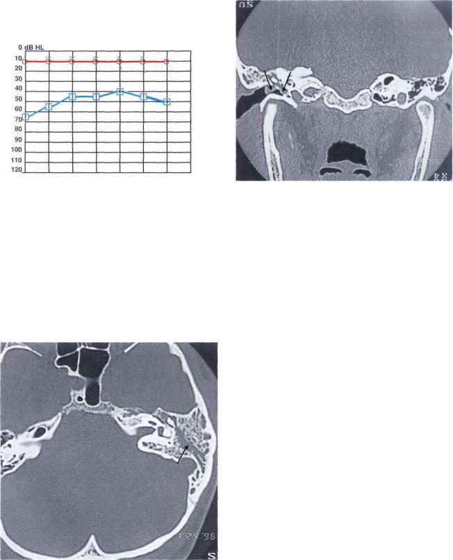
Figure 3.32 This patient complained of hearing loss in the left ear and otalgia of 3 months' duration. Otoscopy revealed a mass occupying the external auditory canal and originating from its anterosuperior region. The inferior part of the tympanic membrane, which is the only visible part, appears whitish due to the presence of a mass in the middle ear. The audiogram (Fig. 3.33) revealed the presence of an ipsilateral conductive hearing loss. The tympanogram was type B. CT scan (Figs. 3.34, 3.35) demonstrated the presence of an isointense soft-tissue mass occupying the middle ear and mastoid with extension into the external auditory canal. No erosion of the ossicular chain, nor of the intercellular septae of the mastoid air cells, was noted. Intraoperatively, a glandu- lar-like tissue was found and a frozen section obtained. The biopsy, confirmed by immmunohistochemical and electron microscopic studies, proved the presence of a carcinoid tumor. A tympanoplasty was performed with total removal of the pathology and the involved malleus and incus.
18 3 Diseases Affecting the External Auditory Canal
125 |
250 |
500 |
1K |
2K |
4K |
8K |
16KHZ |
Figure 3.33 The audiogram shows the presence of significant ipsilateral conductive hearing loss.
Figure 3.34 The CT scan demonstrates a soft-tissue mass occupying the middle ear with extrusion through the tympanic membrane.
Summary
Carcinoid tumors of the middle ear are very rare. They are considered a subgroup of the adenomatous tumors of the middle ear. Clinically, they manifest as hearing loss, tinnitus, aural fullness, facial nerve paresis, vertigo, and otalgia. These tumors require a functional surgery that entails removal of the tympanic membrane and ossicular chain together with the mass. The tympanic membrane is grafted at the same stage, whereas the ossicular chain is reconstructed at a second stage. This strategy ensures that the condition is completely cured.
Figure 3.35 CT scan, axial view. Presence of glue in the mastoid cells without erosion of the intercellular septae.

Histiocytosis X |
19 |
• Histiocytosis X
Histiocytosis X refers to a group of disorders of the reticuloendothelial system characterized by proliferation of cytologically benign histiocytes. The disease can present in three clinical forms, the most benign of which is eosinophilic granuloma, which is usually monostotic. A moderately aggressive form is known as Hand-Schiiller-Christian disease. It is characterized by multifocal lesions that are predominantly osteolytic. The most severe form, Letterer-Siwe disease, occurs in children under 3 years of age and presents with diffuse multiorgan involvement. It has a mortality rate of about 40% despite therapy with cytotoxic drugs and corticosteroids. Survivors suffer from diseases such as diabetes insipidus, pulmonary fibrosis, and vertebral column involvement.
Figure 3.36 A bulging of the posterosuperior wall of the external auditory canal in a 4-year-old child. A similar picture was also seen in the other ear (see CT scan in Fig. 3.37).
Figure 3.37 CT scan of the same case as in Figure 3.36. The middle ear and mastoid are occupied by an isointense mass, A frozen section obtained during surgery revealed the presence of histiocytosis X. The patient was referred to a specialized center for appropriate staging and therapy with cytotoxic drugs and corticosteroids.

20 3 Diseases Affecting the External Auditory Canal
Other Pathologies
Figure 3.38 Polyp in the external canal in a child presenting with continuous otorrhea and hearing loss. A CT scan (Fig. 3.39) shows the presence of a soft-tissue mass eroding the intercellular septae of the mastoid and the ossicular chain, suggestive of cholesteatoma. This was confirmed during surgery.
Figure 3.39 CT scan, axial view. The entire mastoid is occupied by a soft-tissue mass. The intercellular septae of the mastoid and the ossicular chain are absent.
Figure 3.40 Another example of chronic suppurative otitis media with cholesteatoma that manifests with an aural polyp. Though cholesteatoma presents frequently in this manner, it is absolutely essential to abstain from taking a biopsy of the polyp in the outpatient clinic without performing a CT scan of the temporal bone (see Fig. 3.41).
Figure 3.41 The otoscopic view is very similar to that in Figure 3.40. In this case, however, the diagnosis is that of an en-plaque supratentorial meningioma. An outpatient polypectomy in this case might lead to excessive bleeding (see MRI, Figs. 3.42 and 3.43).

Other Pathologies |
21 |
Figure 3.42 MRI with gadolinium enhancement, axial view. The tumor (arrows) is located in the temporal fossa and reaches the area of the petrous apex and Meckel's cavity.
Figure 3.43 MRI with gadolinium, coronal view. The meningioma displaces the temporal lobe upwards (arrows); pathognomonic tails of the dura are visible.
Figure 3.44 Left ear. Glomus jugulare tumor with extension |
Figure 3.45 Left ear. Another example of a glomus tumor. |
|
into the external auditory canal. A biopsy of this lesion might |
||
|
||
lead to severe and often difficult-to-control hemorrhage. |
|

22 3 Diseases Affecting the External Auditory Canal
Figure 3.46 Pulsating neoplasm in the external auditory canal. MR I (Fig. 3.47) revealed the presence of a glomus jugulare tumor involving the vertical internal carotid artery.
•Carcinoma of the External Auditory Canal
Basal cell carcinoma is more frequent in the auricle, particularly in subjects with long exposure to the sun. On the other hand, squamous cell carcinoma accounts for about three quarters of invasive tumors of the external auditory canal and the middle ear. In about 11% of cases, cervical lymph node metastases are present at the time of diagnosis. The most common symptoms include otorrhea, otalgia, hearing loss, facial nerve paralysis, and vertigo. An accurate microscopic examination is important for proper evaluation of the lesion extension. Frequently, an exfoliative lesion is noted, whereas an ulcer is present in other cases. Carcinoma should be suspected in the case of a persistent otitis externa characterized by pain and otorrhea that does not resolve adequately with medical treatment. A biopsy of the lesion will clear any doubts. It is important to perform an accurate examination of the upper deep cervical, postauricular, and parotid lymph nodes (anterior extension of the tumor). The cranial nerves are also evaluated. The facial nerve is the most frequently involved. Involvement of the mandibular nerve indicates tumor extension towards the glenoid fossa. A high-resolution CT scan (bone window) is the most important radiological investigation as it permits the evaluation of bone erosion at the level of the external auditory canal and middle ear. MRI with gadolinium allows the evaluation of tumor extension into the soft tissues.
Figure 3.47 MRI of the same case. A glomus jugulare tumor engulfing the vertical portion of the internal carotid artery is clearly visible.
The tumor should be considered to be T3 or T4 if there is infiltration of the posterior or middle cranial fossae, or invasion of the jugular foramen or glenoid fossa. In such cases, whatever the modality of treatment, the prognosis is almost always poor.
Surgery consists of en-bloc removal of the tumor and a trial to include a safety margin of the surrounding healthy tissue in the specimen. Postoperative radiotherapy should be subsequently performed.

Carcinoma of the External Auditory Canal |
23 |
Figure 3.48 An exfoliative neoplasm that occupies the external auditory canal. The patient complained of otalgia and attacks of bloody otorrhea of 1-month duration. A biopsy was taken and pathologic examination revealed the presence of squamous cell carcinoma. A CT scan (Fig. 3.49) demonstrated erosion of the external auditory canal, particularly its anteroinferior wall, without breaking into the glenoid fossa. En-bloc removal of the tumor was performed, together with a superficial parotidectomy. Radiotherapy was performed postoperatively.
Figure 3.49 CT scan demonstrates erosion of the anteroinferior wall of the external auditory canal. The glenoid fossa is not invaded.
Figure 3.50 Squamous cell carcinoma protruding through the external auditory canal with extension into the glenoid fossa and infiltration of the middle fossa dura (see CT scan, Fig. 3.51 and MRI, Fig. 3.52). Palliative surgery was performed followed by radiotherapy.
Figure 3.51 CT scan. The carcinoma occupies all of the middle ear and the mastoid. The glenoid fossa and the middle fossa plate are eroded.
24 3 Diseases Affecting the External Auditory Canal
Figure 3.52 MRI shows marked anterior extension of the |
Figure 3.53 Squamous cell carcinoma with posterior exten- |
tumor into the infratemporal fossa. |
sion. The mass ifiltrates the skin of the posterior wall of the |
|
external auditory canal (see CT scan, Fig. 3.54) as a result of |
|
which en-bloc resection and subsequent radiotherapy were |
|
performed. |
Figure 3.54 CT scan, axial view. The tumor has eroded the most lateral portion of the posterior bony wall.
Figure 3.55 Nasopharyngeal carcinoma extending into the middle ear and external auditory canal. A polypoid mass infiltrates the tympanic membrane and partially fills the external auditory canal (see CT scan, Fig. 3.56 and MRI, Fig. 3.57). The patient was considered inoperable and was referred to radiotherapy.

Carcinoma of the External Auditory Canal |
25 |
Figure 3.56 The CT scan demonstrates marked infiltration of the nasopharynx, the pterygoid muscles, and the petrous apex.
Summary
A carcinoma arising from the external auditory canal is frequently confused with suppurative otitis. Because of the high incidence of otitis externa and media and because these pathologies are frequently chronic, the diagnosis of carcinoma of the external auditory canal is almost always late. Diagnosis is made by biopsy. A high-resolution CT scan and MRI are necessary for proper evaluation. A high-resolu- tion CT scan determines the osseous erosion caused by the tumor, whereas MRI is superior to CT for the evaluation of soft tissues. MRI shows the presence of dural invasion, intracranial extension, as well as extracranial soft-tissue involvement. Until now there has been no universally accepted system of staging, which is the basis for planning therapy and proper treatment evaluation.
Therapy for carcinoma of the external auditory canal is almost always surgical. Various degrees of resection are utilized according to the extent of the pathology. Very small lesions can be managed by excision biopsy with a safety margin and curettage of the underlying bone.
Lateral en-bloc petrosectomy is the treatment of choice in the majority of carcinomas of the external auditory canal. It entails excision of the external auditory canal (bone and soft tissues), tympanic membrane, and ossicular chain with preservation of the facial nerve. Anteriorly, bone removal extends up
Figure 3.57 MRI with gadolinium confirms the infiltration.
to the level of the temporomandibular joint. The cavity can be exteriorized or obliterated with abdominal fat and the external auditory canal closed as cul-de- sac. When indicated, the resection can include a superficial parotidectomy, resection of the mandibular condyle, and/or neck dissection.
When the tumor has a deeper extension towards the middle ear, en-bloc subtotal resection of the temporal bone is indicated. In such cases, a middle and posterior fossa craniotomy is necessary. Bone removal is performed up to the level of the medial third of the petrous apex and the internal carotid artery. The facial nerve and inner ear are sacrificed.
A more extended procedure is total en-bloc resection of the temporal bone entailing, in addition, the sacrifice of the internal carotid artery, closure of the sigmoid sinus and jugular bulb, and in some cases a total parotidectomy and neck dissection.