

CST 2015 / research-cancer-brochure
.pdfRESEARCH FOCUS
Current Advances
in Cancer Research

CANCER RESEARCH
(Front to Back)
Ariel Twomey with CST since 2012
Christopher Lewis with CST since 2012
Sarah Linscott with CST since 2011 Production Associates
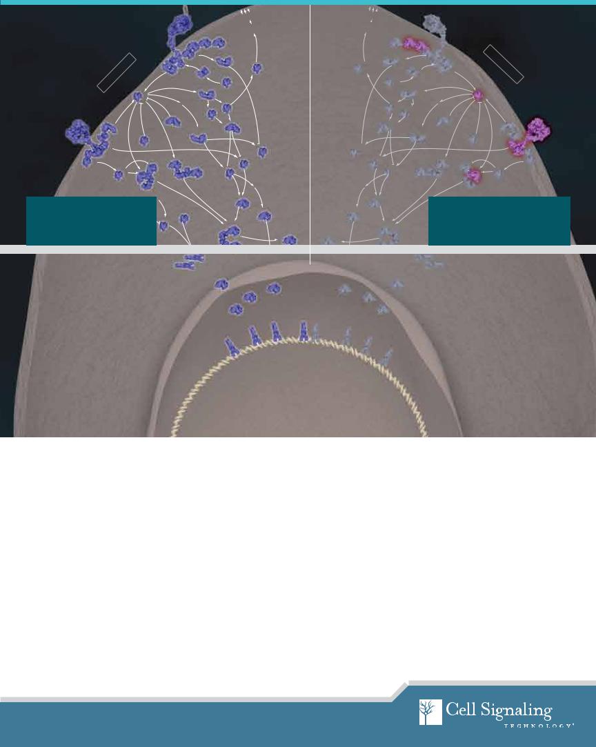
Cancer is a complex disease of unregulated cell growth.
Over a decade ago, Robert Weinberg and Douglas Hanahan defined the six hallmark traits of cancer: sustaining proliferative signaling; evading growth suppressors; activating invasion and metastasis; enabling replicative immortality; inducing angiogenesis; and resisting cell death(1). Since then, a growing body of evidence suggests two additional emerging hallmarks − reprogramming energy metabolism and evading immune destruction − are equally important for the malignant phenotype and may prove to be true hallmarks as research continues in this area(2). These acquired capabilities, which are shared by nearly all cancer cells, are regulated by highly complex protein signaling networks.
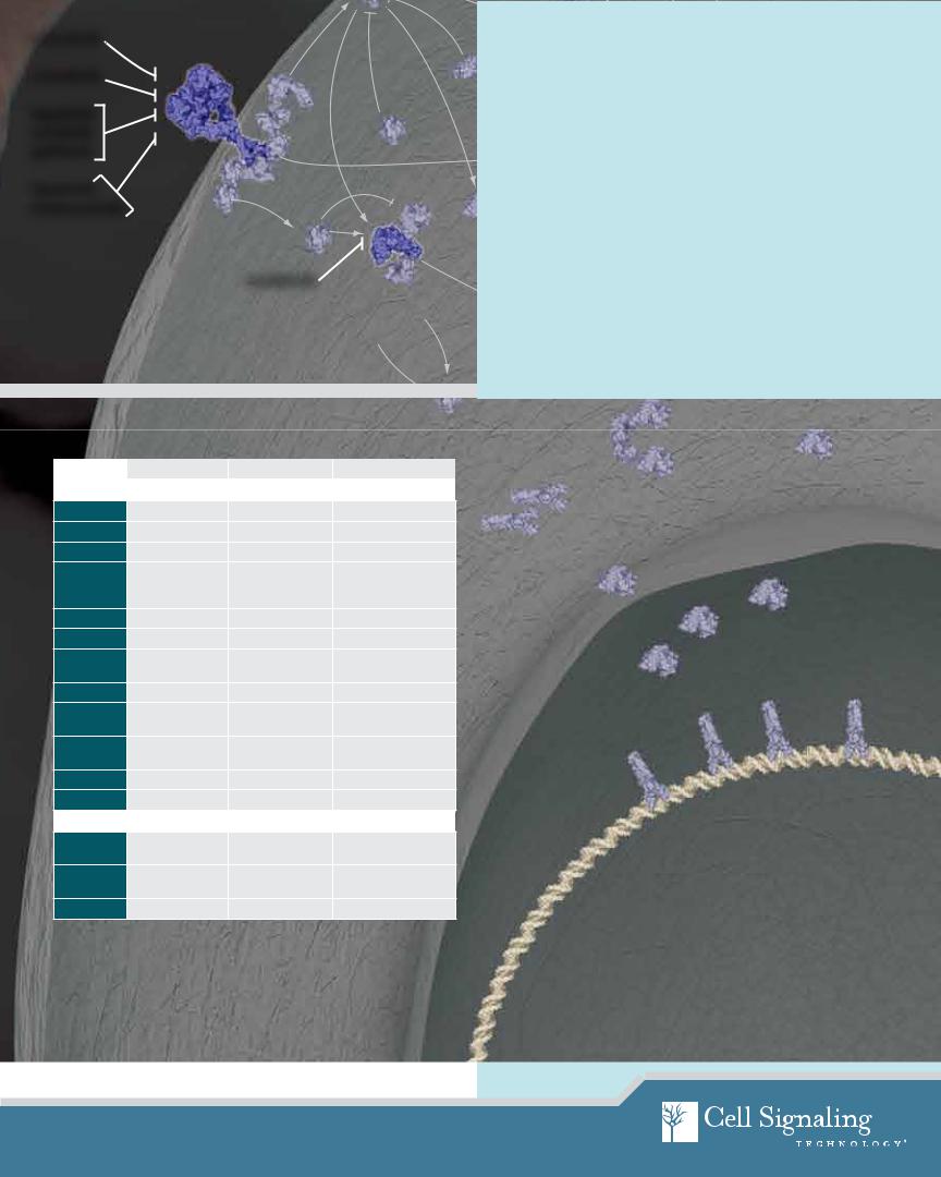
Cover image: Cytochrome c release from the mitochondrion during apoptosis leads to amplification of the caspase signaling cascade via formation of the apoptosome. A radially-symmetric structure, the apoptosome assembles upon binding of cytochrome c (blue) to Apaf-1 (beige), followed by recruitment of caspase-9 (red). Once assembled, the apoptosome’s activated caspase-9 cleaves and activates cytosolic caspase-3 (red, floating) and rapidly amplifies the caspase cascade. This leads to the destruction of numerous intracellular targets by caspase-3 including the actin cytoskeleton (green).
See the molecular movie of the apoptosome forming at www.cellsignal.com/apoptosome
Resources for cancer researchers available at www.cellsignal.com/cancer
Ras
Signaling
Ras
|
|
|
Regulated |
|
|
|
Exocytosis |
Integrin |
|
Exocyst |
|
|
|
|
|
|
|
|
Complex |
|
Src |
PI3K |
ILK |
|
|
|
|
|
FAK |
|
|
GRB2 |
|
|
GRAF |
SOS |
RIAM/Talin |
Rab8a (c-Mel) |
|
|
|||
|
|
||
|
|
|
Rap |
|
RalGEF |
|
|
Regulated |
|
|
Exocytosis |
|
|
Exocyst |
|
Integrin |
Complex |
|
|
ILK |
PI3K |
|
|
Src |
|
|
|
FAK |
GRAF |
|
GRB2 |
|
|
|
Rab8a (c-Mel) |
RIAM/Talin |
SOS |
|
|
|
Rap |
|
|
|
RalGEF |
|
Ras
Signaling
Ras
Receptor Tyrosine Kinase (RTK) |
|
|
|
RalB |
|
|
|
|
|
ALK |
|
|
RasGAP |
|
SOS |
|
|
|
|
ROS1 |
GRB2 |
|
|
RhoGEFs |
EGFR |
NF1 |
|
|
|
HER2 |
|
|
|
|
|
|
|
|
|
Crk |
|
|
|
|
C3G |
|
|
KSR |
|
|
|
Raf-1 |
|
|
|
|
14-3-3 |
c-Raf |
|
|
Rap |
B-Raf |
||
|
|
|
||
|
|
|
|
|
|
|
14-3-3 |
|
|
Normoxia |
|
|
In normal cells, receptor tyrosineHypoxia |
|
|
kinases signal through Ras to acti- |
|
VHL |
vate the MAPK Erk1/2 pathway, lead- |
|
|
HPH |
|
|
ing to highly controlled cell growth |
|
|
|
|
|
and differentiation. |
|
|
RalA
RalBP1
|
Rho |
Rho |
Cdc42 |
Actin |
Actin |
Rac |
Cytoskeleton |
Cytoskeleton |
|
|
|
PAK1 |
|
|
|
JNK |
JNK |
MEK1/2 |
|
|
MP1 |
p90RSK |
p90RSK |
ERK1/2 |
|
|
RalA |
RalB |
|
|
Receptor Tyrosine Kinase (RTK) |
||
|
|
|
||||
|
|
|
RasGAP |
SOS |
|
|
RalBP1 |
|
|
|
|
ALK |
|
|
|
|
|
|
||
|
|
|
|
|
ROS1 |
|
|
|
|
|
|
GRB2 |
|
|
RhoGEFs |
|
NF1 |
EGFR |
||
|
|
|
||||
|
|
|
|
|
HER2 |
|
|
|
|
|
|
|
|
|
|
|
|
|
|
Crk |
Cdc42 |
|
|
KSR |
Raf-1 |
|
C3G |
|
|
|
|
|
||
|
c-Raf |
|
|
|
||
Rac |
14-3-3 |
B-Raf |
Rap |
|
||
|
|
|
||||
|
|
|
|
14-3-3 |
|
|
PAK1 |
|
|
|
Normoxia |
|
|
|
|
|
Hypoxia |
|
|
|
|
|
|
In cancer cells, mutations in various |
|||
|
|
|
oncogenes and |
tumor suppressors |
||
|
|
VHL |
|
|
|
|
|
|
|
result in unregulated MAPK Erk1/2 sig- |
|||
|
|
|
|
HPH |
|
|
MEK1/2 |
|
|
naling, leading to uncontrolled cell pro- |
|||
|
MP1 |
|
liferation and suppression of apoptosis. |
|||
ERK1/2 |
|
|
|
|
|
|
|
HIF-1α(-2α) |
|
|
|
||
Network Signaling
In healthy cells, complex signaling networks continually process input cues from the extracellular and intracellular environment in order to determine cell fate, such as whether or not to grow, proliferate, migrate, or die. This “signal-cue-response” model explains how input cues stimulate the signaling network to govern cell behavior( 3 ). As we continue to understand these signaling networks, it is becoming increasingly clear that the pathways themselves do not follow a static, linear progression, but rather are highly dynamic, multivariate, and can adapt to differing inputs over time. Therefore, genetic mutations that disrupt or change the network nodes may lead to differential input processing resulting in profound effects on cell fate, such as unregulated cell proliferation and ultimately in cancer.
In cancer cells, genetic alterations change the flow of information through the normal signal-cue-response model, resulting in the disease phenotype. Alterations that perturb the network include DNA mutations, amplifications, or changes in copy number, as well as changes in gene expression through epigenetic regulation or mRNA splicing. Understanding the functional consequences of mutations and modeling of disease networks remains a great challenge in the field of cancer research (4).
Systems biology has been fundamental in the study of network signaling in both the normal and disease state. Instead of studying single pathways and nodes, many cancer researchers are now using a systems biology-based approach that combines data from genomics and proteomics to create computational models that can predict cellular response to drug therapy, mutation, or differing input cues ( 5). The data used in these models come from many sources. Genomic information can be generated from next generation and deep sequencing, gene expression arrays, high throughput RNAi screens, and epigenetic profiling to examine methylation state. Proteomic data from mass spectrometry-based assays for phosphorylation and other post-trans- lational modifications are becoming more abundant as technologies advance. This information is combined with data from public repositories such as the one being created by The Cancer Genome Atlas project (also known as the Human Cancer Genome Project), which aims to sequence the genomes from 20 different cancers from various anatomical sites in an attempt to catalog mutations and identify new disease drivers (cancergenome.nih.gov). Together, this data will be invaluable for furthering our understanding of cancer signaling networks and creating predictive models that will help in the design of the most effective drug therapies for cancer patients.
1. |
Hanahan, D and Weinberg, RA. (2000) Cell, 100, 57-70. |
4. |
Creixell, P, Schoof, et al. (2012) Nat. Biotechnol., 30, 842-848. |
2. |
Hanahan, D and Weinberg, RA. (2011) Cell, 144, 646-674. |
5. |
Erler, JT and Linding, R. (2010) J. Pathol., 220, 290-296. |
CANCER RESEARCH
Network Medicine
and Resistance
We now know that treating a complex disease like cancer requires more sophisticated methods than traditional chemotherapy, which indiscriminately targets rapidly dividing cells (both healthy and cancerous) and is accompanied by extensive side effects. The age of personalized medicine is upon us. Clinicians are now beginning to treat patients based on an individual’s specific cancer-causing defect. A new class of molecularly targeted drugs are available that inhibit the action of individual signaling nodes. These targeted drugs are usually directed against kinases and can come in the form of small molecule tyrosine kinase inhibi-
tors (TKIs) or monoclonal antibodies (mAbs) that interfere with signaling. Although these molecularly targeted drugs are effective in inhibiting their intended single target, lessons learned from the complexity of network signaling remind us that these targets do not exist in isolation but rather as part of a complex cancer network. The dynamic nature of the cancer network dictates that although monotherapy with a single agent can be initially effective, the cancer almost always returns. That’s why Dr. Rune Linding at the Technical University of Denmark proposed that future drug strategies target the entire network, a strategy he called “network medicine”( 3 ).
The single biggest challenge clinicians face when using molecularly targeted drugs is resistance. Many researchers are now taking a network medicine approach to combat drug resistance. There are several reasons why relapse occurs. First, one of the enabling characteristics of cancer cells is their genomic instability ( 2). Secondary mutations in the targeted node can prevent inhibitor binding to the target, making it ineffective at inhibiting kinase activity. For example, a secondary mutation in epidermal growth factor receptor ( EGFR), T790M,
results in resistance to the EGFR inhibitors, gefitinib and erlotinib ( 6 ). Secondly, signaling networks have the ability to “rewire”. Targeting activating mutations in receptor tyrosine kinases ( RTKs) using a TKI can cause the cell to “rewire” the network so that signaling bypasses the inhibited kinase. For example, in the HCC827 non-small cell lung cancer ( NSCLC) cell line, resistance to the EGFR inhibitor erlotinib was caused by amplification of c-MET, resulting in continuous signaling through PI3K(7). Resistance to EGFR inhibitors can also occur through activating mutations in PI3K or through amplification of A XL
and its ligand GAS6, which signal through PI3K and may also promote epithelial-mesenchymal transition ( EMT)( 8 ). EMT is a process whereby epithelial cells acquire invasive properties of mesenchymal cells; they display stem cell-like traits, loss of E-cadherin expression, and have a more aggressive, metastatic phenotype.
As we continue to understand and appreciate the complex nature of cancer cells themselves, we must also address the fact that not all cancer cells comprising a tumor are the same. Recent work by Dr. Marco Gerlinger and colleagues at the London Research Institute (Cancer Research UK) describes extensive intratumor heterogeneity in their study of primary renal carcinoma. They performed sequencing and genomic profiling analysis on multiple samples from the same tumor and found that 63% of mutations were not present across all regions of the tumor and that cells within the tumor can be driven by the loss of different tumor suppressor genes ( 9 ). Therefore, clinicians are battling tumors that are not only variable by nature but also heterogeneous in composition, making personalized treatment strategies considerably more challenging.
4
|
|
|
Resources for cancer researchers available at www.cellsignal.com/cancer |
|||
|
|
|
|
|
|
|
|
|
|
|
|
|
|
crizotinib |
|
|
|
|
RalB |
RalA |
|
|
|
|
|
||
crizotinib |
ALK |
SOS |
RasGAP |
|
RalBP1 |
|
lapatinib |
EGFR |
|
|
|
||
GRB2 |
|
|
Combating Resistance: |
|||
|
ROS1 |
|
|
|
Rh GEFs |
|
|
|
|
|
|
|
|
gefitinib |
HER2 |
|
NF1 |
|
Combination Therapies |
|
erlotinib |
|
|
|
|
|
|
|
Crk |
|
|
|
|
Rho |
|
|
|
|
One of the first examples of personalized medicine was the molec- |
||
lapatinib |
C3G |
|
|
|
||
|
|
KSR |
ularly targeted drug imatinib,Cdc42a BCR-ABL kinase inhibitor used to |
|||
trastuzumab |
|
|
|
|||
|
|
Raf-1 |
|
Actin |
||
|
|
|
Cytoskeleton |
|||
|
|
|
|
|
successfully treat patients with chronic myelogenous leukemia |
|
|
|
Rap |
|
14-3-3 c-Raf |
Rac |
|
|
|
B-Raf |
|
(CML). Since then, 17 TKIs and 4 mAbs have been approved in the |
||
|
|
|
|
|||
|
|
|
14-3-3 |
|
U.S. for use in patients ( 8 ). However, although imatinib monotherapy |
|
|
|
sorafenib |
|
is successful in CML, this is not the case for most cancer. Relapse |
||
|
|
|
|
|||
|
|
|
Normoxia |
|
almost always occurs due to rewiring and secondary mutations. |
|
|
|
|
|
Combination therapy may be required for successful treatment. |
||
|
|
|
Hypoxia |
|
|
PAK1 |
|
|
|
|
One strategy for combination therapy is combining a TKI with |
||
|
|
|
|
|
||
|
|
|
|
|
|
JNK |
|
|
|
|
|
traditional chemotherapy. Work from Dr. Michael Yaffe’s lab at |
|
|
|
|
|
VHL |
|
|
|
|
|
HPH |
|
Massachusetts Institute of Technology recently illustrated the poten- |
|
|
|
|
|
tial effectiveness of this strategy in their study on triple negative |
||
|
|
|
|
|
||
Inhibitors |
|
|
|
breast cancer cells, a cancer type for which there are currently no |
||
|
|
|
targeted therapies and overall prognosis is poor(10, 11). Dr. Yaffe used |
|||
|
Inhibitor Target |
Inhibitor Type |
For Research Involving |
|
a systems biology-based approach and advanced computational |
|
|
|
models to predict drug combinations that would result in the great- |
||||
Molecularly Targeted Drugs |
|
|
|
|||
|
|
|
est levels of synergistic killing of tumor cells. His group found that |
|||
gefitinib |
EGFR |
small-molecule |
NSCLC |
|
||
|
sequential application of the TKI erlotinib caused rewiring of the |
|||||
erlotinib |
|
|
|
|
||
EGFR |
small-molecule |
NSCLC; pancreatic cancer |
|
DNA damage response/apoptotic signaling network so that cells |
||
|
|
|
|
|
||
lapatinib |
EGFR, HER2 |
small-molecule |
HER2+ breast cancer |
|
were more vulnerable to the chemotherapeutic agent doxorubicin |
|
cetuximab |
EGFR |
monoclonal antibody |
colorectal cancer |
|
when it was given 8 hours later(11). This study highlights the impor- |
|
|
|
|
(wild type KRAS); |
|
tance of considering the entire signaling network when designing |
|
|
|
|
head and neck cancer |
|
||
|
|
|
|
effective cancer treatments (12). |
||
trastuzumab |
HER2 |
monoclonal antibody |
HER2+ breast cancer |
|
||
|
|
|
||||
per tuzumab |
HER2 dimerization |
monoclonal antibody |
HER2+ breast cancer |
|
Because of the tendency of first generation inhibitors to result in |
|
|
|
|
|
|
||
imatinib |
BCR-ABL |
small-molecule |
CML; gastrointestinal |
|
resistance, newer TKIs, called next generation inhibitors, are being |
|
|
|
|
stromal tumors (GIST) |
|
created to overcome mechanisms of resistance seen in the clinic. |
|
crizotinib |
ALK, ROS, c-Met |
small-molecule |
NSCLC |
|
||
|
Various combinations of molecularly targeted agents, either with or |
|||||
sunitinib |
PDGFR, VEGFR, |
small-molecule |
renal cancer; GIST |
|
||
|
without traditional chemotherapy, are currently being used in clini- |
|||||
|
RET, c-KIT, flt3 |
|
|
|
||
|
|
|
|
cal trials. One strategy is to target the same RTK in more than one |
||
sorafenib |
PDGFR, VEGFR, |
small-molecule |
renal and liver cancers |
|
||
|
way (13 ). For example, HER2-positive breast cancers treated with the |
|||||
|
c-KIT, B-Raf, c-Raf |
|
|
|
||
vandetanib |
VEGFR, EGFR, RET |
small-molecule |
thyroid cancer |
|
mAb trastuzumab often relapse. In June 2012, the FDA approved |
|
ipilimumab |
CTLA-4 |
monoclonal antibody |
melanoma |
|
combination use of trastuzumab and pertuzumab (another HER2- |
|
|
specific mAb that inhibits HER2/3 dimerization and continued sig- |
|||||
Chemotherapeutic Agents |
|
|
|
|||
|
|
|
naling) plus the chemotherapeutic agent docetaxel. Phase 3 clini- |
|||
doxorubicin |
DNA and RNA |
cytotoxic antibiotic |
|
|
||
|
|
cal trials evaluating this combination therapy reported increased |
||||
|
synthesis |
|
|
|
||
docetaxel |
microtubule dynamics |
taxane |
|
|
progression-free survival (14). Other combination strategies include |
|
|
|
“vertical” targeting, which inhibits an RTK and other downstream |
||||
|
|
|
|
|
||
paclitaxel |
microtubule dynamics |
taxane |
|
|
nodes in the same pathway, and “horizontal” targeting, which com- |
|
|
|
|
|
|
bines inhibitors from parallel pathways (13 ). |
|
6. |
Yun, CM. (2008) Proc. Natl. Acad. Sci., 105, |
10. |
Bauer, KR, et al. (2007) Cancer, 109, 1721-1728. |
|
|
2070-2075. |
11. |
Lee, MJ, et al. (2012) Cell, 149, 780-794. |
|
7. |
Engelman, JA, et al. (2007) Science, 316, |
12. |
Erler, JT and Linding, R. (2012) Cell, 149, 731-733. |
|
|
1039-1043. |
13. |
Al-Lazikani, et al. (2012) Nat. Biotechnol., 30, |
|
8. |
Gonzalez de Castro, D, et al. (2013) Clin. Pharmacol. |
|||
|
679-692. |
|||
|
Ther., 93, 252-259. |
14. |
Baselga, J, et al. (2012) N. Engl. J. Med., 366, |
|
9. |
Gerlinger, M, et al. (2012) N. Engl. J. Med., 366, |
|||
|
109-119. |
|||
|
883-892. |
|
|

CANCER RESEARCH
Disease Drivers
Cancer is a genetically based disease, typically due to gain-of-function mutations in oncogenes or loss-of-function mutations in tumor suppressors. A typical tumor can have thousands of genetic mutations, although only some of these will actually drive disease by providing proliferative and survival advantages and are termed oncogenic drivers(15). Oncogene addiction occurs when tumor cells become dependent on a single oncogenic driver in order to maintain their malignant phenotype. The remaining tumor mutations, called passenger mutations, do not affect cell phenotype or contribute to disease. Identification of novel disease drivers remains a primary challenge in cancer biology. Research performed at Cell Signaling Technology (CST) has provided information on some commonly studied oncogenic drivers.
PI3K Phosphoinositide 3-kinase ( PI3K) is a lipid kinase that catalyzes the phosphorylation of phosphatidylinositol-4,5-bisphosphate ( PIP2) to produce phosphatidylinositol-3,4,5-triphosphate ( PIP3), leading to downstream signaling through the Akt/mTOR pathway. The PI K3CA gene, which encodes the p110α catalytic subunit of PI3K, is frequently mutated in cancers of the colon, breast, lung, stomach, and brain (16 ). Specifically, PI K3CA is mutated in 36% of all breast cancers, with higher frequencies found in luminal and HER2+ subtypes (17). Recently, research efforts have focused on the association between activating mutations in PI K3CA and resistance to trastuzumab in HER2+ breast cancer(18 ). Because direct PI3K inhibitors are in early phases of development, inhibition of PI3K activity must be achieved through targeting downstream nodes (19 ). For example, clinical trials are currently underway to test the effectiveness of blocking the PI3K /Akt/ mTOR pathway using the mTOR inhibitor everolimus in patients with trastuzumab-resistant breast cancer(13 ).
HER2 The H ER2 (ErbB2) proto-oncogene encodes a transmembrane, receptor-like glycoprotein with intrinsic tyrosine kinase activity. While HER2 lacks an identified ligand, HER2 kinase activity can be activated in the absence of a ligand when overexpressed and through heteromeric associations with other ErbB family members ( 20 ). Amplification of the H ER2 gene and overexpression of its product are detected in almost 40% of human breast cancers ( 21). HER2 is a key therapeutic target in the treatment of breast cancer and can be inhibited with the small moleculer inhibitor lapatinib and the monoclonal antibodies trastuzumab and pertuzumab ( 22).
15. |
Garraway, LA and Landers, ES. (2013) Cell, 153, |
20. |
Muthuswamy, SK, et al. (1999) Mol. Cell. Biol., 19, |
|
17-37. |
|
6845-6857. |
16. |
Samuels, Y, et al. (2004) Science 304, 554. |
21. |
Dittadi, R and Gion, M. (2000) J. Natl. Cancer Inst., |
17. |
The Cancer Genome Atlas Network (2012) Nature |
|
92, 1443-1444. |
|
490, 61–70. |
22. |
Orphanos, G and Kountourakis, P. (2012) Hematol. |
18. |
Kataoka, Y. et al. (2010) Ann. Oncol. 21, 255–262. |
|
Oncol. Stem Cell Ther., 5, 127-137. |
19. |
Lauring, J, et al. (2013) J. Natl. Compr. Canc. Netw. |
23. |
Van Etten, RA. (1999) Trends Cell Biol. 9, 179-186. |
|
11, 670-678. |
|
|
Abl The c-Abl proto-oncogene encodes a nonreceptor protein tyrosine kinase that is implicated in regulating cell proliferation, differentiation, apoptosis, cell adhesion, and stress responses ( 23 ). c-Abl can fuse with the Bcr gene to form a chimeric Bcr-Abl oncogene. Research studies have shown that the Bcr-Abl fusion results in production of a constitutively active tyrosine kinase, which causes CML( 24). Bcr-Abl activity is inhibited by imatinib, one of the first examples of a successful molecularly targeted drug, which opened the doors to the idea of personalized medicine ( 25). Since the development of imatinib, the 8-year survival rate for patients with CML is 87% compared with a 15% survival rate before imatinib ( 26 ). However, the identification of imatinib-resistant tumor cells, frequently containing Abl point mutations that prevent imatinib binding, have prompted the need for the development of second generation Bcr-Abl inhibitors such as nilotinib and dasatinib ( 27).
EGFR Epidermal growth factor receptor (EGFR) is an RTK that belongs to the HER/ErbB family. Research studies have shown that somatic mutations in the tyrosine kinase domain of EGFR are present in a subset of lung adenocarcinomas that respond to EGFR inhibitors, such as gefitinib and erlotinib (28, 29). Two types of mutations account for approximately 90% of mutated cases: a specific point mutation, L858R, that occurs in exon 21 and short in-frame deletions in exon 19 (30, 31). The most frequent exon 19 deletion is E746-A750, accounting for 90% of lesions at this site, although some rare variants occur. EGFR can also be inhibited with lapatinib and the monoclonal antibody cetuximab.
KRAS is a guanine nucleotide binding protein (G protein) that cycles
24. |
Voncken, JW, et al. (1995) Cell 80, 719-728. |
29. |
Pao, W, et al. (2004) Proc. Natl. Acad. Sci. U S A., |
25. |
Mahon, FX. (2012) Hematology 2012, 122-128. |
|
101, 13306-13311. |
26. |
Kantarjian, H, et al. (2012) Blood 119, 1981-1987. |
30. |
Kosaka, T, et al. (2004) Cancer Res., 64, 8919-8923. |
27. |
Panjarian, S, et al. (2013) J. Biol. Chem. 288, |
31. |
Riely, GJ, et al. (2006) Clin. Cancer Res., 12, |
|
5443-5450. |
|
7232-7241. |
28. |
Lynch, TJ, et al. (2004) N. Engl. J. Med., 350, |
32. |
Boguski, MS and McCormick, F. (1993) Nature 366, |
|
2129-2139. |
|
643-654. |
6

Resources for cancer researchers available at www.cellsignal.com/cancer
between active (GTP-bound) and inactive (GDP-bound) forms to signal through the Raf-MEK-MAPK pathway(33). KRAS mutations in codons 12, 13, and 61 prevent GTPase-activating protein (GAP)-mediated inhibition of KRAS, resulting in constitutive activation(34). KRAS is frequently mutated in metastatic colorectal cancer (mCRC), NSCLC, and pancreatic cancers. In mCRC, KRAS mutations in codons 12 and 13, which are found in approximately 30% of mCRCs, are a predictive biomarker for a negative patient response to EGFR monoclonal antibody therapies, such as cetuximab and panitumumab(8, 35). KRAS mutations are also found in 20-30% NSCLC tumors, although unlike in mCRC, they are not a biomarker for EGFR-targeted therapy(36). Since there are currently no direct inhibitors for KRAS, downstream nodes in the MAPK pathway such as Raf and MEK are being targeted for inhibition in KRAS-driven tumors(36).
RET is an RTK that regulates cell proliferation, migration, and differentiation. Germline mutations in R ET have been found in thyroid cancers, and recently, RET fusion proteins ( KIF5B-RET and CCDC6-RET) have been identified in NSCLC ( 37, 38 ). RET activity can be inhibited with sunitinib, sorafenib, and vandetanib, all of which are multi-targeted RTK inhibitors with specificities for multiple kinases including RET( 36 ).
ALK Anaplastic lymphoma kinase (ALK) is an RTK of the insulin receptor family and is similar in structure to ROS1. Although ALK signaling
Saraswathi Padmanabhan
Research Associate
Development and Antibody
Validation, with CST since 2006
is not very well characterized, it is known to be expressed in the developing central and peripheral nervous system where it signals through MAPK and Akt pathways to promote cell proliferation and survival(39). ALK was originally identified as an oncogenic fusion protein with nucleophosmin (NPM) in anaplastic lymphoma(40). Since then, researchers have discovered ALK to be fused with several other proteins such as EML4, TFG, and KIF5B(41-43). In particular, the EML4-ALK fusion in NSCLC has recently become a promising new therapeutic target, as epidemiological studies suggest it is found in 3%-5% of all NSCLC patients (44). This equates to approximately 10,000 new cases per year in the U.S. ALK is inhibited by crizotinib, and next-generation ALK inhibitors are currently in clinical trials(45).
ROS1 is an RTK of the insulin receptor family that stimulates cell proliferation and survival. Like ALK, ROS1 has been shown to undergo a number of gene rearrangements that result in an oncogenic fusion protein, such as FIG-ROS1 in glioblastoma(46), and SLC34A2-ROS1 and CD74ROS1 in NSCLC(47). In a recent immunohistochemistry (IHC) screening assay of >500 NSCLC samples, researchers found 1.6% of the tumors contained oncogenic ROS1 rearrangements, with the CD74-ROS1 fusion being the most prevalent(48). ROS1 activity can be inhibited with crizotinib, as shown in preclinical and early clinical studies(49).
33. |
Avruch, J. et al. (1994) Trends Biochem. Sci. 19, |
38. |
Takeuchi, K, et al. (2012) Nat. Med., 18, 378-381. |
44. |
Kwak, EL, et al. (2010) N. Engl. J. Med., 363, |
47. |
Stumpfova, M and Jänne, PA. (2012) Clin. Cancer |
|
279-283. |
39. |
Wellstein, A. (2012) Front. Oncol., 2, 192. |
|
1693-1703. |
|
Res., 18, 4222-4224. |
34. |
Bos, JL. (1989) Cancer Res. 49, 4682-4689. |
40. |
Morris, SW, et al. (1994) Science, 263, 1281-1284. |
45. |
Seto, T, et al. (2013) Lancet Oncol., ePub ahead of print. |
48. |
Rimkunas, VM, et al. (2012) Clin. Cancer Res., 18, |
35. |
Dempke, WC and Heinemann, V. (2010) Anticancer |
41. |
Soda, M, et al. (2007) Nature 448, 561-566. |
46. |
Charest, A. et al. (2003) Genes Chromosomes |
|
4449-4457. |
|
Res. 30, 4673-4677. |
42. |
Rikova, K, et al. (2007) Cell, 131, 1190-1203. |
|
Cancer 37, 58–71. |
49. |
Bergethon, K, et al. (2012) J. Clin. Oncol., 30, |
36. |
Villaflor, VM and Salgia, R. (2013) J. Carcinog., 12, 7. |
|
|
|
863-870. |
||
43. |
Takeuchi, K, et al. (2009) Clin. Cancer Res., 15, |
|
|
|
|||
37. |
Eng, C. (1999) J. Clin. Oncol., 17, 380-393. |
|
|
|
|
||
|
3143-3149. |
|
|
|
|

CANCER RESEARCH
The CST Contribution to |
|
Cancer Signaling and |
|
Personalized Medicine |
Matthew Richard |
|
|
|
Intern, Cancer Biology |
Identification of ALK and ROS1 Fusion Proteins in NSCLC and other cancers
Patient with tumor samples that stain positive for ALK expression may be candidates for crizotinib, which was approved in the U.S. in August 2011 for treatment of NSCLC.
Cell Signaling Technology performed an unbiased, large-scale survey of tyrosine kinase activity in lung cancer using PTMScan® Technology. This proprietary technology, developed at CST( 50 ), uses a CST™ phospho-tyrosine motif antibody for immunoaffinity purification of peptides from digested cell extracts combined with LC tandem mass spectrometry to identify and quantify changes in posttranslational modifications such as phosphorylation, acetylation, or ubiquitination.
For this study, we used a phospho-tyro- sine motif antibody to analyze changes in phosphorylation across the proteome in NSCLC cell lines and tissues.
Cell or Tissue Samples
IHC analysis of para"n-embedded human lung carcinoma with high (upper) and low levels (lower) of ALK expression using ALK (D5F3) XP® Rabbit mAb #3633.
A partnership with Pfizer, Inc., creator of the ALK inhibitor crizotinib, and Ventana Medical Systems, Inc., a leader in companion diagnostic testing, was formed to develop the use of ALK ( D5F3) XP® Rabbit mAb in an automated diagnostic IHC screening assay to detect ALK fusion proteins in NSCLC patient samples.
IHC analysis of para"n-embedded human lung carcinoma using ROS1 (D4D6) Rabbit mAb #3287. Note: Staining is of FIG-ROS1 fusion protein(47).
Rabbit monoclonal antibodies specific to ALK and ROS1 [ALK ( D5F3) XP® Rabbit mAb #3633; ROS1 ( D4D6) Rabbit mAb #3287] were developed to detect both full-length and C-terminal fusion proteins. These antibodies have been validated for IHC and can detect ALK and ROS1 fusion protein expression in NSCLC samples ( 52, 47).
8

Resources for cancer researchers available at www.cellsignal.com/cancer
Kinase Family Targeting
P P
P
Immunoprecipitation using motif antibody
Using PTMScan® Technology, we surveyed the phosphotyrosine status of receptor tyrosine kinases (RTK) and non-receptor tyrosine kinases in 41 NSCLC cell lines and over 150 NSCLC tumors.
Over 50 tyrosine kinases and more than 2,500 downstream substrates that play roles in NSCLC growth and progression were identified. Two very exciting findings from this study were the identification of novel ALK and ROS1 C-terminal fusion proteins ( EML4-ALK, CD74ROS, SLC34A2-ROS) in some NSCLC cell lines and tumors (42). Further investigation of ALK and ROS1 in other cancers led to the identification of a novel FN1ALK fusion protein in ovarian cancer and a FIG-ROS1 fusion in cholangiocarcinoma ( 51, 53 ).
|
|
|
FIG-ROS Fusion |
|
|
FIG |
|
ROS |
|
||
|
|
|
|
Extracellular Domain |
|
TM
Coiled Coil
FIG ROS
TM
Coiled Coil
EML4-ALK Fusion
EML4 |
ALK |
WD Repeat Region |
Kinase Domain |
TM
Coiled Coil
|
|
EML4 |
|
ALK |
||||||
|
|
|
|
|
|
|
|
|
|
|
|
|
|
|
|
|
|
|
|
|
Kinase Domain |
|
|
|
|
|
|
|
|
|
TM |
|
Klarisa Rikova |
|
Herbert Haack |
Scientist, Cancer Biology, |
|
Head of Clinical Assay |
with CST since 2000 |
|
with CST since 2003 |
|
|
|
Cancer is one of the leading causes of death worldwide and has touched us all in one way or another.
At Cell Signaling Technology, we are committed to advancing the research efforts directed against this devastating disease. Our Cancer Research Group is focused on unraveling the signaling networks that underlie various cancers. Our PTMScan® Technology Group has developed innovative proteomic technologies for large-scale study and analysis of the cancer proteome with the goal of elucidating complex signaling networks.
At CST, we suppor t the global scientific research community through collaborations between our in-house scientists and key opinion leaders in the field to produce peer-reviewed reference materials, including our pathway diagrams and the PhosphoSitePlus® database. We also sponsor cancer symposiums that bring together exper ts from around the world, help build closeness in research communities, encourage information sharing, and strive to contribute to advancing knowledge through our own research [www.cellsignal.com/CSTcancer]. Our development scientists use all these resources to produce highly specific and sensitive antibodies directed against clinically relevant targets and validate these antibodies in the most meaningful applications for cancer research. We work to make a dif ference, and we want to give you the tools to make a dif ference too.
50.Rush, J, et al. (2005) Nat. Biotechnol., 23, 94-101.
51.Ren H, et al. (2012) Cancer Res 72, 3312-3323
52.Mino-Kenudson, M, et al. (2010) Clin. Cancer Res., 16, 1561-1571.
53.Gu, T.L. et al. (2011) PLoS One 6, e15640.

CANCER RESEARCH
Personalized
Medicine
As we learn more about the specific aberrations that cause cancer, the “one size fits all” approach to cancer therapy using traditional chemotherapy treatment is outdated. Personalized medicine is a reality as we continue to develop molecularly targeted drugs that inhibit the action of faulty signaling nodes in a highly specific way. The key to personalized medicine is identification of patients who may respond positively to specific treatment strategies, allowing the right drug to get to the right patient at the right time.
Identification of Predictive Biomarkers
Predictive biomarkers are the basis of personalized medicine ; biomarker measurements can be taken before treatment begins to characterize a patient’s disease and help predict a positive response to therapy. For example, the presence of the EML4-ALK fusion protein can identif y NSCLC patients who may respond positively to crizotinib therapy, and HER2 expression can identif y breast cancer patients who may benefit from trastuzumab. Based on our continued understanding of network medicine and tumor heterogeneit y, it may become necessar y to examine multiple predictive biomarkers in a single patient sample in order to ef fectively design treatments that combat both primar y cancer-causing mutations and mechanisms of resistance at the same time ( 8 ) .
Development of Companion Diagnostics
Rapid advances in genomic and proteomic technologies are allowing clinicians to assess an individual cancer patient’s molecular profile. Companion diagnostics test for the presence of predictive biomarkers in tumor tissue samples and are used to select patients who may respond positively to a particular drug. Molecular methods currently used in companion diagnostics include real-time PCR, next generation sequencing, multi-gene arrays, methylation screens, immunohistochemistry, and fluorescent in situ hybridization ( FISH)( 54). As more molecularly targeted drugs are approved for patient use, additional diagnostic tests will be necessary to select the appropriate patient populations.
The goal of personalized medicine is to develop more effective treatments with fewer side effects, thereby improving the quality of patient care. As personalized medicine becomes more sophisticated in the future, it is the hope that clinicians will be able to tailor drug dosages and schedules based upon an individual tumor’s unique genetic profile, heterogeneity, and clonal evolution ( 54).
10
The Future.
What’s Next in Cancer Research?
Exciting times are ahead for the field of cancer research. Continued unraveling of cancer signaling networks will undoubtedly lead to advances in combination therapy using small-molecule inhibitors and monoclonal antibodies. However, at the same time, the battle continues on a number of other fronts. In cancer immunotherapy, a new class of therapeutic antibodies are being developed that target immune checkpoints. Tumors use immune checkpoint pathways to evade immunodetection and destruction by T cells ( 55). Ipilimumab, an antibody that blocks the immune checkpoint co-inhibitory receptor CTL A-4, was recently approved for use in patients with melanoma. Antibodies that target a second co-inhibitory receptor, PD-1, are also in development. Recent studies suggest that combined use of CTL A-4 and PD-1 blocking antibodies may prove more effective in causing tumor shrinkage ( 56, 57).
On another front, the development of antibody drug conjugates (ADCs) is raising hopes of new treatment options for cancer patients. ADCs typically combine a tumor-specific antibody with chemotherapeutic agents that deliver toxic drugs to the precise location of the tumor in an attempt to limit side effects. Trastuzumab emtansine (T-DM1) is a conjugate of the HER2-specific monoclonal antibody and the cytotoxic agent mertansine. T-DM1 was approved in February 2013 for use in HER2+ breast cancer patients previously treated with trastuzumab and paclitaxel or docetaxel (58). Cancer vaccination is another strategy being explored for treatment of some types of cancer. Tumor vaccination uses tumor-specific antigens to help the immune system develop immune memory and long-lasting cancer protection. For example, the MAGE-A3 vaccine delivers tumor-specific antigen to patients with NSCLC or melanoma and is currently being studied in Phase III clinical trials (59).
Together, these new therapeutic strategies combined with existing treatments may supply clinicians with an arsenal of weapons to fight this devastating disease and provide hope to patients and their families.
54.Dietel, M, et al. (2013) Cancer Gene Ther., 20, 211-221.
55.Pardoll, DM. (2012) Nat. Rev. Cancer, 12, 252-264.
56.Intlekofer, AM and Thompson, CB. (2013) J. Leukoc. Biol., ePub ahead of print.
57.Callahan, MK and Wolchok. JD. (2013) J. Leukoc. Biol., ePub ahead of print.
58.Ballantyne, A and Dhillon, S. (2013) Drugs , 73, 755-765.
XP®, PTMScan®, PhosphoSitePlus®, CST™, and Cell Signaling Technology® are trademarks of Cell Signaling Technology, Inc.