

Color Atlas of Physiology 2003 thieme
.pdfD. Active and passive components of muscle force (skeletal muscle)
|
|
|
100 |
|
|
|
|
|
II |
|
|
|
|
|
|
|
|
Features of Skeletal Muscle |
|
|
|
|
Muscle force (% of maximum force) |
Active |
|
|
|
|
|
|
|
|
muscle |
|
|
|
|
||
|
|
|
force |
|
|
|
|
||
|
|
|
|
|
|
|
Passive |
||
|
|
|
|
|
|
|
resting tension |
||
|
|
|
|
|
|
|
force |
||
|
|
|
|
|
|
|
Relative |
||
|
|
|
|
|
|
|
muscle length |
||
|
|
|
0 |
|
|
|
|
Mechanical |
|
|
|
|
80 |
90 |
|
100 |
(length at max. |
||
|
|
|
|
|
|
|
|
force = 100%) |
|
|
|
|
|
1.6 |
1.8 |
|
2.2 |
Sarcomere length ( m) |
|
E. Length-force curve for skeletal and cardiac muscle |
|
|
|||||||
|
|
|
|||||||
|
|
1 Striated muscle |
|
|
|
2 Cardiac muscle |
2.15 |
||
|
|
|
|
|
|
|
|
|
|
|
|
|
|
|
|
|
|
Total force |
|
activeforce |
200 |
|
Normal working range |
Total force |
activeforce |
200 |
|
|
|
|
|
|
|
|
|||||
|
Active |
|
|
|
Resting |
||||
|
force |
|
|
|
tension |
||||
|
|
|
|
|
force |
||||
|
|
|
|
Normal rangeworking |
Plate |
||||
maximum |
100 |
|
|
|
maximum |
100 |
|
|
Active |
|
|
|
Resting |
|
|
|
force |
||
|
|
|
tension |
|
|
|
|
||
|
|
|
force |
|
|
|
|
||
% of |
065 |
|
100 |
135 |
% of |
080 |
|
100 |
120 |
|
|
|
|
||||||
|
|
Relative muscle length |
|
|
Relative muscle length |
|
|||
|
(length at max. force, Lmax=100%) |
|
(length at max. force, Lmax=100%) |
||||||
F. Muscle force (or load) and shortening velocity |
|
|
|
||||||
|
100 |
Maximum velocity (Vmax) |
|
1 |
|
|
|
||
|
|
|
|
|
2 |
||||
|
|
|
|
|
|
|
|
|
|
|
|
|
|
|
|
|
|
|
light |
|
|
|
Power with |
|
|
|
Fast |
Load |
|
|
) |
|
|
|
|
|
|||
|
|
|
small load |
|
|
|
|
||
|
max |
|
|
|
|
|
Slow |
|
|
Shorteningvelocity (% of V |
|
|
large load |
|
|
Shortening |
|
heavy |
|
|
|
|
|
|
|
Time |
|||
|
0 |
|
|
Load = muscle force |
|
|
|
69 |
|
|
|
|
|
|
|
|
|||
|
|
|
|
|
|
|
|
||
Despopoulos, Color Atlas of Physiology © 2003 Thieme
All rights reserved. Usage subject to terms and conditions of license.

2 Nerve and Muscle, Physical Work
70
Smooth Muscle
Smooth muscle (SmM) consists of multiple layers of spindle-shaped cells. It is involved in the function of many organs (stomach, intestine, gall bladder, urinary bladder, uterus, bronchi, eyes, etc.) and the blood vessels, where it plays an important role in circulatory control. SmM contains a special type of F- actin-tropomyosin and myosin II filaments (!p. 60), but lacks troponin and myofibrils. Furthermore, it has no distinct tubular system and no sarcomeres (nonstriated). It is therefore called smooth muscle because of this lack of striation (see p. 59 A for further differences in the muscle types). SmM filaments form a loose contractile apparatus arranged approximately longitudinally within the cell and attached to discoid plaques (see B for model), which also provide a mechanical means for cell–cell binding of SmM. Smooth muscle can shorten much more than striated muscle.
The membrane potential of the SmM cells of many organs (e.g., the intestine) is not constant, but fluctuates rhythmically at a low frequency (3 to 15 min– 1) and amplitude (10 to 20 mV), producing slow waves. These waves trigger a burst of action potentials (spikes) when they exceed a certain threshold potential. The longer the slow wave remains above the threshold potential, the greater the number and frequency of the action potentials it produces. A relatively sluggish contraction occurs around 150 ms after a spike (!p. 59 A, left panel). Tetanus occurs at relatively low spike frequencies ( !p. 66). Hence, SmM is constantly in a state of a more or less strong contraction (tonus or tone). The action potential of SmM cells of some organs has a plateau similar to that of the cardiac action potential (!p. 59 A, middle panel).
There are two types of smooth muscles (!A). The cells of single-unit SmM are electrically coupled with each other by gap junctions (!pp. 18 and 50). Stimuli are passed along from cell to cell in organs such as the stomach, intestine, gallbladder, urinary bladder, ureter, uterus, and some types of blood vessels. Stimuli are generated autonomously from within the SmM, partly by pacemaker cells). In other words, the stimulus is innervation-inde-
pendent and, in many cases, spontaneous (myogenic tonus). The second type, multi-unit SmM, contracts primarily due to stimuli from the autonomic nervous system (neurogenic tonus). This occurs in structures such as the arterioles, spermatic ducts, iris, ciliary body, and the muscles at the roots of the hair. Since these SmM cells generally are not connected by gap junctions, stimulation remains localized, as in the motor units of the skeletal muscle.
Smooth muscle tonus is regulated by the degree of depolarization (e.g., through stretch or pacemaker cells) as well as by transmitter substances (e.g., acetylcholine or noradrenaline) and numerous hormones (e.g., estrogens, progesterone and oxytocin in the uterus and histamine, angiotensin II, adiuretin, serotonin and bradykinin in vascular muscle). An increase in tonus will occur if any of these factors directly or indirectly increases the cytosolic Ca2+ concentration to more than 10– 6 mol/L. The Ca2+ influx comes mainly from extracellular sources, but a small portion comes from intracellular stores (!B1). Ca2+ ions bind to calmodulin (CM) (!B2), and Ca2+-CM promotes contraction in the following manner.
Regulation at myosin II (!B3): The Ca2+-CM complex activates myosin light chain kinase (MLCK), which phosphorylates myosin’s regulatory light chain (RLC) in a certain position, thereby enabling the myosin head to interact with actin (!B6).
Regulation at the actin level (!B4). The Ca2+-CM complex also binds with caldesmon (CDM), which then detaches from the actin– tropomyosin complex, thus making it available for filament sliding (!B6). Phosphorylation of CDM by protein kinase C (PK-C) also seems to be able to induce filament sliding (!B5).
Factors that lead to a reduction of tonus are: reduction of the cytosolic Ca2+ concentration to less than 10– 6 mol/L (!B7 ), phosphatase activity (!B8), and PK-C if it phosphorylates another position on the RLC (!B9).
When length–force curves are recorded for smooth muscle, the curve shows that muscle force decreases continuously while muscle length remains constant. This property of a muscle is called plasticity.
Despopoulos, Color Atlas of Physiology © 2003 Thieme
All rights reserved. Usage subject to terms and conditions of license.

A. Smooth muscle fibers according to type of stimulation
1 Single-unit fibers |
Electrical coupling |
2 Multi-unit fibers |
|
|
|
|
(gap junctions) |
Stimulated by |
|
|
|
|
|
autonomic nerve |
Spontaneous stimulation |
|
|
|
|
Local contraction |
General contraction |
|
|
Stomach, intestine, uterus, blood vessels, etc. |
Arterioles, deferent duct, iris, etc. |
|
B. Regulation of smooth muscle contraction |
|
|
|
|
|
|
|
|
Muscle |
||
Depolarization, transmitter, hormones, stretch |
|
|
|
|
|
|
|
|
|||
Intermediate filaments |
|
|
|
|
|
|
|
|
|
|
Smooth |
Adhesive |
|
Nucleus |
|
|
|
|
|
|
|
|
|
plaques |
|
|
|
|
|
|
|
|
|
||
Myocyte |
|
|
|
|
|
|
|
|
|
||
|
|
|
|
|
|
|
|
|
1 |
||
|
|
|
|
|
|
|
|
|
2.16 |
||
|
|
Actin-myosin |
|
|
|
|
|
|
|
|
|
|
|
|
|
|
|
|
|
|
|
Plate |
|
|
7 |
filaments |
|
|
Ca |
2+ |
Ca2+ |
|
Ca2+ |
||
RLC |
Thickening zones |
|
|
|
|
||||||
[Ca2+]i |
|
|
|
|
|
[Ca2+]i |
|
|
|||
Myosin II |
Smooth ER |
|
|
Ca2+ |
|
|
|||||
<10–6 mol/l |
|
|
|
>10 |
–6 |
mol/l |
|
|
|||
(Ca2+ stores) |
|
|
|
|
|
|
|||||
Caldesmon |
|
|
|
|
|
|
|
|
2 |
|
|
(CDM) |
|
|
|
|
|
|
Ca2+ CM |
|
|
||
|
|
|
|
|
|
|
|
|
|
||
Actin-tropomyosin |
|
|
|
|
|
Calmodulin binding |
|
|
|||
|
|
|
|
|
|
|
|
|
|
|
|
Low muscle tone |
|
|
|
|
|
|
|
|
|
|
|
ATP |
9 |
|
Ca2+ |
CM |
|
4 |
CDM |
Ca2+ |
CM |
|
|
|
MLCK |
|
|
||||||||
|
P |
|
3 |
|
|
|
|
|
|
|
|
|
|
|
|
|
|
|
|
|
|
|
|
8 |
|
|
|
|
|
|
|
|
|
|
|
PK-C |
|
|
|
|
|
|
|
|
|
|
|
P |
|
ATP |
P |
|
Release of actin |
|
|
||||
|
|
|
or |
|
|||||||
Phosphatase |
|
ADP |
|
|
|
|
|
|
|
5 |
|
|
|
|
|
|
PK-C |
ATP |
|||||
|
|
Phosphorylation |
|
|
|
|
|||||
|
|
|
|
|
|
|
|
|
|||
|
|
of myosin II |
|
|
|
|
|
|
|
|
|
CDM P
|
6 |
|
P |
Contraction: Increased tone |
71 |
Filaments slide |
Despopoulos, Color Atlas of Physiology © 2003 Thieme
All rights reserved. Usage subject to terms and conditions of license.

Energy Supply for Muscle Contraction
|
Adenosine triphosphate (ATP) is a direct |
||||
|
source of chemical energy for muscle contrac- |
||||
|
tion (!A, pp. 40 and 64). However, a muscle |
||||
|
cell contains only a limited amount of ATP– |
||||
|
only enough to take a sprinter some 10 to 20 m |
||||
|
or so. Hence, spent ATP is continuously re- |
||||
Work |
generated to keep the intracellular ATP con- |
||||
centration constant, even when large quanti- |
|||||
|
|||||
Physical |
ties of it are needed. The three routes of ATP re- |
||||
generation are (!B ): |
|
|
|||
1. Dephosphorylation of creatine phosphate |
|||||
2. Anaerobic glycolysis |
|
|
|||
Muscle, |
3. Aerobic oxidation of glucose and fatty acids. |
||||
Routes 2 and 3 are relatively slow, so creatine |
|||||
|
|||||
|
phosphate (CrP) must provide the chemical |
||||
and |
energy needed for rapid ATP regeneration. ADP |
||||
derived from metabolized ATP is immediately |
|||||
Nerve |
transformed to ATP and creatine (Cr) by mito- |
||||
chondrial creatine kinase (!B1 and p. 40). The |
|||||
|
|||||
2 |
CrP reserve |
of the muscle is |
sufficient |
for |
|
short-term |
high-performance |
bursts |
of |
||
|
|||||
10–20 s (e.g., for a 100-m sprint).
Anaerobic glycolysis occurs later than CrP dephosphorylation (after a maximum of ca. 30 s). In anaerobic glycolysis, muscle glycogen is converted via glucose-6-phosphate to lactic acid (!lactate + H+), yielding 3 ATP molecules for each glucose residue (!B2). During light exercise, lactate! is broken down in the heart and liver whereby H+ ions are used up. Aerobic oxidation of glucose and fatty acids takes place approx. 1 min after this less productive anaerobic form of ATP regeneration. If aerobic oxidation does not produce a sufficient supply of ATP during strenuous exercise, anaerobic glycolysis must also be continued.
In this case, however, glucose must be imported from the liver where it is formed by glycogenolysis and gluconeogenesis (see also p. 282f.). Imported glucose yields only two ATP for each molecule of glucose, because one ATP is required for 6-phosphoryla- tion of glucose.
Aerobic regeneration of ATP from glucose (2 + 34 ATP per glucose residue) or fatty acids is required for sustained exercise (!B3). The cardiac output (= heart rate"stroke volume) and total ventilation must therefore be increased
72to meet the increased metabolic requirements of the muscle; the heart rate then becomes
constant (!p. 75 B). The several minutes that pass before this steady state is achieved are bridged by anaerobic energy production, increased O2 extraction from the blood and depletion of short-term O2 reserves in the muscle (myoglobin). The interim between the two phases is often perceived as the “low point” of physical performance.
The O2 affinity of myoglobin is higher than that of hemoglobin, but lower than that of respiratory chain enzymes. Thus, myoglobin is normally saturated with O2 and can pass on its oxygen to the mitochondria during brief arterial oxygen supply deficits.
The endurance limit, which is some 370 W (!0.5 HP) in top athletes, is mainly dependent on the speed at which O2 is supplied and on how fast aerobic oxidation takes place. When the endurance limit is exceeded, steady state cannot occur, the heart rate then rises continuously (!p. 75 B). The muscles can temporarily compensate for the energy deficit (see above), but the H+-consuming lactate metabolism cannot keep pace with the persistently high level of anaerobic ATP regeneration. An excess of lactate and H+ ions, i.e. lactacidosis, therefore develops. If an individual exceeds his or her endurance limit by around 60%, which is about equivalent to maximum O2 consumption (!p. 74), the plasma lactate concentration will increase sharply, reaching the so-called anaerobic threshold at 4 mmol/L. No significant increase in performance can be expected after that point. The systemic drop in pH results in increasing inhibition of the chemical reactions needed for muscle contraction. This ultimately leads to an ATP deficit, rapid muscle fatigue and, finally, a stoppage of muscle work.
CrP metabolism and anaerobic glycolysis enable the body to achieve three times the performance possible with aerobic ATP regeneration, albeit for only about 40 s. However, these processes result in an O2 deficit that must be compensated for in the post-exercise recovery phase (O2 debt). The body “pays off” this debt by regenerating its energy reserves and breaking down the excess lactate in the liver and heart. The O2 debt after strenuous exercise is much larger (up to 20 L) than the O2 deficit for several reasons.
Despopoulos, Color Atlas of Physiology © 2003 Thieme
All rights reserved. Usage subject to terms and conditions of license.
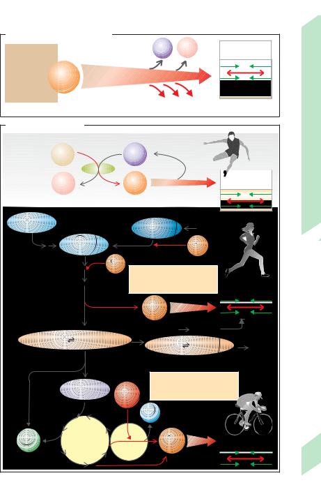
A. ATP as a direct energy source |
|
|
|
||
|
|
ADP |
Pi |
|
Reserve enough for |
Chemical |
|
|
|
|
10 contractions |
|
|
|
|
|
|
energy |
|
|
|
|
|
Reserve: |
ATP |
G ≈ –50 kJ/mol ATP |
|
|
|
ca. 5 mol |
|
|
|
|
Contraction |
per g muscle |
|
|
|
|
|
|
|
|
|
|
|
|
|
|
Heat |
+ |
Mechanical |
|
|
|
energy |
||
|
|
|
|
|
|
B. Regeneration of ATP |
|
|
|
|
|
1 Cleavage of creatine phosphate
Reserve: |
CrP |
ADP |
|
ca. 25 mol |
|
||
per g muscle |
|
Creatine |
|
|
|
kinase |
|
|
|
Short-term |
|
|
Cr |
ATP |
|
|
peak performance |
2 Anaerobic glycolysis
Glycogen |
Reserve: |
|
Blood |
|
|
|
|
|
ca. 100 mol/g muscle |
|
Liver |
|
|
|
|||
|
|
|
glucose |
|
|
|
|
|
|
Glucose-6-P |
|
|
1 |
|
|
|
|
|
|
|
ATP |
|
|
|
||
|
|
|
|
|
|
|
||
anaerobic |
|
1 |
|
|
|
|
|
|
|
ATP |
|
|
|
|
|
|
|
|
|
Net gain: |
|
|
|
|
|
|
|
|
2 mol ATP/mol glucose |
|
|
|
|
||
|
|
|
Long-term high |
|
||||
|
|
(3 mol ATP/mol glucose-6-P) |
|
|
||||
|
|
|
|
|
|
performance |
|
|
|
|
|
4 |
|
|
|
|
|
|
|
|
|
|
|
|
|
|
|
|
|
|
|
|
|
|
|
|
|
|
ATP |
|
|
|
|
|
|
|
|
|
|
|
|
|
|
|
|
Increase in lactic acid |
Drop in pH |
|
|
|
||
|
|
|
|
|
||||
2pyruvic acid 2pyruvate–+2H+ |
2lactic acids |
2H++2 lactate– |
Broken down |
|||||
|
|
|
in liver |
|||||
|
|
|
|
|
|
|
and heart |
|
3 Oxidation of glucose |
|
|
|
|
|
|
|
|
|
2 |
|
Total net gain: |
|
|
|
||
|
|
36 mol ATP/mol glucose |
|
|
||||
|
Acetyl-CoA |
6O2 |
|
|
||||
|
|
|
|
|
|
|
||
aerobic |
|
|
|
|
|
|
|
|
|
|
H2O |
|
|
|
|
|
|
6CO2 |
Krebs |
|
34 |
|
|
|
|
|
|
|
|
Endurance sport |
|
||||
|
ATP |
|
|
|
||||
|
cycle |
Respiratory |
|
|
|
|
|
|
|
|
chain |
|
|
|
|
|
|
|
|
|
|
|
|
|
|
|
|
|
|
|
|
|
|
|
|
|
|
|
|
|
|
|
|
|
|
|
|
|
|
|
|
|
|
Despopoulos, Color Atlas of Physiology © 2003 Thieme
All rights reserved. Usage subject to terms and conditions of license.
Plate 2.17 Energy Supply for Muscle Contraction
73

2 Nerve and Muscle, Physical Work
74
Physical Work
There are three types of muscle work:
Positive dynamic work, which requires to muscles involved to alternately contract and relax (e.g., going uphill).
Negative dynamic work, which requires the muscles involved to alternately extend while braking (braking work) and contract without a load (e.g., going downhill).
Static postural work, which requires continuous contraction (e.g., standing upright). Many activities involve a combination of two or three types of muscle work. Outwardly directed mechanical work is produced in dynamic muscle activity, but not in purely postural work. In the latter case, force!distance = 0. However, chemical energy is still consumed and completely transformed into a form of heat called maintenance heat (= muscle force times the duration of postural work).
In strenuous exercise, the muscles require up to 500 times more O2 than when at rest. At the same time, the muscle must rid itself of metabolic products such as H+, CO2, and lactate (!p. 72). Muscle work therefore requires drastic cardiovascular and respiratory changes.
In untrained subjects (UT), the cardiac output (CO; !p. 186) rises from 5–6 L/min at rest to a maximum of 15–20 L/min during exercise (!p. 77 C). Work-related activation of the sympathetic nervous system increases the heart rate up to ca. 2.5 fold and the stroke volume up to ca. 1.2 fold (UT). In light to moderate exercise, the heart rate soon levels out at a new constant level, and no fatigue occurs. Very strenuous exercise, on the other hand, must soon be interrupted because the heart cannot achieve the required long-term performance (!B). The increased CO provides more blood for the muscles (!A) and the skin (heat loss; !p. 222.). The blood flow in the kidney and intestine, on the other hand, is reduced by the sympathetic tone below the resting value (!A). The systolic blood pressure (!p. 206) rises while the diastolic pressure remains constant, yielding only a moderate increase in the mean pressure.
The smaller the muscle mass involved in the work, the higher the increase in blood pressure. Hence, the blood pressure increase in arm activity (cutting hedges) is higher than that in leg activity (cycling). In patients with coronary artery disease or cerebrovascular sclerosis, arm activity is therefore more dangerous than leg activity due to the risk of myocardial infarction or brain hemorrhage.
Muscular blood flow. At the maximum work level, the blood flow in 1 kg of active muscle rises to as much as 2.5 L/min (!p. 213 A), equivalent to 10% of the maximum cardiac output. Hence, no more than 10 kg of muscle ("1/3 the total muscle mass) can be fully active at any one time. Vasodilatation, which is required for the higher blood flow, is mainly achieved through local chemical influences
(!p. 212). In purely postural work, the increase in blood flow is prevented in part by the fact that the continuously contracted muscle squeezes its own vessels. The muscle then fatigues faster than in rhythmic dynamic work.
During. physical exercise (!C1), the ventilation (VE) increases from a resting value of ca. 7.5 L/min to a maximum of 90 to 120 L/min
(!C3). Both the respiratory rate (40–60 min– 1 max; !C2) and the tidal volume (ca. 2 L max.) contribute. to this increase. Because of the high V.E and increased CO, oxygen consumption (VO2) can increase. from ca. 0.3 L/min at rest to a maximum (Vo2 max) of ca. 3 L/min in UT (!C4 and p. 76). Around 25 L of air has to be ventilated to take up 1 L of O2 at rest, .corresponding.
to a respiratory equivalent. .(VE/VO2) of 25. During physical exercise, VE/VO2 rises beyond the endurance limit to a value of 40–50.
Increased O2 extraction in the tissues. also contributes to the large increase in VO2 during exercise. The decreasing pH and increasing temperature shift the O2 binding curve towards the right (!p. 129 B). O2 extraction is calculated as the arteriovenous difference in O2 concentration (avDo2 in L/L blood) times the blood flow. in L/min. The maximum O2 consumption (VO2 max) is therefore defined as:
.
VO2 max = HRmax · SVmax · avDO2max
where HR. is the heart rate and SV is the stroke volume. VO2 max per body weight is an ideal measure of physical exercise capacity (!p. 76).
Despopoulos, Color Atlas of Physiology © 2003 Thieme
All rights reserved. Usage subject to terms and conditions of license.
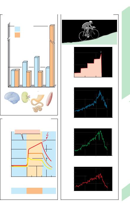
A. Blood supply in organs at rest and |
|
|
|
C. Respiration during physical work |
|
|
|||
during physical work |
|
|
|
|
|
|
|
|
|
|
|
|
|
|
12.5
Physical rest
|
Strenuous work |
|
|
|
|
|
|
|
|
|
|
(submaximal) |
|
|
|
|
|
|
|
|
|
(L/min) |
|
|
|
|
|
|
|
Exhaustion |
|
|
supply |
|
|
|
|
|
|
|
|
||
|
|
|
|
400 |
|
|
|
1 |
|
|
|
|
|
|
|
|
|
|
Physical Work |
||
Blood |
|
|
|
Power (W) |
|
|
|
|
|
|
1.0 |
|
|
|
200 |
|
|
|
|
||
|
|
|
|
|
|
|
|
|||
|
|
|
|
0 |
|
|
|
|
||
|
|
|
|
|
0 |
5 |
10 |
15 min |
2.18 |
|
|
|
|
|
|
|
|
|
|
|
|
0 |
|
|
|
|
|
|
|
|
|
Plate |
CNS |
Kidney |
GI tract |
Muscles |
) |
60 |
|
|
|
|
|
–1 |
|
|
|
|
||||||
|
|
|
|
|
|
|
|
2 |
||
|
|
|
|
(min |
|
|
|
|
|
|
|
|
|
|
40 |
|
|
|
|
|
|
|
|
|
|
rate |
|
|
|
|
|
|
|
|
|
|
|
|
|
|
|
|
|
|
|
|
|
Respiration |
20 |
|
|
|
|
|
|
|
|
|
|
|
|
|
|
|
|
B. Heart rate during physical work |
|
0 |
0 |
5 |
10 |
15 min |
|
|||
|
|
|
||||||||
|
|
|
|
|
|
|
||||
|
|
Maximum rate |
|
|
|
|
(L/min) |
100 |
|
|
|
|
||
|
|
|
|
|
|
|
|
|
|
|
||||
|
|
|
|
|
|
|
|
|
|
|
3 |
|
||
(minrate–1) |
100 |
|
Moderate |
|
|
ventilationTotal |
0 |
0 |
5 |
10 |
|
|||
|
|
|
15 min |
|
||||||||||
|
200 |
|
|
|
|
|
|
|
|
|
|
|
|
|
|
|
|
|
|
|
|
|
|
50 |
|
|
|
|
|
|
150 |
|
Strenuous |
|
|
|
|
|
|
|
|
|
||
Heart |
|
|
Light |
|
|
|
|
|
|
|
|
|
|
|
|
|
|
|
|
|
|
|
|
|
|
|
|
||
|
70 |
|
|
|
|
|
|
|
6 |
|
|
|
|
|
|
|
|
|
|
|
|
|
(L/min) |
|
|
|
|
|
|
|
|
|
|
|
|
|
|
4 |
|
|
|
4 |
|
|
|
|
|
|
|
|
|
|
|
|
|
|
|
||
|
0 |
0 |
5 |
10 |
15 |
20 |
25 |
|
|
|
|
|
|
|
|
|
|
|
|
|
|
|
|||||||
|
|
uptake |
|
|
|
|
|
|
||||||
|
|
|
|
Time (min) |
|
2 |
|
|
|
|
|
|||
|
|
|
|
|
|
|
|
|
|
|
||||
|
|
|
|
|
|
|
|
2 |
|
|
|
|
|
|
|
|
Rest |
Work |
|
Recovery |
O |
0 |
|
|
|
|
|
||
|
|
|
|
0 |
5 |
10 |
15 min |
75 |
||||||
|
|
|
|
|
|
|
|
|
|
|
|
(After J. Stegemann) |
|
|
Despopoulos, Color Atlas of Physiology © 2003 Thieme
All rights reserved. Usage subject to terms and conditions of license.

Physical Fitness and Training
|
The physical exercise capacity can be |
|
|
measured using simple yet standardized tech- |
|
|
niques of ergometry. This may be desirable in |
|
|
athletes, for example, to assess the results of |
|
|
training, or in patients undergoing rehabilita- |
|
|
tion therapy. Ergometry assesses the effects of |
|
Work |
exercise on physiological parameters such as |
|
. |
|
|
O2 consumption (VO2), respiration rate, heart |
||
Physical |
rate (!p. 74), and the plasma lactate concen- |
|
W/kg body weight (BW). |
|
|
|
tration (!A). The measured physical power |
|
Muscle, |
(performance) is expressed in watts (W) or |
|
angle α, exercise performance in watts is calculated |
||
|
In bicycle ergometry, a brake is used to adjust the watt |
|
|
level. In “uphill” ergometry on a treadmill set at an |
|
and |
as a factor of body mass (kg) !gravitational accelera- |
|
tion g (m · s– 2) !distance traveled (m)!sin α !1/ |
||
Nerve |
time required (s– 1). In the Margaria step test, the test |
|
subject is required to run up a staircase as fast as |
||
possible after a certain starting distance. Perform- |
||
2 |
ance is then measured as body mass (kg) !g |
|
|
(m · s– 2)!height/time (m · s– 1). |
|
|
Short-term performance tests |
(10–30 s) |
|
measure performance achieved through the |
|
|
rapidly available energy reserves (creatine |
|
|
phosphate, glycogen). Medium-term perform- |
|
|
ance tests measure performance fueled by an- |
|
|
aerobic glycolysis (!p. 72). The maximum O2 |
|
|
. |
|
|
consumption (VO2 max) is used to measure |
|
|
longer term aerobic exercise performance |
|
|
achieved through oxidation of glucose and free |
|
|
fatty acids (!p. 74). |
|
|
In strenuous exercise (roughly 2/3 the max- |
|
|
imum physical capacity or more), the aerobic |
|
|
mechanisms do not produce enough energy, so |
|
|
anaerobic metabolism must continue as a par- |
|
|
allel energy source. This results in lactacidosis |
|
|
and a sharp increase in the plasma lactate con- |
|
|
centration (!A). Lactate concentrations of |
|
|
up to 2 mmol/L (aerobic threshold) can be |
|
|
tolerated for prolonged periods of exercise. |
|
|
Lactate concentrations above 4 mmol/L (an- |
|
|
aerobic threshold) indicate that the perform- |
|
|
ance limit will soon be reached. Exercise must |
|
|
eventually be interrupted, not because of the |
|
|
increasing lactate concentration, but because |
|
|
of the increasing level of acidosis (!p. 74). |
|
76 |
Physical training raises and maintains the |
|
physical exercise capacity. There |
are three |
|
types of physical training strategies, and most training programs use a combination of them.
Motor learning, which increases the rate and accuracy of motor skills (e.g., typewriting). These activities primarily involve the CNS.
Endurance training, which improves submaximal long-term performance (e.g., running a marathon). The main objectives of endurance training are to increase the oxidative capacity of slow-twitch motor units (!p. 58!, e.g., by increasing the mitochondrial density, increase the.cardiac output and, consequently, to increase VO2 max (!B, C). The resulting increase in heart weight allows higher stroke volumes (!C) as well as higher tidal volumes, resulting in very low resting heart rates and respiratory rates. Trained athletes can therefore achieve larger increases in cardiac output and ventilation. than untrained subjects (!C). The VO2 max of a healthy individual is limited by the cardiovascular capacity, not the respiratory capacity. In individuals who practice endurance training, the exercise-related rise in the lactate concentraton is also lower and occurs later than in untrained subjects (!A).
Strength training improves the maximum short-term performance level (e.g., in weight lifting). The main objectives are to increase the muscle mass by increasing the size of the muscle fibers (hypertrophy) and to increase the glycolytic capacity of type motor units (!p. 58).
Excessive physical exercise causes muscle soreness and stiffness. The underlying cause is not lactic acid accumulation, but sarcomere microtrauma, which leads to muscle swelling and pain. The muscle ache, is a sign of microinflammation (!D).
Muscle fatigue may be peripheral or central. Peripheral fatigue ist caused by the exhaustion of energy reserves and the accumulation of metabolic products in the active muscle. This is particularly quick to occur during postural work (!p. 66). Central fatigue is characterized by work-related pain in the involved muscles and joints that prevents the continuation of physical exercise or decreased the individual’s motivation to continue the exercise.
Despopoulos, Color Atlas of Physiology © 2003 Thieme
All rights reserved. Usage subject to terms and conditions of license.

A. Lactate concentration (phys.exercise)
|
|
|
|
Training-related |
||
(mmol/L) |
10 |
|
|
|
shift |
|
8 |
|
|
|
|
|
|
concentration |
|
|
|
|
|
|
6 |
|
|
|
|
|
|
|
|
|
|
|
|
|
Lactate |
4 |
|
|
|
|
Anaerobic |
|
|
|
|
threshold |
||
|
2 |
|
|
|
|
Aerobic |
|
|
|
|
|
threshold |
|
|
|
|
|
|
|
|
|
00 |
1 |
2 |
3 |
4 |
5 |
|
|
|
Load (W/kg body weight) |
|
||
B. Maximum O2 uptake |
|
|
|
|
· |
|
|
|
Oxygen uptake VO2 |
|
|
|
(mL/min per kg body weight) |
|
|
|
Resting |
· |
|
|
VO2max |
|
|
Women |
|
|
and Training |
Non-athletic |
2.3 |
38 |
|
|
|
|
|
Athletic |
3.3 |
55 |
Fitness |
Men |
|
|
|
|
|
Physical |
|
Non-athletic |
3.2 |
44 |
|
|
|
|
|
|
|
|
2.19 |
Athletic |
4.8 |
67 |
Plate |
|
|
|
|
C. Comparison of non-athletic individuals and endurance athletes
Physiological parameters |
|
|
Non-athletes |
|
Endurance athletes |
||
|
|
|
|
|
|
|
|
(2 men, age 25, 70 kg) |
|
|
|
|
|
|
|
|
|
|
Maximum |
|
|
Maximum |
|
|
|
|
Resting |
|
Resting |
||
Heart weight (g) |
|
|
|
300 |
|
|
500 |
Blood volume (L) |
|
|
|
5.6 |
|
|
5.9 |
Heart rate (min-1) |
|
|
80 |
180 |
|
40 |
180 |
Stroke volume (mL) |
|
|
70 |
100 |
|
140 |
190 |
Cardiac output (L/min) |
|
|
5.6 |
18 |
|
5.6 |
35 |
Total ventilation (L/min) |
|
|
8.0 |
100 |
|
8.0 |
200 |
O2 uptake (L/min) |
|
|
0.3 |
2.8 |
|
0.3 |
5.2 |
(Data partly from H.-J.Ulmer)
D. Post-exercise muscle ache |
|
|
|
Unusually high strain on |
Cracks in Z disks |
Protein breakdown |
|
|
|
||
certain muscles |
|
|
|
|
|
Water influx |
|
|
|
Swelling |
|
|
|
|
Pain |
|
|
Reduced blood flow |
|
Loss of force |
Several hours later |
Reflex tension |
77 |
|
|
||
Despopoulos, Color Atlas of Physiology © 2003 Thieme
All rights reserved. Usage subject to terms and conditions of license.

3 Autonomic Nervous System (ANS)
Organization of the Autonomic
Nervous System
In the somatic nervous system, nerve fibers extend to and from the skeletal muscles, skin and sense organs. They usually emit impulses in response to stimuli from the outside environment, as in the withdrawal reflex (!p. 320). Much somatic nervous activity occurs consciously and under voluntary control. In contrast, the autonomic nervous system (ANS) is mainly concerned with regulation of circulation and internal organs. It responds to changing outside conditions by triggering orthostatic responses, work start reactions, etc. to regulate the body’s internal environment
(!p. 2). As the name implies, most activities of the ANS are not subject to voluntary control.
For the most part, the autonomic and somatic nervous systems are anatomically and functionally separate in the periphery (!A), but closely connected in the central nervous system, CNS (!p. 266). The peripheral ANS is efferent, but most of the nerves containing ANS fibers hold also afferent neurons. These are called visceral afferents because their signals originate from visceral organs, such as the esophagus, gastrointestinal (GI) tract, liver, lungs, heart, arteries, and urinary bladder. Some are also named after the nerve they accompany (e.g., vagal afferents).
Autonomic nervous activity is usually regulated by the reflex arc, which has an afferent limb (visceral and/or somatic afferents) and an efferent limb (autonomic and/or somatic efferents). The afferent fibers convey stimuli from the skin (e.g. nociceptive stimuli; !p. 316) and nocisensors, mechanosensors and chemosensors in organs such as the lungs, gastrointestinal tract, bladder, vascular system and genitals. The ANS provides the autonomic efferent fibers that convey the reflex response to such afferent information, thereby inducing smooth muscle contraction (!p. 70) in organs such as the eye, lung, digestive tract and bladder, and influencing the function of the heart (!p. 194) and glands. Examples of somatic nervous system involvement are afferent
78stimuli from the skin and sense organs (e.g., light stimuli) and efferent impulses to the skeletal muscles (e.g., coughing and vomiting).
Simple reflexes can take place within an organ (e.g., in the gut, !p. 244), but complex reflexes are controlled by superordinate autonomic centers in the CNS, primarily in the spinal cord (!A). These centers are controlled by the hypothalamus, which incorporates the ANS in the execution of its programs (!p. 330). The cerebral cortex is an even higher-ranking center that integrates the ANS with other systems.
The peripheral ANS consists of a sympathetic division and a parasympathetic division (!A) which, for the most part, are separate entities (!also p. 80ff.). The autonomic centers of the sympathetic division lie in the thoracic and lumbar levels of the spinal cord, and those of the parasympathetic division lie in the brain stem (eyes, glands, and organs innervated by the vagus nerve) and sacral part of the spinal cord (bladder, lower parts of the large intestine, and genital organs). (!A). Preganglionic fibers of both divisions of the ANS extend from their centers to the ganglia, where they terminate at the postganglionic neurons.
Preganglionic sympathetic neurons arising from the spinal cord terminate either in the paravertebral ganglionic chain, in the cervical or abdominal ganglia or in so-called terminal ganglia. Transmission of stimuli from preganglionic to postganglionic neurons is cholinergic, that is, mediated by release of the neurotransmitter acetylcholine (!p. 82). Stimulation of all effector organs except sweat glands by the postganglionic sympathetic fibers is adrenergic, i.e., mediated by the release of norepinephrine (!A and p. 84ff.).
Parasympathetic ganglia are situated near or within the effector organ. Synaptic transmissions in the parasympathetic ganglia and at the effector organ are cholinergic (!A).
Most organs are innervated by sympathetic and parasympathetic nerve fibers. Nonetheless, the organ’s response to the two systems can be either antagonistic (e.g., in the heart) or complementary (e.g., in the sex organs).
The adrenal medulla is a ganglion and hormone gland combined. Preganglionic sympathetic fibers in the adrenal medulla release acetylcholine, leading to the secretion of epinephrine (and some norepinephrine) into the bloodstream (!p. 86).
Despopoulos, Color Atlas of Physiology © 2003 Thieme
All rights reserved. Usage subject to terms and conditions of license.