Добавил:
Upload Опубликованный материал нарушает ваши авторские права? Сообщите нам.
Вуз: Предмет: Файл:

Color Atlas of Physiology 2003 thieme

.pdf
Скачиваний:
107
Добавлен:
15.02.2016
Размер:
36.12 Mб
Скачать

A. Characteristics of the vessel segments


Aorta

Greater arteries

Arterial branches

Arterioles

Capillaries

Venules

Venous branches

Greater veins

Venae cavae

Number 1

Increasing


0.16·109

5·109

0.5·109

Decreasing

2

1 Fraction of TPR


19%


47%


27%


7%


2 Mean blood pressure P (mmHg)


100

40


25

20


2–4


–1


·


)


3 Mean flow velocity VA (cm·s


18


5


1.5

0.02–0.1

1,0


6


4 Individual vessel diameter (cm)


3.2


2.6


0.15

1,6


0.8


0.3–0.06


0.00025

–0.7


0.002

0.0009


5 Joint cross-sectional area (cm2)


3500


2700


500


5.3

20


20


100

30

10


6 Joint holding capacity (cm3)


1550


550

900

180

250


250

125

300

250


Total volume of systemic circulation (excluding heart), ca. 4.4 L


Despopoulos, Color Atlas of Physiology © 2003 Thieme

All rights reserved. Usage subject to terms and conditions of license.

Plate 8.2 Blood Vessels and Blood Flow

189

8 Cardiovascular System

190

Cardiac Cycle

The resting heart rate is 60–80 beats per minute. A cardiac cycle (!A) therefore takes roughly 1 s. It can be divided into four distinct phases: (I) contraction phase and (II) ejection phase, both occurring in systole; (III) relaxation phase and filling phase (IV), both occurring in diastole. At the end of phase IV, the atria contract (phase IVc). Electrical excitation of the atria and ventricles precedes their contraction.

The cardiac valves determine the direction of blood flow within the heart, e.g., from the atria to the ventricles (phase IV) or from the ventricles to the aorta or pulmonary artery (phase II). All cardiac valves are closed during phases I and III (!A, top). Opening and closing of the valves is controlled by the pressures exerted on the two sides of the valves.

Cardiac cycle. Near the end of ventricular diastole, the sinoatrial (SA) node emits an electrical impulse, marking to the beginning of the

P wave of

the

ECG (phase IVc,

!A1 and

p. 196ff.).

This

results in atrial

contraction

(!A4) and is followed by ventricular excitation

(QRS complex of the ECG). The ventricular pressure then starts to rise ( !A2, blue line) until it exceeds the atrial pressure, causing the atrioventricular valves (mitral and tricuspid valves) to close. This marks the end of diastole. The mean end-diastolic volume (EDV) in the ventricle is now about 120 mL (!A4) or, more precisely, 70 mL/m2 body surface area.

The isovolumetric contraction phase now begins (phase I, ca. 50 ms). With all valves are closed, the ventricles now contract, producing the first heart sound (!A6), and the ventricular pressure increases rapidly. The slope of this ascending pressure curve indicates the maximum rate of pressure developed (maximum dP/dt). The semilunar valves (aortic and pulmonary valves) now open because the pressure in the left ventricle (!A2, blue line) exceeds that in the aorta (black broken curve) at about 80 mmHg, and the pressure in the right ventricle exceeds that in the pulmonary artery at about 10 mmHg.

The ejection phase (now begins phase II; ca. 210 ms at rest). During this period, the pressure in the left ventricle and aorta reaches a maximum of ca. 120 mmHg (systolic pressure). In the early phase of ejection (IIa or rapid ejec-

tion phase), a large portion of the stroke volume (SV) is rapidly expelled (!A4) and the blood flow rate reaches a maximum (!A5). Myocardial excitation subsequently decreases (T wave of the ECG, !A1) and ventricular pressure decreases (the remaining SV fraction is slowly ejected, phase IIb) until it falls below that of the aorta or pulmonary artery, respectively. This leads to closing of the semilunar valves, producing the second heart sound (!A6). The mean SV at rest is about 80 mL or, more precisely, 47 mL/m2 body surface area. The corresponding mean ejection fraction (SV/ EDV) at rest is about 0.67. The end-systolic volume (ESV) remaining in the ventricles at this point is about 40 mL (!A4).

The first phase of ventricular diastole or isovolumetric relaxation now begins (phase III; ca. 60 ms). The atria have meanwhile refilled, mainly due to the suction effect created by the lowering of the valve plane during ejection. As a result, the central venous pressure (CVP) decreases (!A3, falls from c to x). The ventricular pressure now drops rapidly, causing the atrioventricular valves to open again when it falls short of atrial pressure.

The filling phase now begins (phase IV; ca. 500 ms at rest). The blood passes rapidly from the atria into the ventricles, resulting in a drop in CVP (!A3, point y). Since the ventricles are 80% full by the first quarter of diastole, this is referred to as rapid ventricular filling (phase IVa; !A4). Ventricular filling slows down

(phase IVb), and the atrial systole (phase IVc) and the awave of CVP follows (!A2,3). At a normal heart rate, the atrial contraction contributes about 15% to ventricular filling. When the heart rate increases, the duration of the cardiac cycle decreases mainly at the expense of diastole, and the contribution of atrial contraction to ventricular filling increases.

The heart beats produce a pulse wave (pressure wave) that travels through the arteries at a specific pulse wave velocity (PWV): the PWV of the aorta is 3–5 m/s, and that of the radial artery is 5–12 m/s. PWV is. much higher than the blood flow velocity (V), which peaks at 1 m/s in the aorta and increases proportionally to (a) decreases in the compliance of aortic and arterial walls and (b) increases in blood pressure.

Despopoulos, Color Atlas of Physiology © 2003 Thieme

All rights reserved. Usage subject to terms and conditions of license.

A. Action phases of the heart (cardiac cycle)

Isovolumetric contraction

Atrial systole

Aortic pressure

Left ventricular pressure

Left atrial pressure

Central venous pressure (CVP)

Left ventricular volume

Flow rate

in aortic root

Heart sounds

Duration


Ejection


Isovolumetric


relaxation

Passive


ventricular filling

IVc

I

IIa

IIb

III

IVa

IVb

IVc


Ventricular systole


Ventricular diastole


1

R


T


ECG


P

1mV


Q

Q S


120


Inzisur


mmHg 2

0

a

c


v


2


a


mmHg 3


x


y

0


120


mL


Stroke


End-


volume

Atrial

70


diastolic


(SV)

contraction


volume


4

(EDV)


Residual volume (ESV)

0


500


mL/s

5


0


6

I


II

(III)

(IV)


ms


50

210

60

Considerably heart rate-dependent


(ca. 500 ms at 70 min–1)

Plate 8.3 Cardiac Cycle

191

Despopoulos, Color Atlas of Physiology © 2003 Thieme

All rights reserved. Usage subject to terms and conditions of license.

Despopoulos, Color Atlas of Physiology © 2003 Thieme
All rights reserved. Usage subject to terms and conditions of license.

8 Cardiovascular System

192

Cardiac Impulse Generation and

Conduction

The heart contains muscle cells that generate (pacemaker system), conduct (conduction system) and respond to electrical impulses (working myocardium). Cardiac impulses are generated within the heart (automaticity). The frequency and regularity of pacemaking activity are also intrinsic to the heart (rhythmicity). Myocardial tissue comprises a functional (not truly anatomical) syncytium because the cells are connected by gap junctions (!p. 16ff.). This also includes the atrioventricular junction (!p. 195 A). Thus, an impulse arising in any part of the heart leads to complete contraction of both ventricles and atria or to none at all (all- or-none response).

Cardiac contraction is normally stimulated by impulses from the sinoatrial node (SA node), which is therefore called the primary pacemaker. The impulses are conducted (!A) through the atria to the atrioventricular node (AV node). The bundle of His is the beginning of the specialized conduction system, including also the left and right (Tawara’s) bundle branches and the Purkinje fibers, which further transmit the impulses to the ventricular myocardium, where they travel from inside to outside and from apex to base of the heart. This electrical activity can be tracked in vivo (!C) by electrocardiography (!p. 196ff.).

Pacemaker potential (!B1, top). The cell potential in the SA node is a pacemaker potential. These cells do not have a constant resting potential. Instead, they slowly depolarize immediately after each repolarization, the most negative value of which is the maximum diastolic potential (MDP, ca. –70 mV). The slow diastolic depolarization or prepotential (PP) prevails until the threshold potential (TP) has again been reached. Thus triggering another action potential (AP).

The pacemaker potential (!B1, bottom) is subject to various underlying changes in ion conductance (g) and ionic flow (I) through the plasma membrane (!p. 32ff.). Starting at the MDP, a hyperpolarization-triggered increase in non-selective conductance and influx (If, = “funny”) of cations into the cells lead to slow depolarization (PP). When the TP is reached, gCa increases quickly, and the slope of the

pacemaker potential rises. This upslope is caused by increased influx of Ca2+ (ICa). When the potential rises to the positive range, gK increases sharply, resulting in the efflux of K+ (IK), and the pacemaker cells repolarize to the MDP.

Each action potential in the SA node normally generates one heart beat. The heart rate is therefore determined by the rate of impulse generation by the pacemaker. The impulse generation frequency decreases (!B3, broken curves) when (a) the PP slope decreases (!B3a), (b) the TP becomes less negative (!B3b), (c) the MDP becomes more negative, resulting in the start of spontaneous depolarization at lower levels (!B3c), or (d) repolarization after an action potential occurs more slowly (slope flatter).

The first three conditions extend the time required to reach the threshold potential.

All components of the conduction system can depolarize spontaneously, but the SA node is the natural or nomotopic pacemaker in cardiac excitation (sinus rhythm normally has a rate of 60 to 100 min–1). The intrinsic rhythms of the other pacemakers are slower than the sinus rhythm (!C, table) because the slope of their PPs and repolarizations are “flatter” (see above). APs arising in the SA node therefore arrive at subordinate (“lower”) levels of the conduction system before spontaneous depolarization has reached the intrinsic TP there. The intrinsic rhythm of the lower components come into play (ectopic pacemakers) when (a) their own frequency is enhanced, (b) faster pacemakers are depressed, or (c) the conduction from the SA node is interrupted (!p. 200). The heart beats at the AV rhythm (40 to 55 min–1)/or even slower (25 to 40 min–1) when controlled by tertiary (ventricular) pacemakers.

Overdrive suppression. The automaticity of lower pacemaker cells (e.g., AV node or Purkinje cells) is suppressed temporarily after they have been driven by a high frequency. This leads to increased Na+ influx and therefore to increased activity of the Na+-K+- ATPase. Because it is electrogenic (!p. 28), the cells become hyperpolarized and it takes longer to reach threshold than without prior high-frequency overdrive (!B3c).

The cells of the working myocardium contain voltage-gated fast Na+ channels that permit the brief but rapid influx of Na+ at the beginning of

!

A. Cardiac excitation


I


Conduction

ECG

1mV


Pacemaker


potential


SA node

(spontaneous


and

depolarization)


Atrial


myocardium


Generation

Action potentials


AV node


Bundle of His


100 mV


Purkinje

Impulse


fibers


Ventricular

Cardiac

Stable resting


myocardium

potential


Relative myocardial refractoriness:


8.4

Vulnerable phase

0.1 s

(after Hoffman und Cranefield)

Plate

B. Pacemaker potential and rate of impulse generation


(mV)

1 Pacemaker potential and ionic flow

3 Changes in heart rate due to changes


40


(after Di Francesco)


in pacemaker potential


AP


Membranepotential


Increase in slope

b


–80


–40 mV


c


0


a

of prepotential


(PP)


SP


Threshold-


–40


potential


PP

MDP


(TP)


Due to sympa-


Outward current


thetic stimuli,epi-


0.2 s


IK


nephrine, extra-


cellular K+

,


0


fever, etc.


Inward current


If

ICa


Vagal


potential (MDP)


Maximum diastolic


stimuli, etc.


0.2

0.4 s

4 Factors that affect the conduction


2 Duration of myocardial action


of action potentials (AV node)


potential is dependent on heart rate (f)


dV/dt


(after Trautwein et al.)


Steep:


+30


rapid

Flat:


slow


conduction


0


conduction


Para-


f =


f =


Sympathetic


mV


sympathetic stimuli,


stimuli, etc.


160 min–1

48min–1


temperature

,


quinidine, etc.


–100


0.5 s


193


Despopoulos, Color Atlas of Physiology © 2003 Thieme

All rights reserved. Usage subject to terms and conditions of license.

8 Cardiovascular System

194

!

an AP. The slope of their APs therefore rises more sharply than that of a pacemaker potential (!A). A resting potential prevails between APs, i.e. spontaneous depolarization normally does not occur in the working myocardium. The long-lasting myocardial AP has a characteristic plateau (!p. 59 A). Thus, the firststimulated parts of the myocardium are still in a refractory state when the AP reaches the laststimulated parts of the myocardium. This prevents the cyclic re-entry of APs in the myocardium. This holds true, regardless of whether the heart rate is very fast or very slow since the duration of an AP varies according to heart rate (!B2).

Role of Ca2+. The incoming AP opens voltagegated Ca2+ channels (associated with dihydropyridine receptors) on the sarcolemma of myocardial cells, starting an influx of Ca2+ from the ECF (!p. 63/B3). This produces a local increase in cytosolic Ca2+ (Ca2+ “spark”) which, in turn, triggers the opening of ligand-gated, ry- anodine-sensitive Ca2+ channels in the sarcoplasmic reticulum (Ca2+ store). The influx of Ca2+ into the cytosol results in electromechanical coupling (!p. 62) and myocardial contraction. The cytosolic Ca2+ is also determined by active transport of Ca2+ ions back (a) into the Ca2+ stores via a Ca2+-ATPase, called SERCA, which is stimulated by phospholamban, and

(b) to the ECF. This is achieved with the aid of a

Ca2+-ATPase and a 3Na+/Ca2+ exchange carrier that is driven by the electrochemical Na+ gradient established by Na+-K+-ATPase.

Although the heart beats autonomously, efferent cardiac nerves are mainly responsible for modulating heart action according to changing needs. The autonomic nervous system

(and epinephrine in plasma) can alter the following aspects of heart action: (a) rate of impulse generation by the pacemaker and, thus, the heart rate (chronotropism); (b) velocity of impulse conduction, especially in the AV node (dromotropism); and (c) contractility of the heart, i.e., the force of cardiac muscle contraction at a given initial fiber length (inotropism).

These changes in heart action are induced by acetylcholine (ACh; !p. 82) released by parasympathetic fibers of the vagus nerve (binds with M2 cholinoceptors on pacemaker cells), by norepinephrine (NE) released by sympathetic nerve fibers, and by plasma epi-

nephrine (E). NE and E bind with !1-adreno- ceptors (!p. 84ff.). The firing frequency of the SA node is increased by NE and E (positive chronotropism) and decreased by ACh (negative chronotropism) because these substances alter the slopes of the PP and the MDP in the SA cells (!B3a and c). Under the influence of ACh, the slope of the PP becomes flatter and the MDP becomes more negative gK rises. In versely, slope and amplitude of PP rises under the influence of E or sympathetic stimuli (higher If ) due to a rise in cation (Na+) conductance and, under certain conditions, a decrease in the gK. Only NE and E have chronotropic effects in the lesser components of the impulse conduction system. This is decisive when the AV node or tertiary pacemakers take over.

ACh (left branch of vagus nerve) decreases the velocity of impulse conduction in the AV node, whereas NE and E increase it due to their negative and positive dromotropic effects, respectively. This is mainly achieved through changes in the amplitude and slope of the up-

stroke of the AP (!B3c and B4), gK and gCa.

In positive inotropism, NE and E have a direct effect on the working myocardium. The resulting increase in contractility is based on an increased influx of Ca2+ ions from the ECF triggered by !1-adrenoceptors, resulting in an increased cytosolic Ca2+. This Ca2+ influx can be inhibited by administering Ca2+ channel blockers (Ca2+ antagonists). Other factors that increase cardiac contractility are an increase in AP duration, resulting in a longer duration of Ca2+ influx, and inhibition of Na+-K+-ATPase

(e.g., by the cardiac glycosides digitalis and strophanthin). The consequences are: flatter Na+ gradient across the cell membrane ! decreased driving force for 3 Na+/Ca2+ exchange carriers ! decreased Ca2+ efflux ! increased cytosolic Ca2+ conc.

When the heart rate is low, Ca2+ influx over time is also low (fewer APs per unit time), allowing plenty of time for the efflux of Ca2+ between APs. The mean cytosolic Ca2+ conc. is therefore reduced and contractility is low. Only by this indirect mechanism are parasympathetic neurons able to elicit a negative inotropic effect (frequency inotropism). NE and E can exert their positive inotropic effects either indirectly by increasing the high heart or directly via !1-adrenoceptors of the working

myocardium.

Despopoulos, Color Atlas of Physiology © 2003 Thieme

All rights reserved. Usage subject to terms and conditions of license.

C. Cardiac impulse conduction

Stimulated

SA node


AV node


Un-


stimulated


P

PQ

QRS

+

(mV) 0

ECG (lead II)

QRS

QRS

ST

SA node

AV node

Bundle


T


of His


Bundle


branches


Purkinje fibers


Time

ECG

Conduction

Intrinsic


Normal


velocity

rate

activation sequence


(ms)


(m·s–1)

(min–1)

SA node


Impulse generation


0

P wave

0.05


60–100

Arrival of impulse in

Right atrium

50


0.8–1.0


distal parts of atrium

Left atrium

85


in atrium


AV node


P-Q segment


Arrival of impulse


50

(delayed

0.05


40–55

Relaying of impulse


125

conduction)


His bundle activated


130


1.0–1.5


End of bundle branches activated

145


1.0–1.5


25–40

Purkinje fibers activatedt

150


3.0–3.5


Inner myocardium

Right ventricle

175


completely activated

Left ventricle

190

QRS complex

1.0


None


Outer myocardium

Right ventricle

205


in myo-


completely activated

Left ventricle

225


cardium


Despopoulos, Color Atlas of Physiology © 2003 Thieme

All rights reserved. Usage subject to terms and conditions of license.

Plate 8.5 Cardiac Impulse Generation and Conduction II

195


Electrocardiogram (ECG)

the left side of the thorax in a nearly horizontal


plane (!F). When used in combination with


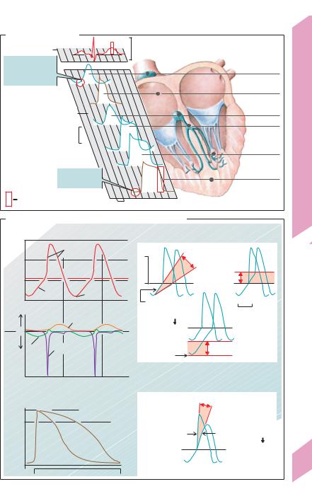
The ECG records potential differences (few

the aforementioned leads in the frontal plane,


m/V) caused by cardiac excitation. This pro-

they provide a three-dimensional view of the


vides information on heart position, relative

integral vector. To make recordings with the


chamber size, heart rhythm, impulse origin/

chest leads (different electrode), the three limb


propagation and rhythm/conduction distur-

leads are connected to form an indifferent elec-


bances, extent and location of myocardial

trode with high resistances (5 kΩ). The chest


ischemia, changes in electrolyte concentra-

leads mainly detect potential vectors directed


tions, and drug effects on the heart. However,

towards the back. These vectors are hardly de-


it does not provide data on cardiac contraction

tectable in the frontal plane. Since the mean


or pumping function.

QRS vector (see below) is usually directed

System


ECG potential differences arise at the inter-

downwards and towards the left back region,


face between stimulated and non-stimulated

the QRS vectors recorded by leads V1–V3 are


myocardium. Totally stimulated or unstimu-

usually negative, while those detected by V5

Cardiovascular


lated myocardial tissue does not generate any

and V6 are positive.


visible potentials The migration of the exci-

segments, and intervals (!B and p. 195 C). By


tatory front through the heart muscle gives

Intraesophageal leads and additional leads positioned


rise to numerous potentials that vary in mag-

in the region of the right chest (Vr3–Vr6) and left back


nitude and direction.

(V7–V9) are useful in certain cases (!F2).


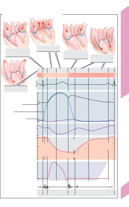
An ECG depicts electrical activity as waves,


These vectors can be depicted as arrows, where the


8


length of the arrow represents the magnitude of the

convention, upward deflection of the waves is


potential and the direction of the arrow indicates the


direction of the potential (arrowhead is +). As in a

defined as positive (+), and downward deflec-


force parallelogram, the integral vector (summa-

tion as negative (!). The electrical activity as-


tion vector) is the sum of the numerous individual

sociated with atrial depolarization is defined


vectors at that moment (!A, red arrow).

as the P wave ("0.3 mV, "0.1 s). Repolariza-


The magnitude and direction of the integral

tion of the atria normally cannot be visualized


vector change during the cardiac cycle, pro-

on the ECG since it tends to be masked by the


ducing the typical vector loop seen on a vector-

QRS complex. The QRS complex ("0.1 s) con-


cardiogram. (In A, the maximum or chief vec-

sists of one, two or three components: Q wave


tor is depicted by the arrow, called the “electri-

(mV " 1/4 of R, "0.04 s), R wave and/or S wave


cal axis” of the heart, see below).

(R+S #0.6 mV). The potential of the mean QRS


Limb and chest leads of the ECG make it

vector is the sum of the amplitudes of the Q, R


possible to visualize the course of the integral

and S waves (taking their positive and negative


vector over time, projected at the plane deter-

polarities into account). The voltage of the


mined by the leads (scalar ECG). Leads parallel

mean QRS vector is higher (in most leads) than


to the integral vector show full deflection (R

that of the P wave because the muscle mass of


wave !1–2 mV), while those perpendicular to

the ventricles is much larger than that of the


it show no deflection. Einthoven leads I, II, and

atria. The R wave is defined as the first positive


III are bipolar limb leads positioned in the fron-

deflection of the QRS complex, which means


tal plane. Lead I records potentials between the

that R waves from different leads may not be


left and right arm, lead II those between the

synchronous. The QRS complex represents the


right arm and left leg, and lead III those be-

depolarization of the ventricles, and the T wave


tween the left arm and left leg (!C1).

represents their repolarization. Although op-


Goldberger leads are unipolar augmented limb

posing processes, the T wave usually points in


leads in the frontal plane. One lead (right arm,

the same direction as the R wave (+ in most


aVR, left arm aVL, or left leg, aVF; !D2) acts as

leads). This means that depolarization and re-


the different electrode, while the other two

polarization do not travel in the same direction

196


limbs are connected and serve as the indiffer-

(!p. 195 C, QRS and T: vector arrows point in


ent (reference) electrode (!D1). Wilson leads

the same direction despite reversed polarity


(V1–V6) are unipolar chest leads positioned on

during repolarization). The PQ (or PR) segment


!

Despopoulos, Color Atlas of Physiology © 2003 Thieme

All rights reserved. Usage subject to terms and conditions of license.

A. Vector loops of cardiac excitation

B. ECG curve


Frontal


mV


R


plane


Sagittal

Vector


T


plane

cardiogram

P


Q S


I


0.08s


(ECG)

R


Wave

P

QRS


T


Electrocardiogram


Segment


PQ

ST


Integral vector


0.12-0.2s

ca.0.35s


Interval

PQ

QT


Horizontal


Heart rate-dependent


plane

(after Antoni)


C. Einthoven leads I, II and III (bipolar)


Plate 8.6

I


I


I


II

III


III


III


II


II

2


3


1


D. Goldberger limb leads (unipolar)


E. Cabrera circle


–120°

–90°

–60°


Right


Left


arm

I

arm


–30°


aVR

aVL


aVL


I


II

III


aVF


aVR


III

aVF

II

+30°


aVR


Foot


+120°

+90°

+60°


2

197

1

Despopoulos, Color Atlas of Physiology © 2003 Thieme

All rights reserved. Usage subject to terms and conditions of license.


!


(complete atrial excitation) and the ST seg-


ment (complete ventricular excitation) lie ap-


prox. on the isoelectric line (0 mV). The PQ (or


PR) interval (!0.2 s) is measured from the


beginning of the P wave to the beginning of the


Q wave (or to the R wave if Q wave is absent)


and corresponds to the time required for atrio-


ventricular conduction (!B). The QT interval is


measured from the start of the Q wave to the


end of the T wave. It represents the overall


time required for depolarization and repolari-


zation of the ventricles and is dependent on

System


the heart rate (0.35 to 0.40 s at a heart rate of


75 min–1).


Figure E

illustrates the six frontal leads

Cardiovascular


(Einthoven

and Goldberger

leads)

on the


Cabrera circle. Synchronous measurement of


the amplitude of Q, R and S from two or more


leads can be used to determine any integral


vector in the frontal plane (!G). The direction


of the largest mean QRS vector is called the QRS

8


axis (!C3 and G, red arrows). If the excitation


spreads normally, the QRS axis roughly corre-


sponds to the anatomic longitudinal axis of the


heart.


The mean QRS axis (“electrical axis”) of the


heart, which normally lies

between + 90


degrees to –30 degrees in adults (!G, H). Right


type (α = + 120" to + 90") is not unusual in


children, but is often a sign of abnormality in


adults. Mean QRS axes ranging from + 90


degrees to + 60 degrees are described as the


vertical type (!G1), and those ranging from


+ 60 degrees to + 30 degrees are classified as


the intermediate type (!G2). Left type occurs


when α = + 30 degrees to –30 degrees (!G3).


Abnormal

deviation: Right

axis

deviation

(#+ 120") can develop due right ventricular hypertrophy, while left axis deviation (more negative than –30") can occur due to left ventricular hypertrophy.

An extensive myocardial infarction (MI) can shift the electrical axis of the heart. Marked Q wave abnormality (!I 1) is typical in transmural myocardial infarction (involving entire thickness of ventricular wall): Q wave duration #0.04 s and Q wave amplitude #25% of total amplitude of the QRS complex. These changes appear within 24 hours of MI and are caused by failure of the dead myocardium to conduct electrical impulses. Preponderance of the exci-

198tatory vector in the healthy contralateral side of the heart therefore occurs while the affected part of the

myocardium should be depolarizing (first 0.04 s of QRS). The so-called “0.04-sec vector” is therefore said to point away from the infarction. Anterior MI is detected as highly negative Q waves (with smaller R waves) mainly in leads V5, V6, I and aVL. Q wave abnormalities can persist for years after MI (!I 2/3), so they may not necessarily be indicative of an acute infarction. ST elevation points to ischemic but not (yet) necrotic parts of the myocardium. This can be observed: (1) in myocardial ischemia (angina pectoris),

(2) in the initial phase of transmural MI, (3) in nontransmural MI, and (4) along the margins of a transmural MI that occurred a few hours to a few days prior (!I 4). The ST segment normalizes within 1 to 2 days of MI, but the T wave remains inverted for a couple of weeks (!I 5 and 2).

Excitation in Electrolyte Disturbances

Hyperkalemia. Mild hyperkalemia causes various changes, like elevation of the MDP (!p. 192) in the SA node. It can sometimes have positive chronotropic effects (!p. 193 B3c). In severe hyperkalemia, the more positive MDP leads to the inactivation of Na+ channels (!p. 46) and to a reduction in the slope and amplitude of APs in the AV node (negative dromotropic effect; !p. 193 B4). Moreover, the K+ conductance (gK) rises, and the PP slope becomes flatter due to a negative chronotropic effect (!p. 193 B3a). Faster myocardial repolarization decreases the cytosolic Ca2+ conc. In extreme cases, the pacemaker is also brought to a standstill (cardiac paralysis). Hypokalemia (moderate) has positive chronotropic and inotropic effects (!p. 193 B3a), whereas hypercalcemia is thought to raise the gK and thereby shortens the duration of the myocardial AP.

ECG. Changes in serum K+ and Ca2+ induce characteristic changes in myocardial excitation.

Hyperkalemia (#6.5 mmol/L): tall, peaked T waves and conduction disturbances associated with an increased PQ interval and a widened QRS. Cardiac arrest can occur in extreme cases.

Hypokalemia (!2.5 mmol/L): ST depression, biphasic T wave (first positive, then negative) followed by a positive U wave.

Hypercalcemia (#2.75 mmol/L total calcium): shortened QT interval due to a shortened ST segment.

Hypocalcemia (!2.25 mmol/L total calcium): prolonged QT interval.

Despopoulos, Color Atlas of Physiology © 2003 Thieme

All rights reserved. Usage subject to terms and conditions of license.

Соседние файлы в предмете [НЕСОРТИРОВАННОЕ]