

Color Atlas of Physiology 2003 thieme
.pdfC. Specific immunity: B-cell activation |
|
|
|
|
|
||
B lymphocytes |
|
|
|
|
|
|
|
|
Antigen binding |
|
|
|
|
|
|
|
|
|
B cell |
|
|
Activation |
|
|
|
|
|
|
|
|
|
|
|
IgD, IgM |
|
|
|
|
|
|
|
monomer |
|
|
|
|
|
“Naive” |
|
Antibody |
|
|
|
|
|
|
network |
|
|
|
|
||
B cell |
|
|
|
|
|
||
|
|
|
|
|
|
III |
|
|
|
|
|
|
|
|
|
|
|
|
|
|
|
Internalization |
System |
|
|
|
|
|
|
|
|
|
1 |
|
|
|
|
|
Immune |
|
|
|
|
|
|
|
|
|
|
|
|
|
|
|
4.6 |
TH2 cell |
|
|
|
|
|
Processing |
Plate |
(see plate B3) |
|
|
|
|
|
|
|
|
|
|
|
|
HLAII |
|
|
|
Cooperation of |
|
|
|
|
|
|
|
TH2 cell and B cell |
|
|
|
|
|
|
TH2 cell |
|
TD |
B cell |
|
|
|
|
|
|
HLAII |
|
|
|
|
|
CD4 |
|
antigen |
|
|
|
|
|
T-cell receptor |
|
|
|
|
|
|
|
CD40 ligand |
|
CD40 |
Presentation |
|
|
|
|
|
|
|
|
|
|
||
2 |
IL-4 |
|
|
|
|
|
|
IL-5 |
|
|
|
|
|
|
|
|
|
|
|
|
|
|
|
|
IL-6 |
|
|
|
|
|
|
|
|
Proliferation |
|
|
|
|
|
|
3 |
|
|
|
Plasma cell |
|
|
|
Differentiation |
|
|
|
|
||
|
|
|
|
|
|
||
|
|
|
|
|
|
According to |
|
|
|
|
|
|
|
class switching |
|
|
|
|
|
|
|
Immun- |
|
|
|
IgM |
IgG |
IgA |
IgE |
globulins |
|
|
|
Specific humoral immune response |
|
99 |
|||
|
|
|
(see plate A) |
|
|
|
|
Despopoulos, Color Atlas of Physiology © 2003 Thieme
All rights reserved. Usage subject to terms and conditions of license.

Hypersensitivity Reactions (Allergies)
|
Allergy is a specific, exaggerated immune re- |
|
|
sponse to a (usually harmless) foreign sub- |
|
|
stance or antigen (!p. 94ff.). Allergens are an- |
|
|
tigens that induce allergies. Small molecules |
|
|
conjugated to endogenous proteins can also |
|
|
have antigenic effects. In this case, they are re- |
|
|
ferred to as incomplete antigens or haptens. |
|
|
The heightened immune response to second- |
|
|
ary antigen contact (!p. 94ff.) normally has a |
|
|
protective effect. In allergies, however, the first |
|
|
contact with an antigen induces sensitization |
|
|
(allergization), and subsequent exposure leads |
|
|
to the destruction of healthy cells and intact |
|
|
tissue. This can also result in damage to endog- |
|
|
enous proteins and autoantibody production. |
|
|
Inflammatory reactions are the main causes of |
|
|
damage. |
|
Blood |
Types of hypersensitivity reactions: Type I |
|
reactions are common. On first contact, the al- |
||
lergen internalized by B cells is presented to |
||
4 |
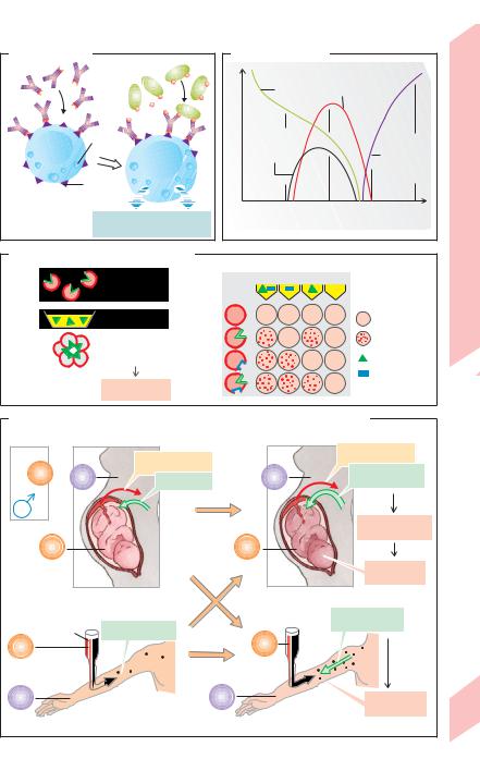
TH2 cells. The B cell then proliferates and differentiates into plasma cells (see p. 98), which release immunoglobulin E (IgE). The Fc fragment of IgE binds to mast cells and basophils. On subsequent contact, the antigens bind to the already available IgE-linked mast cells (!A). Due to the rapid release of mostly vasoactive mediators of inflammation such as histamine, leukotrienes and platelet-activating factor (PAF), an immediate reaction (anaphylaxis) occurs within seconds or minutes: immediate type hypersensitivity. This is the mechanism by which allergens breathed into the lungs trigger hay fever and asthma attacks. The vasodilatory effect of a generalized type I reaction can lead to anaphylactic shock (see p. 218).
In type II reactions, the immune system mainly attacks cells with antigenic properties. This can be attributable to the transfusion of the erythrocytes of the wrong blood group or the binding of haptens (e.g., medications) to endogenous cells. The binding of haptens to platelets can, for example, result in throm-
|
bocytopenia. |
|
|
Type III reactions are caused by antigen-antibody |
|
|
complexes. If more antigen than antibody is availa- |
|
|
ble, soluble antigen-antibody complexes circulate in |
|
100 |
blood for a long time (!B) and settle mainly in the |
|
capillaries, making the capillary wall subject to attack |
||
|
by the complement system. This leads to the development of serum sickness (!B), the main symptoms of which are joint pain and fever.
Type IV reactions are mainly mediated by TH1 cells, TC cells, and macrophages. Since symptoms peak 2 to 4 days after antigen contact, this is called delayed type hypersensitivity. The main triggers are mycobacteria (e.g. Tbc), other foreign proteins, and haptens, such as medications and plant substances, such as poison ivy. Primary transplant rejection is also a type IV hypersensitivity reaction. Contact dermatitis is also a type IV reaction caused by various haptens (e.g., nickel in jewelry).
Blood Groups
A person’s blood group is determined by the type of antigen (certain glycolipids) present on the red blood cells (RBCs). In the AB0 system, the antigens are A and B (!C). In blood type A, antigen A (on RBC) and anti-B antibody (in serum) are present; in type B, B and anti-A are present; in type AB, A and B are present, no antibody; in type 0 (zero), no antigen but anti-A and anti-B are present.
When giving a blood transfusion, it is important that the blood groups of donor and recipient match, i.e. that the RBCs of the donor (e.g. A) do not come in contact with the respective antibodies (e.g. anti-A) in the recipient. If the donor’s blood is the wrong type, agglutination (cross-linking by IgM) and hemolysis (bursting) of the donor’s RBCs will occur (!C1). Donor and recipient blood types must therefore be determined and cross-matched (!C2) prior to a blood transfusion. Since AB0 antibodies belong to the IgM class, they usually do not cross the placenta.
In the Rh system, antibodies against rhesus antigens (C, D, E) on RBCs do not develop unless prior sensitization has occurred. D is by far the most antigenic. A person is Rh-positive (Rh+) when D is present on their RBCs (most people), and Rh-negative (Rh–) when D is absent. Anti-D antibodies belong to the IgG class of immunoglobulins, which are capable of crossing the placenta (!p. 93 D). Rh– individuals can form anti-Rh+ (= anti-D) antibodies, e.g., after sensitization by a mismatched blood transfusion or of an Rh– mother by an Rh+ fetus. Subsequent exposure to the mismatched blood leads to a severe an- tigen-antibody reaction characterized by intravascular agglutination and hemolysis (!D).
Despopoulos, Color Atlas of Physiology © 2003 Thieme
All rights reserved. Usage subject to terms and conditions of license.
A. Anaphylaxis |
Antigen |
lgE |
Granules |
lgE |
receptor |
Mast cell or basophil |
Histamine, PAF, leukotrienes |
and other mediators |
C. ABO blood group incompatibility
1 |
|
2 |
|
|
Red blood cells |
cells |
|
= |
Agglutination |
||
redonblood |
|||
+ |
Antibodies |
|
|
|
Hemolysis |
Antigen |
B. Serum sickness |
|
|
||
in blood |
Antigen |
Symptoms |
|
|
|
|
|
|
|
Concentration |
Antigen- |
|
|
|
antibody |
|
|
Free |
|
complexes |
|
|
||
|
|
|
antibodies |
|
|
|
|
|
|
|
5 |
10 |
15 |
20 |
|
|
Days after first antigen exposure |
||
|
|
|
(After Kownatzki) |
|
Antibody in serum
O A B AB
O |
Compatible |
A |
Incompatible |
|
(agglutination) |
B |
Anti-A |
|
Anti-B |
AB |
|
Plate 4.7 Blood Groups
D. Rh sensitization of mother by child or by Rh-mismatched transfusion |
|
|||||
Father |
Mother |
|
|
|
||
|
|
Rh+ red cells |
|
Rh+ red cells |
|
|
+ |
|
|
High |
|
||
– |
|
– |
|
|||
Rh |
Anti-Rh+ |
anti-Rh+ titer |
|
|||
Rh |
Rh |
|
||||
|
|
|
|
Severe hemolysis |
|
|
|
|
|
|
in fetus |
|
|
Rh+ |
|
Rh+ |
|
|
||
|
|
|
|
Damage |
|
|
|
First Rh+ child |
Subsequent Rh+ children |
|
|||
|
|
|
|
High |
|
|
|
Blood |
Anti-Rh+ antibody |
|
anti-Rh+ titer |
|
|
|
|
|
|
|||
Rh+ |
|
|
Rh+ |
|
|
|
– |
|
|
– |
|
|
|
Rh |
|
|
Rh |
Damage |
|
|
|
|
|
|
101 |
||
First mismatched Rh+ transfusion |
Subsequent mismatched Rh+ transfusion |
|||||
|
||||||
Despopoulos, Color Atlas of Physiology © 2003 Thieme
All rights reserved. Usage subject to terms and conditions of license.

4 Blood
102
Hemostasis
The hemostatic system stops bleeding. Thrombocytes (platelets), coagulation (or clotting) factors in plasma and vessel walls interact to seal leaks in blood vessels. The damaged vessel constricts (release of endothelin), and platelets aggregate at the site of puncture (and attract more platelets) to seal the leak by a platelet plug. The time required for sealing (ca. 2 to 4 min) is called the bleeding time. Subsequently, the coagulation system produces a fibrin meshwork. Due to covalent cross-linking of fibrin, it turns to a fibrin clot or thrombus that retracts afterwards, thus reinforcing the seal. Later recanalization of the vessel can be achieved by fibrinolysis.
Platelets (170–400 · 103 per µL of blood; half-life !10 days) are small non-nucleated bodies that are pinched off from megakaryocytes in the bone marrow. When an endothelial injury occurs, platelets adhere to subendothelial collagen fibers (!A1) bridged by von Willebrand’s factor (vWF), which is formed by endothelial cells and circulates in the plasma complexed with factor VIII. Glycoprotein complex GP Ib/IX on the platelets are vWF receptors. This adhesion activates platelets (!A2). They begin to release substances (!A3), some of which promote platelet adhesiveness (vWF). Others like serotonin, plate- let-derived growth factor (PDGF) and thromboxane A2 (TXA2) promote vasoconstriction. Vasoconstriction and platelet contraction slow the blood flow. Mediators released by platelets enhance platelet activation and attract and activate more platelets: ADP, TXA2, platelet-activating factor (PAF). The shape of activated platelets change drastically (!A4). Discoid platelets become spherical and exhibit pseudopodia that intertwine with those of other platelets. This platelet aggregation (!A5) is further enhanced by thrombin and stabilized by GP IIb/IIIa. Once a platelet changes its shape, GP IIb/IIIa is expressed on the platelet surface, leading to fibrinogen binding and platelet aggregation. GP IIb/IIIa also increases the adhesiveness of platelets, which makes it easier for them to stick to subendothelial fibronectin.
I |
Fibrinogen |
Half-life (h): |
96 |
II K |
Prothrombin |
|
72 |
III |
Tissue thromboplastin |
|
|
IV |
Ionized calcium (Ca2+) |
|
|
V |
Proaccelerin |
|
20 |
VII K |
Proconvertin |
|
5 |
VIII |
Antihemophilic factor A |
|
12 |
IX K |
Antihemophilic factor B; plasma |
|
|
|
thromboplastin component (PTC); |
|
|
|
Christmas factor |
|
24 |
X K |
Stuart–Prower factor |
|
30 |
XI |
Plasma thromboplastin antecedent |
|
|
|
(PTA) |
|
48 |
XII |
Hageman factor |
|
50 |
XIII |
Fibrin-stabilizing factor (FSF) |
250 |
|
–Prekallikrein (PKK); Fletcher factor
–High-molecular-weight kininogen (HMK); Fitzgerald factor
Several coagulation factors are involved in the clotting process. Except for Ca2+, they are proteins formed in the liver (!B and Table). Factors labeled with a “K” in the table (as well as protein C and protein S, see below) are produced with vitamin K, an essential cofactor in posttranslational γ-carboxylation of glutamate residues of the factors. These γ-carboxy- glutamyl groups are chelators of Ca2+. They are required for Ca2+-mediated complex formation of factors on the surface of phospholipid layers (PL), particularly on the platelet membrane (platelet factor 3). Vitamin K is oxidized in the reaction and has to be re-reduced by liver epoxide reductase (vitamin K recycling). Ca2+ ions are required for several steps in the clotting process (!B). When added to blood samples in vitro, citrate, oxalate, and EDTA bind with Ca2+ ions, thereby preventing the blood from clotting. This effect is desirable when performing various blood tests.
Activation of blood clotting (!B). Most coagulation factors normally are not active, or zymogenic. Their activation requires a cascade of events (An “a” added to the factor number means “activated”). Thus, even small amounts of a trigger factor lead to rapid blood clotting. The trigger can be endogenous (within a vessel) or exogenous (external). Endogenous activation (!B2) occurs at an endothelial defect. XII is activated to XIIa by the contact with
!
Despopoulos, Color Atlas of Physiology © 2003 Thieme
All rights reserved. Usage subject to terms and conditions of license.

A. Platelet-mediated hemostasis
|
|
|
|
|
vWF |
Platelet (PLT) |
|
|
|
|
|
|
|
|
|
|
|
|
|
Blood |
|
|
|
|
|
|
|
|
|
|
1 |
|
|
Kollagen |
|
|
Change in shape |
|
|
Endothelial |
|
|
|
|
4 |
|
|||
PLT adhesion |
|
|
|
|
|||||
damage |
|
|
|
|
|
||||
|
|
|
|
2 |
|
|
|
|
|
|
|
|
|
|
PLT activation |
|
|
|
Hemostasis |
|
|
|
|
|
Slowing of |
|
Fibrinogen |
||
|
|
|
|
|
blood flow |
|
|
|
|
|
|
|
|
Vasoconstriction |
Secretion |
|
|
||
|
|
|
|
|
|
|
|||
|
|
|
|
3 |
|
|
Plate 4.8 |
||
|
|
|
vWF |
Serotonin, TXA2 |
ADP, |
5 |
PLT aggregation: |
||
|
|
|
|
||||||
|
|
|
|
PDGF |
PAF |
Clot formation |
|
||
B. Blood clotting |
|
|
|
|
|
|
|
||
|
|
1 |
Exogenous activation |
2 Endogenous activation |
|
|
|||
|
|
|
|
(tissue injury) |
(contact with collagen) |
|
|
||
|
|
|
Ca2+,III |
|
Contact phase |
|
|
||
|
|
|
VII |
HMK KK |
PKK |
|
|
||
|
|
|
|
|
|
XII |
XIIa |
|
|
|
|
|
PL–Ca2+–VIIa |
|
|
|
|
||
|
|
|
|
|
|
XIa |
XI |
|
|
PL |
Phospholipids |
|
|
X |
|
IXa |
IX |
|
|
|
(mainly PLT factor III) |
|
|
|
|
|
|
|
|
|
Zymogen |
|
|
|
PL–Ca2+–IXa–VIIIa |
VIII |
|
|
|
|
Complex |
|
|
|
|
|
|||
|
|
|
|
|
|
|
|
|
|
|
Activates |
|
|
|
|
V |
|
|
|
|
Converts to |
|
|
Xa |
|
|
|
|
|
|
|
Prothrombin (II) |
PL–Ca2+–Xa–Va |
|
|
|
|||
|
|
|
|
|
|
|
|||
|
3 |
Fibrin |
|
|
|
|
|
|
|
|
formation |
|
|
|
|
|
|
||
|
|
|
|
|
|
Thrombin (IIa) |
PLT aggregation |
|
|
|
|
Fibrinogen (I) |
|
XIII |
|
|
|
||
|
|
|
|
|
|
|
|
|
|
|
|
|
|
|
|
XIIIa |
|
|
|
|
|
|
|
|
Fibrin monomer |
Fibrin mesh-work |
103 |
||
|
|
|
|
|
|
||||
Despopoulos, Color Atlas of Physiology © 2003 Thieme
All rights reserved. Usage subject to terms and conditions of license.

|
|
! |
|
|
negative charges of subendothelial collagen |
|
|
and sulfatide groups. This stimulates the con- |
|
|
version of prekallikrein (PKK) to kallikrein |
|
|
(KK), which enhances XII activation (positive |
|
|
feedback). Next, XIIa activates XI to XIa, which |
|
|
converts IX to IXa and, subsequently, VIII to |
|
|
VIIIa. Complexes formed by conjugation of IXa |
|
|
and VIIIa with Ca2+ on phospholipid (PL) layers |
|
|
activate X. Exogenous activation now merges |
|
|
with endogenous activation (!B1). In rela- |
|
|
tively large injuries, tissue thrombokinase (fac- |
|
|
tor III), present on of nonvascular cells, is ex- |
|
|
posed to the blood, resulting in activation of |
|
|
VII. VII forms complexes with Ca2+ and phos- |
|
|
pholipids, thereby activating X (and IX). |
|
|
Fibrin formation (!B3). After activation of |
|
|
X to Xa by endogenous and/or exogenous acti- |
|
|
vation (the latter is faster), Xa activates V and |
|
|
conjugates with Va and Ca2+ on the surface of |
Blood |
|
membranes. This complex, called prothrombi- |
|
(IIa). In the process, Ca2+ binds with phos- |
|
|
|
nase, activates prothrombin (II) to thrombin |
4 |
|
pholipids, and the N-terminal end of pro- |
|
|
|
|
|
thrombin splits off. The thrombin liberated in |
|
|
the process now activates (a) fibrinogen (I) to |
|
|
fibrin (Ia), (b) fibrin-stabilizing factor (XIII), |
|
|
and (c) V, VIII and XI (positive feedback). The |
|
|
single (monomeric) fibrin threads form a |
|
|
soluble meshwork (fibrinS; “s” for soluble) |
|
|
which XIIIa ultimately stabilizes to insoluble fi- |
|
|
brin (fibrini). XIIIa is a transamidase that links |
|
|
the side chains of the fibrin threads via |
|
|
covalent bonds. |
|
|
Fibrinolysis and Thromboprotection |
|
|
To prevent excessive clotting and occlusion of |
|
|
major blood vessels (thrombosis) and em- |
|
|
bolisms due to clot migration, fibrinS is re-dis- |
|
|
solved (fibrinolysis) and inhibitory factors are |
|
|
activated as soon as vessel repair is initiated. |
|
|
Fibrinolysis is mediated by plasmin (!C). |
|
|
Various factors in blood (plasma kallikrein, |
|
|
factor XIIa), tissues (tissue plasminogen acti- |
|
|
vator, tPA, endothelial etc.), and urine (uroki- |
|
|
nase) activate plasminogen to plasmin. Strep- |
|
|
tokinase, staphylokinase and tPA are used |
|
|
therapeutically to activate plasminogen. This |
|
|
is useful for dissolving a fresh thrombus lo- |
104 |
|
cated, e.g., in a coronary artery. Fibrin is split |
|
into fibrinopeptides which inhibit thrombin |
formation and polymerization of fibrin to prevent further clot formation. Alpha2-antiplas- min is an endogenous inhibitor of fibrinolysis. Tranexamic acid is administered therapeutically for the same purpose.
Thromboprotection. Antithrombin III, a serpin, is the most important thromboprotective plasma protein (!D).
It inactivates the protease activity of thrombin and factors IXa, Xa, XIa and XIIa by forming complexes with them. This is enhanced by heparin and heparinlike endothelial glucosaminoglycans. Heparin is produced naturally by mast cells and granulocytes, and synthetic heparin is injected for therapeutic purposes.
The binding of thrombin with endothelial thrombomodulin provides further thromboprotection. Only in this form does thrombin have anticoagulant effects (!D, negative feedback). Thrombomodulin activates protein C to Ca which, after binding to protein S, deactivates coagulation factors Va and VIIIa. The synthesis of proteins C and S is vitamin K-dependent. Other plasma proteins that inhibit thrombin are α2-macroglobulin and α1-an- titrypsin (!D). Endothelial cells secrete tissue thromboplastin inhibitor, a substance that inhibits exogenous activation of coagulation, and prostacyclin (= prostaglandin I2), which inhibits platelet adhesion to the normal endothelium.
Anticoagulants are administered for thromboprotection in patients at risk of blood clotting. Injected heparin has immediate action. Oral coumarin derivatives (phenprocoumon, warfarin, acenocoumarol) are vitamin K antagonists that work by inhibiting liver epoxide reductase, which is necessary for vitamin D recycling. Therefore, these drugs do not take effect until the serum concentration of vitamin K-dependent coagulation factors has decreased. Cyclooxygenase inhibitors, such as aspirin (acetylsalicylic acid), inhibit platelet aggregation by blocking thromboxane A2 (TXA2) synthesis (!p. 269).
Hemorrhagic diatheses can have the following causes: a) Congenital deficiency of certain coagulation factors. Lack of VIII or IX, for example, leads to hemophilia A or B, respectively. b) Acquired deficiency of coagulation factors. The main causes are liver damage as well as vitamin K deficiency due to the destruction of vitamin K-producing intestinal flora or intestinal malabsorption. c) Increased consumption of coagulation factors, by disseminated intravascular coagulation. d) Platelet deficiency (thrombocytopenia) or platelet defect (thrombocytopathy). e) Certain vascular diseases, and f) excessive fibrinolysis.
Despopoulos, Color Atlas of Physiology © 2003 Thieme
All rights reserved. Usage subject to terms and conditions of license.

C. Fibrinolysis |
|
|
|
Plasminogen |
Fibrin mesh-work |
|
|
|
|
Streptokinase |
Thromboprotection |
Plasma |
|
Staphylokinase |
|
kallikrein |
|
||
|
|
||
(PKK) |
|
|
|
|
|
Plasmin |
|
XIIa |
tPa |
|
|
|
|
Aprotinin, etc. |
|
|
|
and |
|
Kallikrein |
Tranexamic acid |
||
|
|
||
|
|
Fibrinolysis |
|
Urokinase |
α2-Antiplasmin |
||
|
|
||
Activates |
|
|
|
Converts to |
|
|
|
Inhibits |
|
Soluble fibrinopeptides |
4.9 |
Medication |
|
||
D. Suppression of coagulation |
|
Plate |
|
|
|
||
Heparin |
Exogenous |
Endogenous |
|
|
activation |
activation |
|
|
PL– Ca2+– VIIa |
XI |
|
|
|
|
|
|
|
XIIa |
|
|
|
IX |
S |
|
|
Protein |
|
|
|
XIa |
|
|
|
|
|
|
X |
|
|
AntithrombinIII |
|
PL– Ca2+– IXa – VIIIa |
|
V |
|
Protein Ca |
|
|
|
|
|
|
PL– Ca2+– Xa –Va |
Negative |
|
|
|
feedback |
|
Prothrombin |
Thrombin |
|
|
|
|
Binding to endothelial |
|
|
|
thrombomodulin |
Protein C |
Activates |
α2-Macroglobulin |
Fibrin |
|
Converts to |
α1-Antitrypsin |
|
|
|
|
||
|
Fibrinopeptides |
|
|
Inhibits |
|
105 |
|
|
|
||
|
|
|
|
Despopoulos, Color Atlas of Physiology © 2003 Thieme
All rights reserved. Usage subject to terms and conditions of license.

5
106
Respiration
Lung Function, Respiration |
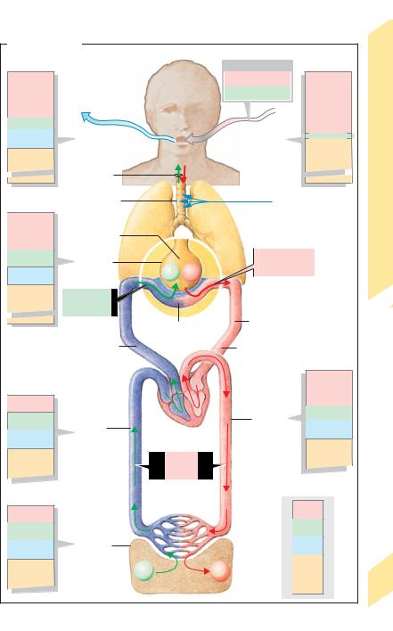
The human body contains around 300 mil- |
||||
lion alveoli, or thin-walled air sacs (ca. 0.3 mm |
|||||
|
|
|
|||
The lung is mainly responsible for respiration, |
in diameter) located on the terminal branches |
||||
but it also has metabolic functions, e.g. con- |
of the bronchial tree. They are surrounded by a |
||||
version of |
angiotensin I to |
angiotensin II |
dense network of pulmonary capillaries and |
||
(!p. 184). In addition, the pulmonary circula- |
have a total surface area of about 100 m2. Be- |
||||
tion buffers the blood volume (!p. 204) and fil- |
cause of this and the small air/blood diffusion |
||||
ters out small blood clots from the venous |
distances of only a few µm (!p. 22, Eq. 1.7), |
||||
circulation before they obstruct the arterial |
sufficient quantities of O2 can diffuse across the |
||||
circulation (heart, brain!). |
|
alveolar wall into the blood and CO2 towards |
|||
External respiration is the exchange of |
the alveolar space (!p. 120ff.), even at a ten- |
||||
gases between the body and the environment. |
fold increased O2 demand (!p. 74). The oxy- |
||||
(Internal or tissue respiration involves nutrient |
gen-deficient “venous” blood of the pulmonary |
||||
oxidation, !p. 228). Convection (bulk flow) is |
artery is thus oxygenated (“arterialized”) and |
||||
the means by which the body transports gases |
pumped back by the left heart to the periphery. |
||||
over long distances (!p. 24) along with the |
The cardiac output (CO) is the volume of blood |
||||
flow of air and blood. Both flows are driven by a |
|||||
pumped through the pulmonary and systemic circu- |
|||||
pressure difference. Diffusion is used to trans- |
|||||
lation per unit time (5–6 L/min at rest). CO times the |
|||||
port gases over short distances of a few µm— |
arterial–venous O2 difference (avDO2)—i.e., the differ- |
||||
e.g., through cell membranes and other physi- |
ence between the arterial O2 fraction in the aorta |
||||
ological barriers (!p. 20ff.). The exchange of |
and in mixed venous blood of the right atrium (ca. |
||||
gas between the atmosphere and alveoli is |
0.05 L of O2 per L of blood)—gives the O2 volume |
||||
transported per unit time from the lungs to the periph- |
|||||
called ventilation. Oxygen (O2) in the inspired |
|||||
ery. At rest, it amounts to (6 !0.05 = ) 0.3 L/min, a |
|||||
air is convected to the alveoli before diffusing |
. |
(see above). Inversely, if |
|||
value matching that of VO2 |
|||||
across the alveolar membrane into the blood- |
. |
|
|||
VO2 and avDO2 have been measured, CO can be cal- |
|||||
stream. It is then transported via the blood- |
culated (Fick’s principle): |
|
|||
stream to the tissues, where it diffuses from |
. |
|
|||
CO " VO2/avDO2 |
[5.1] |
||||
the blood into the cells and, ultimately, to the |
The stroke volume (SV) is obtained by dividing CO by |
||||
intracellular |
mitochondria. |
Carbon dioxide |
the heart rate (pulse rate). |
|
|
(CO2) produced in the mitochondria returns to |
According to Dalton’s law, the total pressure |
||||
the lung by the same route. |
. |
(Ptotal) of a mixture of gases is the sum of the |
|||
The total ventilation per unit time, VT (also |
partial pressures (P) of the individual gases. |
||||
called minute volume) is the volume (V) of air |
The volume fraction (F, in L/L; !p. 376), of the |
||||
inspired or expired per time. As the expiratory |
individual gas relative to the total volume |
||||
volume. is usually measured, it is also abbre- |
times Ptotal gives the partial pressure—in the |
||||
viated VE. (The dot means “per unit time”). At |
case of O2, for example, PO2 = FO2 ! Ptotal. The |
||||
|
. |
|
|||
rest, the body maintains a VE of about 8 L/min, |
atmospheric partial pressures in dry ambient |
||||
with .a corresponding oxygen consumption |
air at sea level (Ptotal = 101.3 kPa = 760 mmHg) |
||||
rate (VO2 ) of.about 0.3 L/min and a CO2 elimina- |
are: FO2 = 0.209, FCO2 = 0.0004, and FN2 + noble |
||||
tion rate (VCO2) of about 0.25 L/min. Thus, |
gases = 0.79 (!A, top right). |
||||
about 26 L of air have to be inspired and ex- |
If the mixture of gases is “wet”, the partial pressure |
||||
pired to supply 1 L of O2 (respiratory equivalent |
|||||
of water, PH2O has to be subtracted from Ptotal (usu- |
|||||
= 26). The tidal volume (VT) is the volume of air |
|||||
ally = atmospheric pressure). The other partial pres- |
|||||
moved in and out during one respiratory cycle. |
|||||
sures will then be lower, since Px = Fx (Ptotal – PH2O). |
|||||
. |
|
|
|||
VE is the product of VT (ca. 0.5 L at rest) and res- |
When passing through the respiratory tract (37 #C), |
||||
piration rate f (about 16/min at rest) (see p. 74 |
inspired air is fully saturated with water. As a result, |
||||
for values during physical work). .Only around |
PH2O rises to 6.27 kPa (47 |
mmHg), and PO2 drops |
|||
1.32 kPa lower than the |
dry atmospheric air |
||||
5.6 L/min (at f = 16 min-1) of the VE of 8 L/min |
|||||
reaches the alveoli; this is known as alveolar |
(!p. 112). The partial pressures in the alveoli, arter- |
||||
ies, veins (mixed venous blood), tissues, and expira- |
|||||
|
. |
|
|||
ventilation (VA). The rest fills airways space not |
tory air (all “wet”) are listed in A. |
||||
contributing to gas exchange (dead space ven-
.
tilation, VD; !pp. 114 and 120).
Despopoulos, Color Atlas of Physiology © 2003 Thieme
All rights reserved. Usage subject to terms and conditions of license.
A. Gas transport |
|
|
|
|
|
|
|
Partial pressures |
|
|
|
|
Partial pressures |
|
|
kPa (mmHg) |
|
|
|
Fraction (L/L) |
kPa (mmHg) |
|
|
|
|
|
|
FO2 |
≈ 0.21 |
|
|
15.33 (115) |
|
|
|
FCO2 ≈ 0.0003 |
|
|
|
|
|
|
|
|
|
21.17 (158.8) |
|
4.43 (33) |
|
|
|
|
|
|
Respiration |
6.27 (47) |
Expired air (wet) |
|
|
Inspired air (dry) |
0.03 (0.23) |
||
|
|
|
|
|
|
||
|
|
|
|
|
|
80.10 (601) |
|
75.33 (565) |
. |
|
|
|
|
|
|
|
|
|
|
|
|
LungFunction, |
|
|
VE ≈ 8L/min |
|
|
|
|
|
|
∑p= 101.3 (760) . |
|
|
Humidification |
∑p= 101.3 (760) |
|||
|
VD ≈ 2.4L/min |
|
|
|
|
(Sea level) |
|
13.33 (100) |
. |
|
|
|
|
|
|
VA ≈ 5.6L/min |
|
|
|
|
|
|
|
5.2 (39) |
Alveoles |
|
|
|
. |
|
5.1 |
|
|
|
|
Plate |
|||
|
CO2 |
O2 |
|
VO2 ≈ 0.3L/min |
|||
6.27 (47) |
|
|
|
|
|||
|
. |
|
|
|
|
|
|
76.5 (574) |
VCO2 |
|
|
|
|
|
|
|
≈ 0.25L/min |
|
|
|
|
|
|
|
|
Pulmonary capillaries |
Cardiac output (CO) |
|
|||
∑p= 101.3 (760) |
|
≈ 6L/min |
|
|
|||
|
|
|
|
|
|
||
|
Pulmonary artery |
|
|
Pulmonary veins |
|
|
|
|
|
|
|
|
|
12.66 (95) |
|
5.33 (40) |
|
|
|
|
|
5.27 (41) |
|
6.0 (45) |
|
|
|
|
Arteries |
|
|
Veins |
Right |
|
Left |
6.27 (47) |
|
||
|
|
|
|
||||
6.27 (47) |
(venae cavae) |
heart |
|
heart |
|
|
|
|
|
avDO2 |
|
|
|
|
|
|
|
≈ 0.05LO2/Lblood |
|
|
|
||
≤ 5.33 (40) |
|
Capillaries |
|
|
O2 |
|
|
|
|
|
|
|
|
|
|
≥ 6.0 (45) |
|
|
|
|
|
CO2 |
|
|
|
|
|
|
|
|
|
6.27 (47) |
Tissue |
|
|
|
|
H2O |
|
|
|
|
|
|
|
|
|
|
|
CO2 |
|
|
|
N2 |
|
|
|
|
O2 |
|
+ |
|
|
|
|
|
|
|
|
Noble |
107 |
|
|
|
|
|
|
gases |
|
|
|
|
|
|
|
|
|
Despopoulos, Color Atlas of Physiology © 2003 Thieme
All rights reserved. Usage subject to terms and conditions of license.

5 Respiration
108
Mechanics of Breathing
Pressure differences between the alveoli and the environment are the driving “forces” for the exchange of gases that occurs during ventilation. Alveolar pressure (PA = intrapulmonary pressure; !B) must be lower than the barometric pressure (PB) during inspiration (breathing in), and higher during expiration (breathing out). If PB is defined as zero, the alveolar pressure is negative during inspiration and positive during expiration (!B). These pressure differences are created through coordinated movement of the diaphragm and chest (thorax), resulting in an increase in lung volume (Vpulm) during inspiration and a decrease during expiration (!A1,2).
The inspiratory muscles consist of the diaphragm, scalene muscles, and external intercostal muscles. Their contraction lowers (flattens) the diaphragm and raises and expands the chest, thus expanding the lungs. Inspiration is therefore active. The external intercostal muscles and accessory respiratory muscles are activated for deep breathing. During expiration, the diaphragm and other inspiratory muscles relax, thereby raising the diaphragm and lowering and reducing the volume of the chest and lungs. Since this action occurs primarily due to the intrinsic elastic recoil of the lungs (!p. 116), expiration is passive at rest. In deeper breathing, active mechanisms can also play a role in expiration: the internal intercostal muscles contract, and the diaphragm is pushed upward by abdominal pressure created by the muscles of the abdominal wall.
Two adjacent ribs are bound by internal and external intercostal muscle. Counteractivity of the muscles is due to variable leverage of the upper and lower rib (!A3). The distance separating the insertion of the external intercostal muscle on the upper rib (Y) from the axis of rotation of the upper rib (X) is smaller than the distance separating the insertion of the muscles on the lower rib (Z!) and the axis of rotation of the lower rib (X!). Therefore, X!–Z! is longer and a more powerful lever than X–Y. The chest generally rises when the external intercostal muscles contract, and lowers when the opposing internal intercostal muscles contract.
To exploit the motion of the diaphragm and chest for ventilation, the lungs must be able to follow this motion without being completely attached to the diaphragm and chest. This is achieved with the aid of the pleura, a thin fluid-covered sheet of cells that invests each lung (visceral pleura), thereby separating it from the adjacent organs, which are covered by the pleura as well (parietal pleura).
In its natural state, the lung tends to shrink due to its intrinsic elasticity and alveolar surface tension (!p. 118). Since the fluid in the pleural space cannot expand, the lung sticks to the inner surface of the chest, resulting in suction (which still allows tangential movement of the two pleural sheets). Pleural pressure (Ppl) is then negative with respect to atmospheric pressure. Ppl, also called intrapleural (Pip) or intrathoracic pressure, can be measured during breathing (dynamically) using an esophageal probe (!Ppl). The intensity of suction (negative pressure) increases when the chest expands during inspiration, and decreases during expiration (!B). Ppl usually does not become positive unless there is very forceful expiration requiring the use of expiratory muscles. The difference between the alveolar and the pleural pressure (PA - Ppl) is called transpulmonary pressure (!p. 114).
Characterization of breathing activity. The terms hyperpnea and hypopnea are used to describe abnormal increases or decreases in the depth and rate of respiratory movements. Tachypnea (too fast), bradypnea (too slow), and apnea (cessation of breathing) describe abnormal changes in the respiratory rate. The terms hyperventilation and hypoventilation imply that the volume of exhaled CO2 is larger or smaller, respectively, than the rate of CO2 production, and the arterial partial pressure of CO2 (PaCO2) decreases or rises accordingly (!p. 142). Dyspnea is used to describe difficult or labored breathing, and orthopnea occurs when breathing is difficult except in an upright position.
Despopoulos, Color Atlas of Physiology © 2003 Thieme
All rights reserved. Usage subject to terms and conditions of license.