

SAOphthalmicAtlas&Guide
.pdfFigure 8.15 Multifocal areas of retinal degeneration in a 2-year-old, castrated male Bernese mountain dog. The degenerate areas appear white and are the result of previous chorioretinitis. Decreased pigmentation in the nontapetal fundus is discussed on page 133. Retinal degeneration is discussed on pages 136 and 137.
Figure 8.16 Preretinal hemorrhage in the ventral, nontapetal fundus. The keel shape indicates the hemorrhage is anterior to the retina. The preretinal location is also known because the hemorrhage obscures visualization of the RPE. Retinal hemorrhage is discussed on page 133.
Figure 8.17 Multiple red, ovoid lesions consistent with intraretinal hemorrhage in a 5-year-old, castrated male cocker spaniel. This patient was diagnosed with optic neuritis and chorioretinitis, but a diagnostic workup was declined. Retinal hemorrhage is discussed on page 133.
Chapter 8 Posterior segment 51
Chapter 8

52 Section I Atlas
Figure 8.18 Complete, serous retinal detachment and hemorrhage in a 16-year-old, castrated male DSH diagnosed with systemic hypertension. Detachment is recognized by multiple planes of focus in this image and an obscured view of the optic nerve (due to retina suspended anterior to the nerve). On the day of this photograph, the blood pressure was 180 mm Hg. Hypertensive retinopathy is discussed on pages 140 and 141.
Figure 8.19a Complete, serous retinal detachment of unknown cause in a 4-year-old, castrated male German shepherd. The only areas of retinal attachment are at the ciliary body and optic nerve. When a significant portion of the retina is detached, the retina is usually visible on gross, external ophthalmic examination. The detached retina is visible within the pupil and looks like a white membrane containing blood vessels. Folds within the detached retina are usually visible, as in this patient. The detachments in this dog resolved with systemic steroid therapy. Retinal detachment is discussed on pages 138 to 140.
Figure 8.19b Fundic photograph of the same eye as in the previous figure. Note that the dorsal retina is in focus, while the remainder of the retina and the optic nerve are out of focus, indicating that these structures are at different planes. The folds that were visible on external examination are also visible in this photograph (arrowheads). Retinal detachment is discussed on pages 138 to 140.
Chapter 8

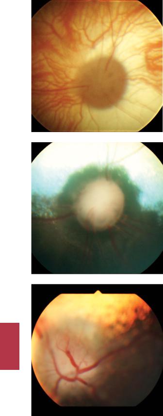
Figure 8.20 Rhegmatogenous retinal detachment. The optic nerve is at 12 o’clock in the photograph. The optic nerve cannot be seen clearly because the retina has torn along its attachment along the dorsal aspect of the ciliary body, allowing it to fall ventrally and anterior to the optic nerve. The retina appears as a white “veil.” Two retinal blood vessels are visible within the detached retina. Retinal detachment is discussed on pages 138 to 140.
Figure 8.21 Optic nerve atrophy in a subalbinotic canine fundus. This atrophied nerve has less myelin than a normal nerve and is therefore smaller and darker than a normal nerve (see Figure 8.2 for normal). Loss of myelin is particularly noticeable from 6 to 11 o’clock, where the edge of the nerve appears black. The blood vessels do not form a robust vascular ring on the optic nerve head in contrast to the normal nerve. Careful examination shows subtle bending of the vessels at the nerve edge and darker retina immediately around the optic nerve. These changes at the edge of the nerve occur because the optic nerve head is recessed (more posterior to the retina) and reflect the transition of planes between the retina and the optic nerve. Pathologic changes to the optic nerve are discussed on page 133.
Figure 8.22 Optic nerve atrophy and retinal degeneration as a sequela to severe chorioretinitis. The degeneration is more severe in this photograph than in the previous figure. This optic nerve is small and dark due to demyelination. In addition, the vessels no longer cross over the optic nerve head, reaching only the nerve periphery. The tapetum immediately around the optic nerve is hyperreflective
(seen as yellow in this photograph), indicating retinal degeneration.
Chapter 8 Posterior segment 53
Chapter 8
54 Section I Atlas
Figure 8.23 Optic disc cupping in a subalbinotic canine fundus. This is the result of chronic glaucoma. The retina immediately around the optic nerve appears darker than normal. Retinal blood vessels reach the edge of the optic nerve, but do not cross over the optic nerve head. It is difficult to distinguish the edges of the nerve from surrounding retina because both tissues are dark. Glaucoma is discussed in Chapter 18.
Figure 8.24 Typical appearance of a cupped optic nerve. When compared with a normal optic nerve (see Figure 8.2), the edges of the cupped nerve are dark, the cupped nerve is misshapen, the retinal vessels only reach its edge (rather than crossing over the nerve head), and the nerve is located more posteriorly than normal. Near the nerve head, there is subtle bending of the retinal vessels because they must change planes when transitioning between the retina and the more posteriorly located optic nerve. The peripapillary tapetum is also darker than in the normal dog. Glaucoma is discussed in Chapter 18.
Figure 8.25 Canine optic neuritis. The edges of the optic nerve are indistinct. Because it is in focus while the remainder of the fundus is out of focus, it must be in a different plane than the retina. This is due to the severe swelling of the nerve and protrusion into the vitreous. Optic neuritis is discussed on pages 141 and 142.
Chapter 8

Figure 8.26 Optic neuritis and retinal hemorrhage in a cat suspected of having FIP. Inflammation of the optic nerve is inferred by its swelling and indistinct borders. Focal hemorrhage is present around the optic nerve as well. Optic neuritis is discussed on pages 141 and 142.
Figure 8.27 The typical appearance of asteroid hyalosis is multiple small, refractile objects suspended throughout the vitreous. Because of their location, they will be visible anterior to the retina and optic nerve on funduscopy but rarely interfere significantly with visualization of the fundus. Asteroid hyalosis is discussed on page 133.
Figure 8.28 Choroidal hypoplasia in the right eye of a collie dog. The choroid lateral to the optic nerve is less pigmented than normal, and its vessel pattern is disorganized. Normal choroid, which is seen ventral to the area of hypoplasia, is more melanotic and the vessels are arranged relatively parallel to one another. The round, white object in the bottom right-hand corner of the photograph is a flash artifact. Collie eye anomaly is discussed on pages 133 and 134.
Chapter 8 Posterior segment 55
Chapter 8

56 Section I Atlas
Figure 8.29 Choroidal hypoplasia and optic nerve coloboma in the left eye of a collie dog. The lateral aspect of the optic head, which appears gray and is devoid of blood vessels, is colobomatous and depressed. The hypoplastic choroid is lateral to the optic nerve; it is the less pigmented tissue to the right of the nerve in the photograph. Choroidal vessels are present within the area of choroidal hypoplasia, but they are not arranged in a regular, linear pattern. Collie eye anomaly is discussed on
pages 133 and 134.
Chapter 8

9 Glaucoma
Please see Chapter 18 for more information about diseases, diagnostic testing, and treatment plans related to the glaucoma.
Figure 9.1 The typical presentation of primary glaucoma is acute onset of unilateral marked episcleral congestion, diffuse corneal edema, and mydriasis. Because this photograph was taken following administration of latanoprost, mydriasis is not present in this photograph. This patient, a 3-year-old, male Boston terrier, also has deep corneal vessels because he presented six days after the acute IOP spike. Glaucoma is discussed on pages 144 to 148.
Figure 9.2 Haab’s striae (arrowheads) are curvilinear corneal opacities seen with chronic glaucoma. They are breaks within Descemet’s membrane that occur as the eye stretches. Glaucoma is discussed on pages 144 to 148.
Small Animal Ophthalmic Atlas and Guide, First Edition. Christine C. Lim.
© 2015 John Wiley & Sons, Inc. Published 2015 by John Wiley & Sons, Inc.
57

Section II
Guide

10 Orbit
Please see Chapter 1 for images of the orbit.
The bony orbit is a relatively enclosed space. In addition to the globe, the orbit contains the following:
•Extraocular muscles
○○Dorsal, medial, ventral, and lateral rectus muscles
○○Dorsal and ventral oblique muscles
○○Retractor bulbi muscle
•Third eyelid
•Glands
○○Orbital lacrimal gland
○○Gland of the third eyelid
•External ophthalmic artery and its branches
•An ophthalmic venous plexus and branches of the external ophthalmic vein
•Cranial nerves II through VI
•Orbital fat pad
•Conjunctiva
•Lymphatics
•Connective tissue
The walls of the orbit are composed of the following:
•Lateral aspect
○○Orbital ligament
○○Temporal muscle
•Dorsal aspect
○○Frontal bone
•Medial aspect
○○Frontal bone
•Ventral floor
○○Sphenoid bone
○○Palatine bone
○○Medial pterygoid muscle
○○Zygomatic salivary gland
•Apex
○○Presphenoid bone
•Ventral orbital rim
○○Lacrimal bone
○○Frontal bone
○○Maxillary bone
○○Zygomatic bone
Structures adjacent to the orbit include the following:
•Frontal sinus (dorsomedial to orbit)
•Maxillary sinus (anteroventral to orbit)
•Nasal cavity (ventromedial to orbit)
•Roots of the fourth maxillary premolars and maxillary molars
•Ramus of the mandible
•Masticatory muscles
Diseases of any structures within or adjacent to the orbit tend to cause a space-occupying effect and displacement of the orbital structures. This predominantly occurs through the path of least resistance, meaning anteriorly through the palpebral fissure. Therefore, orbital structures tend to become more visible in the presence of orbital disease. General and common signs of orbital disease include the following:
•Exophthalmos (Figure 1.1)
○○That is, the eye is pushed anteriorly and is more prominent than normal.
○○Subtle exophthalmos can be missed if the eyes are not viewed from above the head and from the side of the head (in addition to the examination conducted while facing the patient).
○○Do not confuse this with buphthalmos.
■■The latter is an enlargement of the eye, which also gives it a more prominent appearance.
■■Exophthalmos can be differentiated from buphthalmos by measurement of the horizontal corneal diameter.
■■The horizontal corneal diameter of the buphthalmic eye is greater than the measurement for the fellow eye, whereas diameters are equal if an eye is exophthalmic.
•Third eyelid elevation (Figures 1.2 and 1.3)
•Chemosis (Figure 4.4)
•Conjunctival and episcleral vascular engorgement
•Periocular tissue swelling
•Decreased retropulsion of the globe
The aforementioned clinical signs are almost always present in orbital cellulitis/abscess and orbital neoplasia.
Enophthalmos (Figure 1.3) can also indicate orbital disease, although this is less common than exophthalmos. It is seen
Small Animal Ophthalmic Atlas and Guide, First Edition. Christine C. Lim.
© 2015 John Wiley & Sons, Inc. Published 2015 by John Wiley & Sons, Inc.
61

Chapter 10
62 Section II Guide
with Horner’s syndrome, in some cases of orbital neoplasia, in association with facial fractures, as a sequela to severe orbital inflammation, and in diseases where orbital volume decreases (e.g., atrophy of the orbital fat pad, which accompanies weight loss).
Diseases of the orbit
Brachycephalic ocular syndrome
What it is
•Brachycephalic ocular syndrome refers to a set of conformational abnormalities affecting the eyelids and the orbit.
•This syndrome is usually accompanied by chronic surface ocular irritation and trauma occurring secondary to the conformational abnormalities.
Predisposed individuals
•The syndrome occurs most often in brachycephalic dogs and cats, but some nonbrachycephalic dogs also exhibit the same orbit and eyelid abnormalities.
•Commonly affected dog breeds include the pug, shih tzu, lhasa apso, and Pekingese. Affected cat breeds include the Persian, Himalayan, and Burmese.
Defining characteristics
Affected dogs exhibit the following facial characteristics: 1 Bilateral exophthalmos (Figure 1.4)
○○Due to bony orbits that are shallower than those of mesocephalic and dolichocephalic individuals.
○○The result of exophthalmos is increased ocular exposure. The exposure in turn makes the eyes more susceptible to trauma.
2 Enlarged palpebral fissures (Figures 1.4 and 1.5)
○○Excessively large eyelid openings.
○○Also referred to as macroblepharon or macropalpebral fissure.
○○Contributes to lagophthalmos and corneal exposure, leading to chronic corneal drying and irritation.
3 Medial trichiasis (Figure 1.5)
○○Ventromedial entropion of varying severity is present. Epiphora is a common secondary effect; the entropion can misalign nasolacrimal puncta and prevent tears from entering the nasolacrimal drainage system.
○○Often, hair follicles are present within the medial canthus. The hairs usually are directed onto the cornea.
○○In breeds with prominent nasal folds, the folds may lie against the cornea.
○○Constant corneal contact with hairs results in chronic irritation to the medial cornea.
Clinical significance
•The aforementioned abnormalities chronically irritate the cornea and allow the ocular surface to become exposed and dry.
•The exposure and dryness are irritating.
•This exposure predisposes to keratitis and corneal ulceration, especially of the medial and central cornea.
○○Therefore, pathologic changes to the cornea are predominantly visible in the medial and central portions of the cornea.
•In addition to chronic ocular surface inflammation, the abnormalities associated with brachycephalic ocular syndrome impair or even prevent healing of corneal ulcers.
•In dogs, pigmentary keratitis (see pages 103 and 104) is a common consequence of brachycephalic ocular syndrome.
○○This condition causes vision impairment and, if progressive, even blindness.
•In cats, corneal sequestra (see pages 113 and 114) commonly develop as a result of brachycephalic ocular syndrome.
○○This condition is painful and predisposes the cornea to infection.
Diagnosis
Diagnosis of brachycephalic ocular syndrome is made by observation of the conformational abnormalities.
Treatment
•The goals of treatment are to
○○reduce surface ocular irritation and
○○prevent development of corneal changes or, if changes are already present, slow progression of corneal lesions.
•Treatment may be limited to medical management, may include surgery, or may be a combination of medications and surgery.
•For eyes that have not yet developed keratitis, and when the severity of conformational abnormalities is mild, ophthalmic lubrication alone may be sufficient to prevent development of surface ocular inflammation.
•However, for eyes with mild keratitis, addition of an ophthalmic anti-inflammatory medication is likely needed.
•When keratitis is present, or when conformational abnormalities are pronounced, medications should be started and the patient referred for potential surgery.
1 Medical management of brachycephalic ocular syndrome
○○Tear film replacements (ophthalmic lubricating solutions or ointments)
■■These are prescribed to reduce exposure and drying of the central cornea.
■■Apply two to four times daily, or more often, if needed.
■■Preservative-free preparations are preferred when application is frequent, since ophthalmic preservatives themselves can irritate the ocular surface.
○○Ophthalmic anti-inflammatory medications (corticosteroids, CsA, or both in combination)
■■These are used to decrease corneal inflammation (i.e., to treat keratitis), often concurrently with tear film replacements.