

SAOphthalmicAtlas&Guide
.pdf7 Lens
Please see Chapter 16 for more information about diseases, diagnostic testing, and treatment plans related to the lens.
Figure 7.1 Nuclear sclerosis in the right eye of a 12-year-old, castrated male Gordon setter. Due to the angle of light, the sclerotic nucleus appears opaque. However, changes to the angle of viewing and the angle of light will reveal a clear, unobstructed tapetal reflection similar to that in the next photograph. Nuclear sclerosis is discussed on
pages 125 and 126.
Figure 7.2 Nuclear sclerosis in the left eye of a 10-year-old, spayed female miniature schnauzer. The nuclear density is circular and central within the pupil. Outer edges of the nucleus are marked with arrows. Colors are visible near the outer nucleus (also indicated by arrows) due to difference in refraction of various wavelengths of light. The appearance of these colors is also typical of the appearance of nuclear sclerosis. Nuclear sclerosis is discussed on pages 125 and 126.
Small Animal Ophthalmic Atlas and Guide, First Edition. Christine C. Lim.
© 2015 John Wiley & Sons, Inc. Published 2015 by John Wiley & Sons, Inc.
41

42 Section I Atlas
Chapter 7
Figure 7.3 Incipient cataract. The cataract, visible in the central pupil, appears bright yellow due to the reflection of light from the light source. Signs of visual compromise were absent. Routine monitoring was recommended. Cataracts are discussed on pages 126 to 128.
Figure 7.4 Incipient cataract (arrow). Note the bright appearance of the cataract, resulting from scattering of light from the light source. The patient did not show any signs of vision compromise. No treatment was instituted but monitoring was recommended. Cataracts are discussed on pages 126 to 128.
Figure 7.5 Incomplete cataract in the right eye of a DSH. No change in vision was noticed by the owners. The tapetal reflection (green to yellow color) is partially blocked by the white cataract, which is densest in the dorsal one-third of the lens, but which also affects the lens more ventrally. Melanin is adhered to the dorsal anterior lens capsule. Topical anti-inflammatory medications were started, and routine monitoring was advised. Cataracts are discussed on pages 126 to 128.

Figure 7.6 Unilateral incomplete cataract. This dog was still able to navigate around the home because the other eye was unaffected. The tapetal reflection (red) is visible throughout the lens, but is less bright in areas where the cataract is more dense. This owner elected topical anti-inflammatory therapy and periodic monitoring. Cataracts are discussed on pages 126 to 128.
Figure 7.7 In this photograph, the lens appears diffusely opaque, with only a small amount of tapetal reflection visible (faint yellow visible in portions of the lens). Because some tapetal reflection is still visible, the cataract is classified as incomplete. This dog was bilaterally affected and severely visually impaired. Topical anti-inflammatory medications were initiated, and phacoemulsification with intraocular lens implantation was performed. Cataracts are discussed on pages 126 to 128.
Figure 7.8 Complete diabetic cataracts. Both eyes were similarly affected. This dog was blind. The cataracts are considered complete because no tapetal reflection is visible. Note the conjunctival and episcleral hyperemia, resulting from LIU. Topical anti-inflammatories were started, and phacoemulsification with intraocular lens implantation was performed. Cataracts are discussed on pages 126 to 128.
Chapter 7 Lens 43
Chapter 7

44 Section I Atlas
Chapter 7
Figure 7.9 This is a left eye viewed from the left side of the dog, highlighting the contours of the anterior lens capsule. Note the uneven, wrinkled surface of the anterior lens capsule, which occurs with leakage of lens protein from the lens. The tapetal reflection is not visible. The cataract was therefore classified as complete and resorbing. Cataracts are discussed on pages 126 to 128.
Figure 7.10 Incomplete, resorbing cataract. In this photograph, resorption is indicated by the sparkly appearance to the lens. Ophthalmic examination also detected wrinkling of the lens capsule. The tapetal reflection is yellow and can be seen in the peripheral lens, particularly ventrolaterally. Mild episcleral hyperemia is a reflection of LIU. Superficial corneal vascularization and corneal melanosis are visible extending from the 9 o’clock limbus into the cornea. Cataracts are discussed on pages 126 to 128.
Figure 7.11 Lens subluxation, incipient cataracts (arrowheads), and nuclear sclerosis. The equator of the lens is indicated by the arrow. The aphakic crescent is the space between the lens equator and the pupillary margin, in this case extending from approximately 8 to 12 o’clock. At the tapered edges of the crescent, zonules extend radially across the aphakic cresent (from lens equator to the ciliary body). Nuclear sclerosis is the clear, circular shape within the central lens. Lens subluxation is discussed on pages 128 and 129.
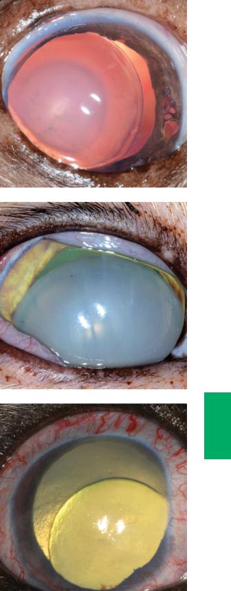
Figure 7.12 Nuclear sclerosis, incipient cataracts, and lens subluxation in the right eye of a 14-year-old, castrated male Chihuahua. The aphakic crescent is between the medial lens equator and pupillary margin from approximately 12 to 5 o’clock.
The nuclear sclerosis is the cloudy circle within the central lens, and the incipient cataracts are the gray streaks within the ventrolateral nucleus. Iris atrophy is also visible in the medial iris. Lens subluxation is discussed on pages 128 and 129.
Figure 7.13 Complete cataract and anterior lens luxation in the right eye of a cat. The opaque lens prevents complete visualization of the iris. Green tapetal reflection is visible through the aphakic crescent (between the dorsal iris and the lens). Anterior uveitis and IOP elevation were also present. Note the chemosis, conjunctival and episcleral hyperemia, and superficial corneal vascularization (dorsolateral limbus). Lens luxation is discussed on pages 128 and 129.
Figure 7.14 Posterior lens luxation secondary to glaucoma. The IOP was 38 mm Hg. Buphthalmos led to zonular breakdown and luxation of the lens into the vitreous cavity. Note the episcleral congestion and mydriasis, also consistent with a diagnosis of glaucoma. Lens subluxation is discussed on pages 128 and 129.
Chapter 7 Lens 45
Chapter 7

8 |
Posterior segment |
|
Please see Chapter 17 for more information about diseases, diagnostic testing, and treatment plans related to the posterior segment.
Figure 8.1 Normal appearance of the canine fundus. The canine tapetum is usually yellow, green, or orange. The canine optic nerve is white, due to myelin, and is usually round to triangular in shape. It is located near the tapetal–nontapetal junction near the central fundus. Three to four larger vessels cross the optic nerve head and extend into the peripheral fundus. Of these, the dorsal retinal venule is most visible; the others are more difficult to see against the brown nontapetal fundus. The normal fundus is discussed on page 132.
Figure 8.2 More magnified photograph of the normal canine fundus. For orientation purposes, please note that dorsal is the upper, left-hand corner of this photograph (ie, the photo is slightly tilted). In this dog, the tapetal fundus is orange while the nontapetal fundus is brown. The transition from tapetal to nontapetal fundus can be
smooth, but is often irregular, as in this photo. The optic nerve is white, and vessels can be seen crossing the optic nerve head. The vessels extend to the periphery of the fundus. The normal fundus is discussed on page 132.
Small Animal Ophthalmic Atlas and Guide, First Edition. Christine C. Lim.
© 2015 John Wiley & Sons, Inc. Published 2015 by John Wiley & Sons, Inc.
46

Figure 8.3 Normal appearance of the feline tapetal fundus. The ventral, nontapetal fundus is not pictured. The feline tapetum is usually green or yellow. The nontapetum is usually brown. The optic nerve is round and is gray due to the lack of myelin. Typically, three to four larger vessels extend from the periphery of the optic nerve to the peripheral fundus. The vessels do not cross the anterior surface of the optic nerve. Smaller retinal vessels are also present. The normal fundus is discussed on page 132.
Figure 8.4 Subalbinotic, atapetal canine fundus. This is a normal variant. Instead of a typical tapetal fundus and a brown nontapetal fundus, choroidal vessels are visible. In contrast to retinal vessels, these do not arise from the optic nerve, tend to be wider than retinal vessels, and are arranged parallel to one another. The normal fundus is discussed on page 132.
Figure 8.5 Normal appearance of a subalbinotic feline fundus. For orientation purposes, please note that dorsal is the upper, left-hand corner of this photograph (ie, the photo is slightly tilted). The tapetum is present in this cat. Because the RPE contains little to no melanin in the nontapetal fundus, choroidal vessels are visible throughout the nontapetal fundus. Choroidal vessels are wider than retinal vessels, do
not extend from the optic nerve, and appear parallel to each other. The normal fundus is discussed on page 132.
Chapter 8 Posterior segment 47
Chapter 8

48 Section I Atlas
Figure 8.6 Diffuse tapetal hyperreflectivity and marked retinal vascular attenuation in a 1-year-old, castrated male Bengal. The changes were bilaterally symmetrical. This kitten had always been quieter and clumsier than his littermates and 3 months prior to presentation stopped chasing his toys and could no longer navigate the house without bumping into furniture. This patient was diagnosed with PRa, which is discussed on pages 134 and 135.
Figure 8.7 Diffuse tapetal hyperreflectivity and marked retinal vascular attenuation in a 4-year-old, castrated male mixed breed dog. The patient experienced progressive vision loss over a 6-month period and exhibited severe visual deficits at presentation. The changes were present bilaterally. This patient was diagnosed with PRa, which is discussed on pages 134 and 135.
Figure 8.8 Typical appearance of a chorioretinal scar/focal retinal degeneration (far right edge of photo). The peripheral portion of the oval lesion is hyperreflective
(seen as bright yellow to orange), and the center of the lesion is dark. Hyperreflectivity is due to thinning of the degenerate retina, and the hyperpigmentation is due to melanin clumping. Retinal degeneration, excluding PRa and SARDS, is discussed on pages 136 and 137.
Chapter 8

Figure 8.9 Two foci of retinal degeneration, which were incidental findings during ophthalmic examination. These areas, also referred to as chorioretinal scars, are flat, round, and hyperreflective (orange to yellow in the photograph) with black centers. For orientation, please note the optic nerve, outside of the photo, is to the lower right-hand side of this photo. Retinal degeneration, excluding PRa and SARDS, is discussed on pages 136 and 137.
Figure 8.10 Multifocal tapetal hyporeflectivity in a 12-year-old, spayed female DSH with a blood pressure measurement of 220 mm Hg. For orientation, please note the optic nerve, outside of the photo, is to the lower right-hand side of this photo. The normal areas of the tapetal fundus are yellow, while the hyporeflective areas are green to gray. The hyporeflectivity is due to retinal edema and fluid in the subretinal space, resulting from systemic hypertension. The central, circular lesion is retina that has detached as a result of fluid in the subretinal space. Tapetal hyporeflectivity is discussed on page 132.
Figure 8.11 Multifocal tapetal hyporeflectivity in a 4-month-old, female mixed breed dog. For orientation, please note the optic nerve is immediately outside of the frame at the bottom, left-hand corner of the photograph. This patient had many congenital ocular abnormalities in both eyes, including cataract, lens subluxation, and retinal detachment. The hyporeflective areas are pinpoint and gray. Tapetal hyporeflectivity is discussed on page 132.
Chapter 8 Posterior segment 49
Chapter 8

50 Section I Atlas
Figure 8.12 Left fundus of a golden retriever diagnosed with blastomycosis and chorioretinitis. The optic nerve is the lower right-hand corner of the photograph and is not clearly visible. There are two round areas of tapetal hyporeflectivity, one larger than the other, dorsal and medial to the optic disc. They are both gray compared with the surrounding yellow–green tapetum. Both of these areas were presumed to be infiltrates of Blastomyces organisms and white blood cells. Tapetal hyporeflectivity is discussed on page 132.
Figure 8.13 Multifocal areas of increased pigmentation and decreased pigmentation in the nontapetal fundus. The patient was a 6-year-old, castrated male Labrador retriever that was diagnosed with a brain tumor. The darker areas were thought to be clumps of melanin, while the whiter areas were thought to be accumulation of fluid and cells. Decreased pigmentation in the nontapetal fundus is discussed on page 133.
Figure 8.14 Decreased pigmentation of the nontapetal fundus in a 2-year-old, castrated male Doberman pinscher. This dog had undergone months of treatment for blastomycosis. The center of the white lesion is a thicker, creamier white than the periphery. The center of the lesion was due to white cell infiltrate, while the white areas at the periphery of the lesion were due to post-inflammatory thinning and depigmentation of the RPE and choroid. Decreased pigmentation in the nontapetal fundus is discussed on page 133.
Chapter 8