

Gale Encyclopedia of Genetic Disorder / Gale Encyclopedia of Genetic Disorders, Two Volume Set - Volume 1 - A-L - I
.pdfDiabetes Mellitus, Type I
Diabetes
(Gale Group)
Demographics
Worldwide, diabetes mellitus represents a large proportion of the common, chronic diseases caused by multiple factors. Between 5% and 10% of adults in the Western world are affected by some form of diabetes. About 1/10,000 people have IDDM. The incidence of NIDDM is about three-fold that of IDDM—up to 5% of the U.S. population age 20-74. Up to an additional 11% have impaired glucose tolerance (IGT), which can represent an early stage of NIDDM.
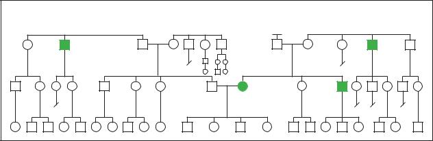
Incidence rates of all types of diabetes vary among ethnic groups—a result of differing genetic and environmental backgrounds. For IDDM, incidence rates range from less than 1/100,000 among Japanese to greater than 25/100,000 among Scandinavians. Ethnic variation follows a different pattern for diabetes overall, which consists primarily of those with NIDDM and IGT. While NIDDM rates are very low among the Eskimo, IGT is very common.
Population studies suggest that there may be one or more major genes that influence diabetes susceptibility, particularly NIDDM susceptibility, in certain populations with high to very high incidence rates of clinical diabetes. These include Mexican Americans, Pima Indians, Oklahoma Seminoles, and several populations in the South Pacific including the Nauruans.
In other populations, increased incidences of NIDDM suggest the role of environmental factors in the disease’s development. Changes in diet and lifestyle are implicated as contributing factors in the increased incidence seen by members of ethnic groups who have experienced Westernization due to immigration patterns or other cultural changes. Such factors may play a role in the increased incidence of NIDDM seen in African Americans, Japanese Americans, certain Native Ameri-
can groups, South Pacific Nauruans, and recently Westernized aboriginal Australians. Differences in incidence rates among various populations is a reflection of the multiple underlying genetic and environmental factors that contribute to the development of all types of diabetes mellitus.
Signs and symptoms
The onset of IDDM is marked by the sudden, dramatic appearance of one or more of the following symptoms:
•Frequent urination
•Extreme thirst and/or hunger
•Rapid weight loss
•Irritability
•Weakness and exhaustion
•Nausea and vomiting
NIDDM usually develops much more gradually. Symptoms can be subtle and include any of the above symptoms, in addition to the following:
•Itching
•Blurry vision
•Obesity
•Tingling or numbness in feet
•Slow healing of the skin or gums
•Recurrent bladder infections
Diabetes can affect many of the body’s organs and systems. Individuals with IDDM are prone to a potentially life-threatening complication called ketosis, in which elevated tissue and fluid levels of ketones may lead to toxic results. People with diabetes are also prone
G A L E E N C Y C L O P E D I A O F G E N E T I C D I S O R D E R S |
333 |
Diabetes
to infections. Infections of the kidney can lead to kidney disease and failure. A specific type of infection by an organism called Mucormycosis tends to occur following ketosis events in individuals with IDDM. This infection usually begins in the nasal passages and can become quite serious if it spreads to the soft tissues and bones of the face, the eye, the skull, or the brain. Gangrene can occur in individuals with poorly controlled disease and has the potential to result in limb amputation. There is an increased risk for cataracts, as well as glaucoma. Left untreated, such complications can lead to blindness. Vascular disease is common in both IDDM and NIDDM. Atherosclerosis—hardening of the arteries—can occur early and advance quickly, increasing the risk for stroke, kidney disease, and heart disease. Heart attack is the most common cause of death in diabetes. Disease of the peripheral blood vessels occurs commonly, particularly when kidney disease is also present. This can lead to increased bruising and development of ulcers, particularly in the leg.
MODY
Clinical severity is determined in part by the specific gene associated with disease within a family. MODY3 mutations result in the most severe clinical presentation, with 97% of cases having NIDDM, as opposed to impaired glucose tolerance. Individuals with MODY1 commonly experience vascular complications and require insulin in one-third of cases. Glucokinase (GCK) gene mutations, although the most common cause of MODY, tend to result in the mildest clinical picture. Approximately 46% have NIDDM, and the remaining individuals have IGT. Individuals with GCK-related MODY rarely need insulin and usually don’t experience vascular complications.
Diagnosis
Diagnosis of diabetes can be based on the presence of suggestive symptoms, together with lab results that support the specific diagnosis.
IDDM is a distinct disease that, in most all cases, is easy to diagnose based on clinical symptoms and lab values. The identification of certain autoantibodies (immune system proteins directed against ‘self’ tissues) is particularly helpful in diagnosing IDDM. The onset of IDDM is almost always rapid and dramatic. Rarely, onset can be gradual and result in a diagnostic dilemma in which it is difficult to distinguish from NIDDM, particularly in an individual who is age 35-50 and not obese. Testing for autoantibodies in such individuals can help distinguish the two diseases. Although not typically done, testing for the presence of HLA-DR3 and/or HLA-DR4 may also be
informative. Individuals who have a relative with IDDM are also at increased risk of a variety of other autoimmune diseases—notably thyroid disease, autoimmune gastritis, and adrenal disease.
For individuals at increased risk of diabetes, a screening glucose tolerance test is recommended periodically and may identify diabetes before symptoms become obvious. Since gestational diabetes is such a common pregnancy complication, and the impact of unmanaged disease on the fetus is serious, all pregnant women are screened between the 24th and 28th weeks of pregnancy. For those at increased risk for NIDDM due to an affected relative, increased screening for risk factors for cardiovascular disease is also recommended.
Genetic testing
IDDM The fact that only about one-half of one percent of individuals with DR3 or DR4 develop IDDM is one indicator that HLA-typing on all individuals in the population is not a useful approach for determining IDDM risk. When there is a family history of IDDM, however, HLA-typing may have a role. When considering the risk for someone with a family history to develop IDDM, using the risk figures generated from large population studies based on family history alone (not HLA typing) is most appropriate. However, these risks could potentially be modified by HLA typing results. For example, there is a 1/14 risk for IDDM in the sibling of an affected individual. If HLA typing reveals that the sibling has inherited a completely different set of HLA types, the risk can be more accurately given as 1/100. On the other hand, if there are shared DR3/DR4 HLA types, this increases the risk to 1/5-1/4. Given HLA typing results or not, an individual with a sibling with IDDM is at sufficiently increased risk to warrant increased screening and education about early signs of the disease.
NIDDM NIDDM genetic susceptibility is highly heterogeneous. There are no single genes that alone increase susceptibility to a significantly high degree that testing should be considered. Like in IDDM, it is even more appropriate in NIDDM to discuss genetic susceptibility relative to population studies that determine risk based on family history alone (not based on genetic testing). These studies indicate that individuals with a parent, sibling, or child with NIDDM is at a 10-15% risk to develop NIDDM and a 20-30% risk for IGT, which may be an early sign of developing NIDDM. Symptoms that suggest a diagnosis of NIDDM can occur in younger individuals or those that do not fit the typical profile of someone with NIDDM in other ways (i.e. not obese). In these cases, genetic testing may play a role to help determine the true diagnosis of that individual and/or allow for a more accurate risk assessment.
334 |
G A L E E N C Y C L O P E D I A O F G E N E T I C D I S O R D E R S |
Diabetes Mellitus, Type II |
Diabetes |
|
(Gale Group)
MODY As discussed previously, there is a unique form of NIDDM called MODY. MODY is caused primarily by mutations in the glucokinase gene. Genetic testing for this form of diabetes is available and can be very helpful in diagnosis and risk assessment for other family members, if a glucokinase mutation is detected.
NIDDM DUE TO INSULIN GENE MUTATIONS In families with late onset of NIDDM, characteristic lab values, and a dominant pattern of inheritance, insulin gene testing is available. Other lab techniques are able to distinguish variant forms of insulin that result from known mutations. A positive genetic diagnosis of this type of NIDDM can be very helpful in risk assessment for other family members.
SYNDROMES WITH DIABETES AS A FEATURE There are also several underlying syndromes and diseases of which NIDDM, IDDM, and/or IGT are potential complications. These are generally accompanied by several other signs and symptoms. If one of these syndromes is suspected, the availability, benefits, and limitations of genetic testing can be considered. Mitochondrial DNA testing may be indicated in families that show NIDDM and/or IDDM transmitted only from mothers to children together with other features characteristic of mitochondrial syndromes. In some cases, genetic testing may be appropriate and can assist in diagnosis, medical management for other potential complications, and risk assessment for other family members.
Treatment and management
Management approaches for all types of diabetes are aimed at controlling blood glucose levels, preventing complications through lifestyle changes, and treating complications symptomatically as they arise.
The first step toward controlling blood glucose levels is monitoring the levels, which is done for all types
of diabetes. This can be done daily with home glucose tests, as well as every few months through a physician using a test called the hemoglobin A1c test. When levels are abnormal, adjustments can be made in the timing and or quantity of dosages of insulin for IDDM and in oral glucose-lowering medications in NIDDM. Management of blood glucose levels is particularly important when diabetes occurs in pregnancy, to avoid the potential damaging effects on the developing fetus. Increased fetal monitoring and education is also a part of this management.
Lifestyle changes include changes in diet aimed at maintaining ideal body weight, lowering blood glucose levels, and preventing heart and blood vessel disease. Exercise also helps to maintain ideal body weight and helps the cardiovascular system remain healthy. In addition, exercise is important for helping insulin to function more efficiently in some forms of diabetes.
The acute and chronic complications of diabetes should be recognized and managed properly. Ketosis is an acute, potentially life-threatening complication that can be identified in its early stages by the presence of ketones in the urine. Home urine ketone tests are available and should be used—particularly in individuals with IDDM—when a person is sick or has a highly elevated blood glucose level prior to eating. Other medical com- plications—including infection, cataracts, and cardiovascular disease—are treated with conventional medicine as they arise.
Since diabetes can affect multiple body systems and has an impact on lifestyle on a daily basis, the disease is best managed by a multidisciplinary approach to care. Such an approach may involve many types of specialists, including physicians, dieticians, psychologists, high-risk obstetricians, genetic counselors, ophthalmologists, cardiologists, kidney specialists, and others.
GALE ENCYCLOPEDIA OF GENETIC DISORDERS |
335 |

Diastrophic dysplasia
Potential future treatments may include the longrange goal of gene therapy, particularly for IDDM. This therapy may be aimed at preventing or repairing damage to the insulin-producing pancreas, or restoring insulin production by some other means. There are several significant technical challenges that must be overcome, however, before gene therapy could become a reality.
Prognosis
As with many common chronic diseases, early diagnosis and treatment is very important to prevent diabetesassociated complications. Particularly for NIDDM, recognizing and modifying risk factors related to lifestyle plays a very important role and can often lead to the avoidance of complications or even the development of disease. With all types of diabetes, appropriate management can lead to increased quality of life and health.
Resources
BOOKS
Jorde, L.B., et al. Medical Genetics. 2nd ed. St. Louis: Moseby, 1999.
Raffel, L.J., et al. “Diabetes Mellitus.” Emery and Rimoin’s Principles and Practice of Medical Genetics. 3rd ed. Ed. D.L. Rimoin, J.M. Connor, and R.E. Pyeritz, 479–504. New York: Churchill Livingston, 1997.
PERIODICALS
Efrat, S. “Prospects for gene therapy of insulin-dependent diabetes mellitus.” Diabetologia 41 (1998): 1401–09.
Lynn, S., et al. “Mitochondrial diabetes: investigation and identification of a novel mutation.” Diabetes 47 (1998): 1800–02.
Permutt, M.A., et al. “Genetics of type II diabetes.” Recent
Progress in Hormone Research. 53 (1998): 201–16. Sankaranarayanan, K., et al. “Ionizing radiation and genetic
risks. VI. Chronic multifactorial diseases: a review of epidemiological and genetical aspects of coronary heart disease, essential hypertension and diabetes.” Mutation Research 436, no. 1 (1999): 21–57.
She, J.X., and M.P. Marron. “Genetic susceptibility factors in type 1 diabetes: linkage, disequilibrium and functional analyses.” Current Opinion in Immunology 10 (1998): 682–89.
Velho, G., and P. Froguel. “Genetic, metabolic and clinical characteristics of maturity onset diabetes of the young.”
European Journal of Endocrinology. 138 (1998): 233–39. Zimmet, P.Z. “The pathogenesis and prevention of diabetes in adults: genes, autoimmunity, and demography.” Diabetes
Care 18, no. 7 (1995): 1050–64.
ORGANIZATIONS
American Diabetes Association. 1701 N. Beauregard St.,
Alexandria, VA 22311. (703) 549-1500 or (800) 342-2383.
http://www.diabetes.org .
Diabetes Action Research and Education Foundation. 426 C St.
NE, Washington, DC 20002. http://www.daref.org .
Juvenile Diabetes Foundation International (JDF). 120 Wall St., New York, NY 10005. (212) 785-9500 x708 or (800) 5332873. http://www.jdf.org .
WEBSITES
“Ask NOAH About: Diabetes.” New York Online Access to Health. http://www.noah-health.org/english/illness/ diabetes/diabetes.html .
“Diabetes in Pregnancy.” Fact sheet from March of Dimes.http://www.modimes.org/HealthLibrary2/FactSheets/ DiabetesInPregnancy.htm .
“Diabetes Public Health Resource.” Center for Disease Controlhttp://www.cdc.gov/diabetes/faqs.htm .
National Diabetes Information Clearinghouse of the National Institute of Diabetes & Digestive & Kidney Diseases.http://www.niddk.nih.gov/health/diabetes/pubs/dmover/ dmover.htm .
Jennifer Denise Bojanowski, MS, CGC
I Diastrophic dysplasia
Definition
Diastrophic dysplasia (DTD) is a rare genetic disorder of bone growth and formation that is evident at birth.
Description
Diastrophic dysplasia is one of the genetic osteochondrodysplasias, a group of disorders characterized by abnormal growth and formation of bone and cartilage. The main features of DTD include: malformed ears, cleft palate, short limbs, short stature, spinal and joint deformities, and abnormalities of the bones of the hands and feet. Although children with DTD may experience delays in motor development (e.g. walking at a later age than expected), they are of normal intelligence. The syndrome derives its name from the Greek word, diastrophos, meaning twisted or crooked. Maroteaux and Lamy first used the term diastrophic dysplasia in 1960 to describe three of their patients and eleven other cases already reported in the literature. Since then, at least 300 cases of DTD have been described. Diastrophic dysplasia is also known as diastrophic nanism or diastrophic dwarfism and is abbreviated as DTD or DD.
Genetic profile
The gene responsible for DTD, known as the diastrophic dysplasia sulfate transporter gene (DTDST gene), is located at the end of the long arm of chromosome 5, at position 5q32-33. The DTDST gene produces a protein
336 |
G A L E E N C Y C L O P E D I A O F G E N E T I C D I S O R D E R S |
that functions as a channel and transports sulfate across the cell membrane. DTD is inherited in an autosomal recessive manner. Affected individuals have a mutation in both copies of their DTDST gene; they inherit one mutation from each parent. Parents of affected individuals are carriers; they have a mutation in one copy of their DTDST gene and are without symptoms of the disorder.
Most bone in the body begins as cartilage and later hardens (ossifies) to form bone. In certain parts of the body such as the rib, auricle, and joints, cartilage does not ossify; it remains as cartilage and functions as loadbearing or shock-absorbing tissue. Cartilage contains sul- fur-containing compounds, known as proteoglycans. It is thought that abnormal function of the DTD sulfate transporter leads to insufficient sulfate uptake by proteogycans in the cartilage. This undersulfation results in weakness and distortion of the cartilage. The exact mechanism by which this occurs is not fully understood.
Three other genetic skeletal dysplasias: recessively inherited multiple epiphyseal dysplasia (rMED), atelosteogenesis type 2 (AO-2), and achondrogenesis type IB (ACG-IB), are also due to mutations in the DTDST gene. When compared to DTD, both AO-2 and ACG-1B are more severe skeletal dysplasias, with the latter being a lethal disorder. Recessively inherited MED is a relatively mild condition. This broad range in severity, from mild to fatal, is attributed to the different types and combinations of genetic mutations within the DTDST gene that are responsible for these four related diseases.
Demographics
Diastrophic dysplasia is a rare disorder in most parts of the world except in Finland where the incidence of the disease is estimated at one in every 32,600 live births. Approximately 1–2% of Finnish people are DTD carriers. Most Finnish DTD gene carriers possess the same ancestral mutation, known as DTDST (Fin). The high frequency of this single mutation in Finland is attributed to a founder effect.
Signs and symptoms
Diastrophic dysplasia is a variable condition that tends to become more severe with age. Many manifestations of the disorder are prenatal in onset and are therefore apparent at birth.
Growth
Diastrophic dysplasia is considered a short-limbed skeletal dysplasia because the limbs are disproportionately short for the overall height of the individual. The
newborn with DTD tends to be short with an average birth length of 16.5 in (42 cm). This growth failure continues throughout childhood and is progressive in nature. The degree of deformity caused by orthopedic complications of this disorder can influence overall height. A wide range of final adult heights has been reported with lower limits at 2 ft 10 in (86 cm) and 3 ft 5 in (104 cm) and upper limits at 4 ft 5 in (135.7 cm) and 4 ft 3 in (129 cm) for males and females respectively. On x ray, the limb bones appear short and thick with broad metaphyses and flattened, irregular epiphyses.
Craniofacial
One of the most distinct features of DTD is the socalled “cauliflower ear.” In over 80% of infants with DTD, fluid-filled cysts appear on the outer ear (pinnae) during the first few weeks of life. These cysts later calcify and may eventually ossify to form bone. In as many as 75% of individuals with DTD, some form of cleft palate is present. Although individuals with DTD may have a small chin (micrognathia), the head is otherwise normal in size.
Thoracic
Occasionally there may be abnormalities of cartilage in the trachea, larynx, and bronchi, which may lead to a life-threatening complication—collapse of the airways— especially in early infancy.
Spinal
Spina bifida occulta in the neck (cervical) and upper back (thoracic) region is the most common spinal abnormality found in DTD and is present in over 50% of cases. In spina bifida occulta there is incomplete closure of bones of the spinal column. Other common spinal abnormalities include progressive curvature of the spine, either from front to back (kyphosis) or from side to side (scoliosis). Kyphosis in the neck region (cervical kyphosis) is present in at least 30% of affected individuals and is usually evident at birth. This type of spine curvature usually resolves over time without treatment. In severe cases however, cervical kyphosis can lead to respiratory problems. Scoliosis, which is generally not present at birth, may appear at an early age and become problematic in early adolescence. Nearly 50% of females and at least 20% of males will develop scoliosis.
Joint
Joint changes in diastrophic dysplasia are progressive in nature and can be a painful complication of this disorder. Individuals with DTD may experience limited mobility and/or permanent immobility (contractures),
dysplasia Diastrophic
G A L E E N C Y C L O P E D I A O F G E N E T I C D I S O R D E R S |
337 |

Diastrophic dysplasia
K E Y T E R M S
Amniocentesis—A procedure performed at 16-18 weeks of pregnancy in which a needle is inserted through a woman’s abdomen into her uterus to draw out a small sample of the amniotic fluid from around the baby. Either the fluid itself or cells from the fluid can be used for a variety of tests to obtain information about genetic disorders and other medical conditions in the fetus.
Cartilage—Supportive connective tissue which cushions bone at the joints or which connects muscle to bone.
Chondrocyte—A specialized type of cell that secretes the material which surrounds the cells in cartilage.
Chorionic villus sampling (CVS)—A procedure used for prenatal diagnosis at 10-12 weeks gestation. Under ultrasound guidance a needle is inserted either through the mother’s vagina or abdominal wall and a sample of cells is collected from around the fetus. These cells are then tested for chromosome abnormalities or other genetic diseases.
Chromosome—A microscopic thread-like structure found within each cell of the body and consists of a complex of proteins and DNA. Humans have 46 chromosomes arranged into 23 pairs. Changes in either the total number of chromosomes or their shape and size (structure) may lead to physical or mental abnormalities.
Cleft palate—A congenital malformation in which there is an abnormal opening in the roof of the mouth that allows the nasal passages and the mouth to be improperly connected.
Clubfoot—Abnormal permanent bending of the ankle and foot. Also called talipes equinovarus.
Collagen—The main supportive protein of cartilage, connective tissue, tendon, skin, and bone.
especially in the knees and shoulders. The joints in an individual with DTD are also prone to partial or complete dislocations in the shoulders, hips, kneecaps, and elbows.
Hands and feet
The hands of a child with diastrophic dysplasia are distinct. The fingers are short (brachydactyly) and there may be fusion of the joints between the bones of the fin-
Deoxyribonucleic acid (DNA)—The genetic material in cells that holds the inherited instructions for growth, development, and cellular functioning.
DNA mutation analysis—A direct approach to the detection of a specific genetic mutation or mutations using one or more laboratory techniques.
Dysplasia—The abnormal growth or development of a tissue or organ.
Epiphyses—The growth area at the end of a bone.
Fibroblast—Cells that form connective tissue fibers like skin.
Founder effect—increased frequency of a gene mutation in a population that was founded by a small ancestral group of people, at least one of whom was a carrier of the gene mutation.
Gene—A building block of inheritance, which contains the instructions for the production of a particular protein, and is made up of a molecular sequence found on a section of DNA. Each gene is found on a precise location on a chromosome.
Linkage analysis—A method of finding mutations based on their proximity to previously identified genetic landmarks.
Metacarpal—A hand bone extending from the wrist to a finger or thumb.
Metaphyses—The growth zone of the long bones located between the epiphyses the ends (epiphyses) and the shaft (diaphysis) of the bone.
Mutation—A permanent change in the genetic material that may alter a trait or characteristic of an individual, or manifest as disease, and can be transmitted to offspring.
Nanism—Short stature.
Sulfate—A chemical compound containing sulfur and oxygen.
Vertebra—One of the 23 bones which comprise the spine. Vertebrae is the plural form.
gers (symphalangism). The metacarpal bone of the thumb is short and oval-shaped; these bony deformations cause the thumb to deviate away from the hand and assume the appearance of the so-called “hitchhiker thumb,” a classic feature of DTD. The bony changes in the feet are similar to those found in the hands. The great toes may deviate outward, much like the thumbs. Clubfoot deformity (talipes), due to abnormal formation
338 |
G A L E E N C Y C L O P E D I A O F G E N E T I C D I S O R D E R S |
and limited mobility of the bones of the feet, is a common birth defect found in newborns with DTD.
Diagnosis
At birth the diagnosis of diastrophic dysplasia is based on the presence of the characteristic physical and radiologic (x ray) findings. DNA mutation analysis may be helpful in confirmation of a suspected diagnosis. In those rarer cases where DNA mutation analysis does not detect changes, a laboratory test that measures the uptake of sulfate by fibroblasts or chondrocytes may be useful in making a diagnosis.
If there is a family history of diastrophic dysplasia and DNA is available from the affected individual, then prenatal diagnosis using DNA methods, either mutation analysis or linkage analysis, may be possible. DNA mutation analysis detects approximately 90% of DTDST mutations in suspected patients. In patients where the mutations are unknown or undetectable, another DNA method known as linkage analysis may be possible and, if so, it can usually distinguish an affected from an unaffected pregnancy with at least 95% certainty. In linkage analysis, DNA from multiple family members, including the person with DTD, is required. DNA-based testing can be performed through chorionic villus sampling or through amniocentesis.
If DNA-based testing is not possible, prenatal diagnosis of diastrophic dysplasia in an at-risk pregnancy may be made during the second and third trimesters through ultrasound. The ultrasound findings in an affected fetus may include: a small chin (micrognathia), abnormally short limbs, inward (ulnar) deviation of the hands, the “hitchhiker” thumb, clubfeet, joint contractures, and spinal curvature.
General population carrier screening is not available except in Finland where the frequency of a single ancestral mutation is high.
Treatment and management
There is currently no treatment that normalizes the skeletal growth and development in a child with diastrophic dysplasia. The medical management and treatment of individuals with DTD generally requires a multidisciplinary team of specialists that should include experts in orthopedics. At birth it is recommended that a neonatologist be present because of the potential for respiratory problems. Surgery may be indicated in infancy if congenital abnormalities such as open cleft palate and/or clubfoot deformity are present. Throughout childhood and adulthood, bracing, surgery, and physical therapy are measures often used to treat the spinal and joint deformi-
ties of DTD. Such measures, however, may not fully correct these deformities.
Due to the significant short-limbed short stature associated with diastrophic dysplasia, certain modifications to home, school, and work environments are necessary in order for a person with DTD to perform daily tasks. Occupational therapy may help affected individuals, especially children, learn how to use assistive devices and to adapt to various situations.
Prognosis
In infancy there is an increased mortality rate, as high as 25%, due to respiratory complications caused by weakness and collapse of the cartilage of the wind pipe (trachea) and/or the voice box (larynx), conditions which may require surgical intervention. Some forms of cleft palate and micrognathia may be life threatening in early life as they can result in respiratory obstruction. Severe spinal abnormalities such as cervical kyphosis may also cause respiratory problems. After the newborn period, the life span of an individual with DTD is usually normal with the exception of those cases where spinal cord compression occurs as a result of severe cervical kyphosis with vertebrae subluxation. Spinal cord compression is a significant medical problem that can lead to muscle weakness, paralysis, or death. In a susceptible individual, spinal cord compression may occur for the first time during surgery due to the hyperextended neck position used during intubation. Other anesthetic techniques may be indicated for such cases.
People with diastrophic dysplasia are of normal intelligence and are able to have children. Since many of the abnormalities associated with DTD are relatively resistant to surgery, many individuals with DTD will have some degree of physical handicap as they get older. They may continue to require medical management of their spinal and joint complications throughout adult life.
Resources
BOOKS
Bianchi, Diana W., et al. Fetology: Diagnosis and Management of the Fetal Patient. New York: McGraw-Hill, 2000.
Jones, Kenneth Lyons. Smith’s Recognizable Patterns of Human Malformation. Philadelphia: W.B. Saunders Company, 1997.
PERIODICALS
Makitie, Outi, et al. “Growth in Diastrophic Dysplasia.” The Journal of Pediatrics 130 (1997): 641–6.
Remes, Ville, et al. “Cervical Kyphosis in Diastrophic Dysplasia.” Spine 24, no. 19 (1999): 1990–95.
Rossi, Antonio, et al. “Mutations in the Diastrophic Dysplasia Sulfate Transporter (DTDST) gene (SLC26A2): 22 Novel
dysplasia Diastrophic
G A L E E N C Y C L O P E D I A O F G E N E T I C D I S O R D E R S |
339 |

Distal arthrogryposis syndrome
Mutations, Mutation Review, Associated Skeletal Phenotypes, and Diagnostic Relevance.” Human Mutation 17 (2001): 159–71.
Satoh, Hideshi, et al. “Functional analysis of Diastrophic Dysplasia Sulfate Transporter.” The Journal of Biological Chemistry 273, no. 20 (1998): 12307–15.
ORGANIZATIONS
National Organization for Rare Disorders (NORD). PO Box 8923, New Fairfield, CT 06812-8923 (203) 746-6518 or (800) 999-6673. Fax: (203) 746-6481. http://www
.rarediseases.org .
WEBSITES
Diastrophic Help Web Site. http://pixelscapes.com/ddhelp/ . The Kathryn and Alan C. Greenberg Center for Skeletal Dysplasias Web Page. http://www.med.jhu.edu/
Greenberg.Center/Greenberg.htm .
Dawn Cardeiro, MS, CGC
Diffuse angiokeratomia see Fabry disease
Disorder of cornification 10 see Sjögren
Larsson syndrome
I Distal arthrogryposis syndrome
Definition
Distal arthrogryposis syndrome is a rare genetic disorder in which affected individuals are born with a characteristic bending at the joints of the hands and feet. A contracture is the word used to describe what happens at the joints to cause this bending. In addition to contractures of the hand and feet, individuals with distal arthrogryposis are born with a tightly clenched fist and overlapping fingers.
Description
The word arthrogryposis means a flexed (bent) or curved joint. Distal means the furthest from any one point of reference or something that is remote. Therefore, distal arthrogryposis syndrome causes the joints at the most remote parts of our limbs, the hands and feet, to be flexed.
Consistent fetal movement during pregnancy is necessary for the development of the joints. Without regular motion, the joints become tight resulting in contractures.
The first cases of arthryogryposis were identified in 1923. Arthryogryposis multiple congenital (AMC) is also referred to as fetal akinesia/hypokinesia sequence that is not a disorder, but describes what happens when there is no fetal movement during fetal development. The reasons for lack of fetal motion include neurologic, muscular, connective tissue, or skeletal abnormalities or intrauterine crowding. There are various disorders that involve some form of arthrogryposis.
Distal arthrogryposis was identified as a separate genetic disorder in 1982. Two types of distal arthrogryposis have been identified. Type 1 or typical distal arthrogryposis, is used to describe individuals with distal contractures of the hands and feet, characteristic positioning of the hands and feet, and normal intelligence. Type 2 distal arthrogryposis is known as the atypical form. It is characterized by additional birth defects and mild intellectual delays.
There are other syndomes which include arthrogryposis, however distal arthrogryposis has been characterized as its own syndrome by its inheritance pattern. In addition to the inheritance pattern, there are other features that differentiate this type of arthrogryposis from other forms. Some of these features include a characteristic position of the hands at birth; the fists are clenched and the fingers are bent and overlapping. In addition, problems with the positioning of the feet, called clubfoot is often seen in these individuals. Another distinguishing characteristic is an extremely wide variability in the severity and number of joint contractures someone may exhibit. This variability is often noticed between two affected individuals from the same family.
Genetic profile
Distal arthrogryposis syndrome is inherited in an autosomal dominant manner. Autosomal dominant inheritance patterns only require one genetic mutation on one of the chromosome pairs to exhibit symptoms of the disease. Chromosomes are the structures that carry genes. Genes are the blueprints for who we are and what we look like. Humans have 23 pairs, or 46 total chromosomes in every cell of their body. The first 22 chromosomes are numbered 1–22 and are called autosomes. The remaining pair is assigned a letter either an X or a Y and are the sex determining chromosomes. A typical male is described as 46, XY. A typical female is 46, XX.
Each parent contributes one of their paired chromosomes to their children. Before fertilization occurs, the father’s sperm cell divides in half and the total number of chromosomes reduces from 46 to 23. The mother’s egg cell undergoes the same type of reduction as well. At the
340 |
G A L E E N C Y C L O P E D I A O F G E N E T I C D I S O R D E R S |

time of conception, each parent contributes 23 chromosomes, one of each pair, to their children. All of the genetic information is contained on each chromosome.
If either the father or the mother is affected with distal arthrogryposis, there is a 50% chance they will pass on the chromosome with the gene for this disease to each of their children. The specific gene for distal arthrogryposis is not known, however we do know that it is located on chromosome number 9.
The symptoms of distal arthrogryposis can be different between two affected relatives. For example, a mother may have contractures in all of her joints, but her child may only be affected with contractures in the hands. Because of this variability in the symptoms of this disease, it is believed there is more than one gene mutation that causes distal arthrogryposis. As of 2001, the only gene thought to cause this disease is on chromosome number 9. The exact location and type of genetic mutation on chromosome 9 is not known and therefore, the only genetic testing available as of 2001 is research based.
Demographics
Distal arthrogryposis can affect individuals from all types of populations and ethnic groups. This disease can affect both males and females. There have been only a handful of individuals described with this type of arthrogryposis. The physician, Dr. Hall, who named the disorder in 1982, had initially identified 37 patients with type 1 and type 2 distal arthrogryposis syndrome. She identified 14 individuals with type 1 and 23 individuals with type 2. Since then, numerous other individuals have been diagnosed with distal arthrogryposis. The exact incidence has not been reported in the literature.
Signs and symptoms
At birth, many individuals have been diagnosed based on their characteristic hand positioning. Virtually all individuals with distal arthrogryposis are born with their hands clenched tightly in a fist. The thumb is turned inwards lying over the palm, called abduction. The fingers are also overlapping on eachother. This hand positioning is also characteristic of a more serious condition called trisomy 18. The majority of patients with distal arthrogryposis will also have problems with the positioning of their feet. Many patients will have some form of clubfoot, where the foot is twisted out of shape or position. Another word for clubfoot is talipes.
In addition to the hand and foot involvement, a small percentage of patients will have a dislocation or separa-
K E Y T E R M S
Amniotic fluid—The fluid which surrounds a developing baby during pregnancy.
Cell—The smallest living units of the body which group together to form tissues and help the body perform specific functions.
Flexion—The act of bending or condition of being bent.
Inheritance pattern—The way in which a genetic disease is passed on in a family.
Neurologic—Pertaining the nervous system.
Trisomy 18—A chromosomal alteration where a child is born with three copies of chromosome number 18 and as a result is affected with multiple birth defects and mental retardation.
Ultrasound evaluation—A procedure which examines the tissue and bone structures of an individual or a developing baby.
tion of the hip joint as well as difficulty bending at the hips and tendency for there to be a slight degree of unnatural bending at the hip joints. The knees may also exhibit similar problems of being slightly bent and fixed at that point. Few individuals are born with stiff shoulders.
Type 2 distal arthrogryposis syndrome includes other birth defects not seen in type 1 individuals. For example, type 2 distal arthrogryposis involves problems with the closure of the lip called cleft lip or an opening in the roof of the mouth called cleft palate.
Other abnormalities seen in type 2 distal arthrogryposis include a small tongue, short stature, a curvature of the spine, more serious joint contractures, and mental delays.
Diagnosis
The diagnosis of distal arthrogryposis can sometimes be made during pregnancy from an ultrasound evaluation. An ultrasound may detect the characteristic hand finding as well as the flexion deformities of both the hands and the feet. An affected fetus may have difficulty swallowing and this is exhibited on an ultrasound evaluation as extra amniotic fluid surrounding the baby called polyhydramnios. Another very important and specific diagnositic sign for distal arthrogryposis during a pregnancy is no fetal movement. Ultrasound findings have been detected as early as 17 weeks of a pregnancy.
syndrome arthrogryposis Distal
G A L E E N C Y C L O P E D I A O F G E N E T I C D I S O R D E R S |
341 |

DNA (deoxyribonucleic acid)
After birth, a diagnosis is made by a physician performing a physical examination of a baby suspected of having this disorder. If a baby is affected with type 2 distal arthrogryposis, they may have a difficult time eating properly. As of 2001, the only type of genetic testing available is research based. Because there is likely more than one gene that causes the disease, the genetic testing being performed at this time is not yet offered to affected individuals in order to confirm a diagnosis.
Treatment and management
The treatment for individuals with distal arthrogryposis is adjusted to the needs of the affected child. With therapy after birth to help loosen the joints and retrain the muscles, most individuals do remarkably well. The hands do not remain clenched an entire lifetime, but will eventually unclench. Sometimes the fingers will remain bent to some degree. Clubfoot can usually be corrected so that the feet can be positioned to be straight.
Prognosis
The prognosis depends on how severely affected an individual is and how many joints are involved. Some of the more severe cases may be associated with an early death due to sudden respiratory failure and difficulty breathing properly. The majority of individuals with distal arthrogryposis do very well after receiving the necessary therapies and sometimes surgery to correct severe joint contractions.
Resources
BOOKS
Fleischer, A., et al. Sonography in Obstetrics and Gynecology, Principles & Practice. Stamford, Conn.: 1996.
Jones, Kenneth. Smith’s Recognizable Patterns of Human Malformation. 5th ed. Philadelphia: W.B. Saunders Company, 1997.
PERIODICALS
Sonoda, T. “Two brothers with distal arthrogryposis, peculiar facial appearance, cleft palate, short stature, hydronephrosis, retentio testis, and normal intelligence: a new type of distal arthrogryposis?” American Journal of Medical
Genetics. (April 2000): 280–85.
Wong, V. “The spectrum of arthrogryposis in 33 Chinese children.” Brain Development. (April 1997): 187–96.
WEBSITES
“Arthrogryposis Multiplex Congenita, Distal, Type 1.” Online Mendelian Inheritance in Man. http://www.ncbi.nlm
.gov/Omim/ . Limb Anomalies.
http://www.kumc.edu/gec/support/limb.html .
Katherine S. Hunt, MS
I DNA (deoxyribonucleic acid)
Genetics is the science of heredity that involves the study of the structure and function of genes and the methods by which genetic infomation contained in genes is passed from one generation to the next. The modern science of genetics can be traced to the research of Gregor Mendel (1823–1884), who was able to develop a series of laws that described mathematically the way hereditary characteristics pass from parents to offspring. These laws assume that hereditary characteristics are contained in discrete units of genetic material now known as genes.
The story of genetics during the twentieth century is, in one sense, an effort to discover the gene itself. An important breakthrough came in the early 1900s with the work of the American geneticist, Thomas Hunt Morgan (1866–1945). Working with fruit flies, Morgan was able to show that genes are somehow associated with the chromosomes that occur in the nuclei of cells. By 1912, Hunt’s colleague, American geneticist A. H. Sturtevant (1891–1970) was able to construct the first chromosome map showing the relative positions of different genes on a chromosome. The gene then had a concrete, physical referent; it was a portion of a chromosome.
During the 1920s and 1930s, a small group of scientists looked for a more specific description of the gene by focusing their research on the gene’s molecular composition. Most researchers of the day assumed that genes were some kind of protein molecule. Protein molecules are large and complex. They can occur in an almost infinite variety of structures. This quality is expected for a class of molecules that must be able to carry the enormous variety of genetic traits.
A smaller group of researchers looked to a second family of compounds as potential candidates for the molecules of heredity. These were the nucleic acids. The nucleic acids were first discovered in 1869 by the Swiss physician Johann Miescher (1844–1895). Miescher originally called these compounds “nuclein” because they were first obtained from the nuclei of cells. One of Miescher’s students, Richard Altmann, later suggested a new name for the compounds, a name that better reflected their chemical nature: nucleic acids.
Nucleic acids seemed unlikely candidates as molecules of heredity in the 1930s. What was then known about their structure suggested that they were too simple to carry the vast array of complex information needed in a molecule of heredity. Each nucleic acid molecule consists of a long chain of alternating sugar and phosphate fragments to which are attached some sequence of four of five different nitrogen bases: adenine, cytosine, guanine, uracil and thymine (the exact bases found in a molecule depend slightly on the type of nucleic acid).
342 |
G A L E E N C Y C L O P E D I A O F G E N E T I C D I S O R D E R S |