

Gale Encyclopedia of Genetic Disorder / Gale Encyclopedia of Genetic Disorders, Two Volume Set - Volume 1 - A-L - I
.pdfEmery-Dreifuss muscular dystrophy
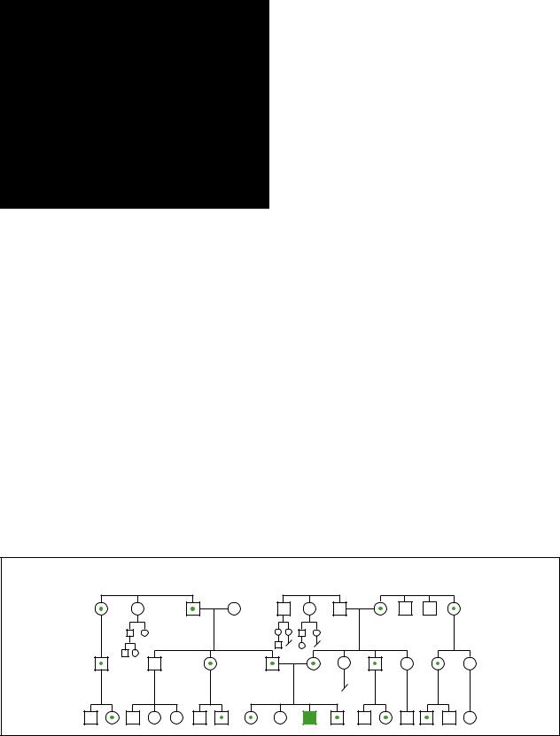
Symptoms and disease severity may vary between individuals. Three modes of inheritance exist: X- linked, autosomal dominant, and autosomal recessive. The symptoms of the autosomal dominant and X-linked forms of the disease are identical, however the autosomal dominant form appears to have a later onset of symptoms.
Genetic profile
Emery Dreifuss muscular dystrophy is inherited in different ways in different families. Most commonly EDMD is inherited in an X-linked recessive manner. Autosomal dominant inheritance of EDMD is also well characterized. As of early 2001 only one case of autosomal recessive inheritance of EDMD has been reported.
Rarely a new mutation causing EDMD can also occur, causing disease in a person with no family history. This is called a sporadic occurrence and is the ressult of a new change in a gene (new mutation) in that individual. New mutations account for approximately 10% of cases of EDMD.
X-linked recessive form
Emery-Dreifuss muscular dystrophy is usually inherited in an X-linked recessive manner. EDMD is the third most common type of X-linked muscular dystrophy. Symptoms begin in the first decade of life. A tendency to walk on the toes is often one of the first signs of EDMD. Muscle weakness first affects the lower extremities usually at age four or five.
X-linked diseases map to the human X chromosome, a sex chromosome. Females have two X chromosomes, whereas males have one X chromosome and one Y chromosome. Because males have only one X chromosome, they require only one X-linked disease gene to display disease. Since females have two X chromosomes, the effect of one X-linked recessive disease gene is masked by the disease gene’s normal counterpart on the other X chromosome.
In classic X-linked inheritance all males are affected, presenting full clinical symptoms of the disease. Females are usually not affected. Affected fathers can never pass X-linked diseases to their sons. However, affected fathers always pass X-linked disease genes to their daughters. Females who inherit the faulty gene but do not show the disease are known as carriers. Female carriers of X- linked EDMD have a 50% chance to pass the diseasecausing gene to each of their children.
It is unusual for female carriers of an X-linked disease to show symptoms of the disease. In X-linked EDMD, carrier females can exhibit certain symptoms of
the disease. Females have two X chromosomes in each of their body cells. Very early on in fetal development, one X chromosome in each cell of a female is inactivated. The pattern of inactivation is random, so carrier females may express the disease-causing gene in some of their cells. An estimated 10–20% of female carriers of X- linked EDMD display varying symptoms of the disease. Female carriers can display the dangerous heart symptoms of EDMD. Less commonly, carrier females may show late-onset muscle weakness.
In 1994 it was recognized that the X-linked recessive form of Emery-Dreifuss muscular dystrophy is caused by changes, or mutations, in a gene now known as EMD or STA. This gene is located on the long arm of the human X chromosome at a location designated as Xq28. The STA gene is approximately 2,100 base pairs in length. This gene codes for emerin, an amino acid protein.
Emerin is an important protein normally found on the inner nuclear membrane of skeletal, cardiac, and smooth muscle cells as well as in other tissues. Emerin is missing from the nuclear membranes of males affected with X-linked EDMD. Emerin is not altered in other neuromuscular disorders.
Autosomal dominant form
In some families, Emery-Dreifuss muscular dystrophy may be inherited in an autosomal dominant pattern. Autosomal dominant EDMD is known as Emery-Dreifuss muscular dystrophy 2 (EDMD2), Hauptmann-Thannhauser muscular dystrophy, and Scapuloilioperoneal atrophy with cardiopathy. Autosomal dominant disorders affect both sexes equally. In autosomal dominant conditions a person, male or female, requires only one faulty gene to produce disease. There are no unaffected carriers of EDMD2. In families with EDMD2, both males and females can be affected and father to son inheritance of the disease can occur. Every child of a person affected with EDMD2 has a 50% chance of inheriting the disease.
In families with EDMD2, affected members exhibit a later onset of the same symptoms as someone affected with X-linked EDMD. Symptoms begin between the ages of 17 and 42. EDMD2 and X-linked EDMD are caused by changes in different genes on different chromosomes.
Muscle biopsy of people with EDMD2 are found to have normal emerin levels. In families with EDMD2, the disease is caused by changes, or mutations, in a gene known as Lamin A/C, or LMNA. Lamin A/C is located in a specific area on the long arm of chromosome 1 known as 1q21.2.
Lamin A/C codes for two proteins, lamins A and C. Like emerin, these lamins are associated with the nuclear
384 |
GALE ENCYCLOPEDIA OF GENETIC DISORDERS |

membrane. People with autosomal dominant EDMD2 have normal levels of emerin and low levels of these lamin proteins. Emerin and these lamins form an important protein complex in a cell’s nuclear membrane. As of early 2001, the exact role of this complex is unclear. Scientists theorize that this important complex of proteins stabilizes the nuclear membrane and plays a role in regeneration of muscle fibers.
Autosomal recessive form
As of early 2001 a single case of autosomal recessively inherited EDMD has been documented. EDMD of autosomal recessive inheritance has been named EmeryDreifuss muscular dystrophy 3 (EDMD3). For someone to be affected with an autosomal recessive disease they must inherit two copies of a disease-causing gene, one from each parent. A parent who has only one gene associated with autosomal recessive EDMD is not affected by the disease and is known as a carrier of the disease. Two carriers of autosomal recessive EDMD have a 25% chance to have a child affected with the disorder in each pregnancy.
Like EDMD2, EDMD3 is caused by mutations in the Lamin A/C gene located on the long arm of chromosome 1 at an area designated as 1q21.2. As of early 2001, the single known mutation associated with EDMD3 has not been found to also lead to EDMD2.
The single known patient with autosomal recessively inherited EDMD (EDMD3) displayed symptoms similar to those of X-linked and autosomal dominant EDMD without any heart involvement. He had difficulties when he started walking at 14 months of age. At five years of age, his contractures were so severe that he could not stand. At age 40, he was confined to a wheelchair and exhibited severe widespread muscle wasting. He displayed normal intelligence and did not have any heart problems. His carrier parents had no heart, skeletal, or muscle abnormalities.
Demographics
X-linked EDMD is estimated to occur in one in 100,000 births. EDMD2 and EDMD3 are far less common. As of early 2001, only one case of EDMD3 has been documented.
Only males exhibit full symptoms of X-linked EDMD. EDMD2 and EDMD3 may occur in males and females. X-linked EDMD and EDMD2 have been documented in many countries. There does not appear to be a single founder of these diseases, as many families have distinctly different backgrounds and different diseasecausing mutations.
KEY TERMS
Amniocentesis—A procedure performed at 16-18 weeks of pregnancy in which a needle is inserted through a woman’s abdomen into her uterus to draw out a small sample of the amniotic fluid from around the baby. Either the fluid itself or cells from the fluid can be used for a variety of tests to obtain information about genetic disorders and other medical conditions in the fetus.
Autosomal—Relating to any chromosome besides the X and Y sex chromosomes. Human cells contain 22 pairs of autosomes and one pair of sex chromosomes.
Chorionic villus sampling (CVS)—A procedure used for prenatal diagnosis at 10-12 weeks gestation. Under ultrasound guidance a needle is inserted either through the mother’s vagina or abdominal wall and a sample of cells is collected from around the fetus. These cells are then tested for chromosome abnormalities or other genetic diseases.
Contracture—A tightening of muscles that prevents normal movement of the associated limb or other body part.
Sporadic—Isolated or appearing occasionally with no apparent pattern.
Signs and symptoms
Emery-Dreifuss muscular dystrophy is recognized by a classic triad of symptoms: contractures at a young age, progressive muscle weakness and degeneration involving the upper arms and lower legs, and cardiac (heart) muscle disease.
Contractures
Contractures, or frozen joints, are a hallmark of all forms of EDMD. A contracture is the abnormal shortening of a body part, usually a muscle or a tendon. This shortening creates joint deformity. Contractures usually begin in childhood or adolescence before any muscle weakness is evident. In most cases, contractures are recognized before patients reach 10 years of age.
Contractures may display as flexion or extension deformities. In a flexion contracture a muscle or tendon remains abnormally flexed, permanently bending a body part at a joint. In an extension contracture a muscle or tendon remains abnormally extended, not allowing a body part to bend at a joint. Affected persons cannot con-
dystrophy muscular Dreifuss-Emery
GALE ENCYCLOPEDIA OF GENETIC DISORDERS |
385 |
Emery-Dreifuss muscular dystrophy
trol these contractures and cannot release them at will. Contractures are treated with stretching, physical therapy, bracing, and surgery.
People affected with EDMD often have flexion contractures of the elbows and ankles. Elbow contractures force the elbow to remain bent at an angle. Contractures of the Achilles tendons, or heel cords, force the feet to remain in a pointed toe position. Children with EDMD often walk on their toes due to heel cord contractures. Neck and trunk contractures may also occur, restricting movement of the neck or the entire spine. Scoliosis is commonly found in patients with EDMD.
Muscle weakness and degeneration
Muscle weakness and degeneration are slowly progressive, affecting a distinct pattern of muscles. This pattern includes the muscles of the upper arms and the muscles of the lower legs. The biceps (inner upper arm), triceps (outer upper arm), tibialis anterior (inner lower leg), and peroneal (outer lower leg) muscles are commonly involved. Later, the muscles of the shoulder girdle and pelvic girdle, the shoulder and hip area muscles that stabilize and support the attachment of the arms and legs, may also be affected. Additionally, the highly specialized muscle of the heart is at risk for weakness and degeneration.
Heart disorders
Heart disease associated with EDMD may be life threatening. It is, however, potentially treatable. Not all patients with EDMD develop heart involvement. Any heart involvement often becomes apparent in the second to third decade of life. In rare cases heart problems may be the first symptom of EDMD. Early recognition of heart involvement is of utmost importance as surgical placement of a pacemaker may be life saving.
EDMD is associated with cardiac conduction defects (electrical impulse problems), heart muscle degeneration, and unusual tissues (abnormal fatty and fibrous tissues) growing into the heart. Conduction abnormalities can manifest as heart rhythm disturbances known as arrhythmias or, more seriously, heart block. Heart block is a dangerous situation where the heart is unable to respond correctly to its own electrical system. Arrhythmias and heart block can lead to fainting or even sudden death.
One uncommon type of heart conduction problem, total permanent auricular paralysis (TPAP), is relatively specific to EDMD. Scientists have found that 33% of 109 published cases of TPAP were due to EDMD.
The level of skeletal involvement in a patient with EDMD is not indicative of their level of heart involve-
ment. Heart problems can be unpredictable, occasionally leading to sudden death without any prior symptom. In a review of 73 cases of X-linked EDMD, scientists found that 30 patients died suddenly between ages 25 and 39. Frequent careful checkups with a cardiologist (heart specialist) are necessary. Preventive surgical implantation of a pacemaker is often considered.
Female carriers of X-linked EDMD
Female carriers of X-linked EDMD may display some symptoms of disease. They can have the dangerous heart problems or, less commonly, muscle weakness. One case of sudden death of a female carrier of X-linked EDMD has been reported. It is recommended that female carriers of X-linked EDMD have regular examinations by a cardiologist.
Diagnosis
Diagnosis of EDMD is based on the classic triad of distinctive clinical symptoms seen in this disease. A diagnosis based on careful neuromuscular examination may be confirmed with muscle biopsy or DNA testing. Other special laboratory tests and neuromuscular tests may help physicians to confirm or rule out EDMD.
Creatine kinase (CK), a muscle enzyme, is often measured when symptoms of muscular dystrophy are present. CK levels are only mildly elevated in EDMD. Muscle biopsy can show microscopic changes in muscle fibers. Muscle biopsy also allows for a very practical test for X-linked EDMD where muscle tissue is stained with a chemical that binds specifically to emerin. If emerin is present, X-linked EDMD can be ruled out. If emerin is reduced or absent, X-linked EDMD is diagnosed.
Genetic testing and prenatal diagnosis for X-linked Emery-Dreifuss muscular dystrophy is available on a clinical basis. To perform DNA testing for X-linked EDMD a blood sample is required. This method of testing can diagnose female carriers of X-linked EDMD. Prenatal testing requires fetal cells obtained via amniocentesis or chorionic villus sampling. Once the specific alteration in the gene is identified in an affected family member, female relatives at risk to be carriers can be tested and prenatal diagnosis can be offered. Prenatal testing is performed on DNA extracted from fetal cells obtained by amniocentesis or chorionic villus sampling.
Treatment and management
The muscle and skeletal symptoms of EDMD are treated as they appear. People with EDMD should see a neurologist at least once a year. Stretching and working with a physical therapist is useful in preventing or delay-
386 |
GALE ENCYCLOPEDIA OF GENETIC DISORDERS |

ing contractures. Occupational therapy can help patients adapt their activities and environment to their own particular needs. Ankle and foot braces are used to prevent leg deformity. Surgery may be necessary to release contractures. Exercise can help maintain muscle use and overall good health. Affected individuals may eventually require a wheelchair or other adaptive equipment.
Persons affected with EDMD require frequent, at least annual, heart checkups with a cardiologist. Heart symptoms can appear suddenly with disastrous consequences, so patients often have a pacemaker implanted before they have had any serious heart problem. Antiarrhythmia drugs, diuretics, ACE inhibitors, and blood thinners may help with some of the cardiovascular symptoms associated with EDMD. Heart transplant has been successful. Relatives of patients with EDMD, especially female carriers of X-linked EDMD, should also be offered yearly screening for heart involvement via electrocardiography and echocardiography.
Scientists are currently researching gene therapy as a possible treatment for EDMD. STA, the gene known to be involved in the X-linked form of EDMD, is a relatively small, less complicated gene. A small gene with a widespread product, such as STA, shows great promise for gene therapy.
Prognosis
Without serious heart involvement, most people with EDMD are expected to survive at least into middle age. Slow progression of muscle involvement allows most patients to walk and work until middle age or late adult life. Intellect is not affected.
Resources
BOOKS
Emery, Alan E. H. Muscular Dystrophy: The Facts. New York:
Oxford University Press, Inc., 2000.
ORGANIZATIONS
Muscular Dystrophy Association. 3300 East Sunrise Dr., Tucson, AZ 85718. (520) 529-2000 or (800) 572-1717.http://www.mdausa.org .
WEBSITES
Gene Clinics. http://www.geneclinics.org .
Online Mendelian Inheritance in Man.
http://www3.ncbi.nlm.nih.gov/Omim .
Judy C. Hawkins, MS
Emery-Dreifuss syndrome see
Emery-Dreifuss muscular dystrophy
I Encephalocele
Definition
An encephalocele is a defect characterized by the herniation of brain tissue and membranes through an opening in the cranium.
Description
Encephlaoceles are classified as neural tube defects, which are a group of disorders occurring due to the failure of closure of the neural tube at about week four of fetal development.
Other neural tube defects include anencephaly and spina bifida. Anencephaly results from failure of closure of the cranial end of the neural tube. This is a lethal condition. Spina bifida results from failure of neural tube closure in the spine. Spina bifida is a variable condition that is usually not lethal, but causes problems with bladder and bowel control and ambulation. It is usually associated with hydrocephalus (water on the brain), which can be treated with a shunt to drain the fluid into the body cavity. Encephalocele is the most rare neural tube defect.
Encephaloceles are classified according to their location. Occipital (arising at the back of the head where the head meets the neck) encephaloceles occur in 75% of cases, parietal encephaloceles in 10%, and anterior encephaloceles (arising from the base of the nose) in 15%. AnterioPosterior encephaloceles have a poorer prognosis.
Genetic profile
The genetics of neural tube defects, including encephalocele, are not well understood.
Most encephaloceles are sporadic, following a multifactorial pattern (genetic and environmental factors involved) of inheritance. It is known that there is a genetic basis to encephaloceles and other neural tube defects, and it is believed that neural tube defects may be caused by different genetic factors in different subsets of families. Proof that genetic factors contribute to encephaloceles is that it is known to run in families, and it has been seen in association with some chromosome abnormalities. The number of genes and their location is still not known.
Occipital encephaloceles are associated with several single gene syndromes, including Meckle syndrome, dyssegmental dwarfism, Knobloch syndrome, Warburg syndrome, cryptophthalmos, and Voss syndrome. Anterior encephalocele may occur with frontonasal dys-
Encephalocele
GALE ENCYCLOPEDIA OF GENETIC DISORDERS |
387 |

Encephalocele
This 16 week old fetus has developed an encephalocele. The formation of the brain outside of the skull is visible.
(Custom Medical Stock Photo, Inc.)
plasia. Encephalocele can also be seen in the amniotic band syndrome.
Demographics
The frequency of encephalocele has been reported to be between one in 2,000 to one in 5,000 live births. Anterior encephalocele is more common in Africa, Thailand, and India. Females outnumber males for occipital encephalocele but not other types.
The incidence of all neural tube defects is different in different parts of the world. It is highest in northern Europe, specifically the British Isles and especially South Wales. In the United States, it is higher on the East Coast than the West Coast.
The rate of sporadic neural tube defects in the general population is about one in 1,000. The rate is higher in areas with higher incidence. The chance for a recurrence of a neural tube defect after having an affected child is 2%. After two affected children the risk is 10%. The chance for an affected person to have an affected child is 4%. The chance for a second degree relative to have an affected child is 0.5%. Third degree relatives do not have an increased risk. Recurrence risks are given for neural tube defects as a group. A family with a previous child with anencephaly could have a child with spina bifida or encephalocele (the types do not “breed true” in families).
Care must be taken to be sure that the neural tube defect in the family was sporadic and not associated with
a genetic syndrome, which would have a higher risk of recurrence.
Signs and symptoms
Symptoms of encephalocele may include hydrocephalus, spastic quadriplegia (paralysis of all four limbs), developmental delay, mental and growth retardation, uneven gait (ataxia), or seizures.
The size of the cerebral and skull abnormalities associated with encephaloceles are variable. Large encephaloceles are usually associated with microcephaly (abnormally small head). Microcephaly is usually associated with mental retardation.
Occipital encephalocele may be asymptomatic. If the ventricles are involved, hydrocephalus may occur. Anterior encephalocele may progress in size and may be solid, cystic, or both. There may be microcephaly and/or hydrocephaly, ocular hypertelorism (wide-spaced eyes), and cleft palate. There may be problems with vision, breathing, and feeding in patients with anterior encephaloceles. Many patients have mental retardation.
Diagnosis
Encephalocele can be diagnosed by ultrasound examination. Ultrasound examination is a screening test, the quality of which is affected by many factors including the machine used, skill of the operator, size and location of the lesion, and position of the fetus.
It is not likely that maternal serum alpha-fetoprotein testing (AFP) or amniocentesis would detect encephalocele. Alpha fetoprotein is a normal serum protein produced by the fetal liver. The AFP normally stays within the fetus, with a small amount present in the amniotic fluid from the fetal urine. When there is an “open” neural tube defect, there is a high amount of AFP in the amniotic fluid and the maternal serum. Although encephalocele is a neural tube defect, AFP testing on maternal blood or amniotic fluid only detects open neural tube defects. Encephaloceles are closed neural tube defects, meaning they are covered by a thick covering. This covering does not allow the AFP to leak into the maternal blood or the amniotic fluid in increased amounts that would be detected by the aforementioned tests. Pregnancies in which an encephalocele is diagnosed should be offered an amniocentesis and amniotic fluid biochemistry to better understand the cause of the abnormality.
CT scan can be used to determine the contents of the encephalocele once the baby is born. Some centers offer fetal MRI to attempt to classify the encephalocele prior to deliver. This is usually done at 22 weeks gestation.
388 |
GALE ENCYCLOPEDIA OF GENETIC DISORDERS |

Treatment and management
Nutrition, specifically defeciency of folic acid, has been implicated as causing an increased risk for neural tube defects. All women of childbearing age should take 0.4 mg of folic acid to reduce the risk of birth defects. Women with a previous child with a neural tube defect should take 4.0 mg of folic acid. This amount has been shown to reduce the recurrence risk for neural tube defects by 50%.
Prognosis
Size, location, and contents of the encephalocele determine the outcome for the child. Anterior encephaloceles have a much better prognosis than posterior. Mortality due to occipital encephalocele is reported as about 30% if hydrocephalus is present, and 2% if it is not. For all types of encephalocele with hydrocephalus, the mortality rate is 60%. Most patients with parietal encephalocele have associated brain malformations, and mental retardation occurs in 40%. Massive occipital encephalocele with microcephaly have a mortality rate of nearly 100%. Patients with encephaloceles that contain a single frontal lobe are more likely to have normal intelligence without hydrocephalus. Posterior have a poorer prognosis if they contain large amounts of the contents of the posterior fossa (an area of the brain at the back of the head), especially the brain stem. Complications such as hemorrhage or air embolism (stroke) can occur.
Resources
BOOKS
Goodman, Richard M., and Robert J. Gorlin. Encephalocele.
New York: Oxford University Press, 1983.
ORGANIZATIONS
Association of Birth Defects in Children. 930 Woodcock Rd., Suite 225, Orlando, FL 32803. (407) 895-0802.http://www.biethdefects.org .
March of Dimes Birth Defects Foundation. 1275 Mamaroneck Ave., White Plains, NY 10605. (888) 663-4637. resourcecenter@modimes.org. http://www.modimes
.org .
WEBSITES
National Institute of Neurological Disorders and Stroke.http://www.ninds.nih.gov/health_and_medical/ disorders/encephaloceles .
Online Mendelian Inheritance in Man.http://www.ncbi.nlm.nih.gov/htbin-post/OMIM .
Amy Vance, MS, CGC
I Engelmann disease
Definition
Engelmann disease is a rare genetic condition that causes the long bones in the legs to become abnormally wide and may change the structure of other bones in the body. Its effects include bone pain (especially in the legs), skeletal disorders, and weak, underdeveloped leg muscles.
Description
Despite their strength and durability, human bones are living organisms. Throughout the life span, bones are constantly being broken down and rebuilt again without losing their proper size and shape. Diseases that interfere with this delicately orchestrated process (called bone remodeling) can produce pain and restrict our freedom of movement. In Engelmann disease, which was first described in 1920, the shafts of the long bones in the legs become thicker than normal. The femur (thigh bone) and tibia (shin bone) are primarily affected. These changes often cause severe bone pain and weak muscles in the legs. The weak, aching muscles associated with Engelmann disease may result in an unusual walk that resembles a “waddle.” People with Engelmann may be bow-legged and have thin, elongated legs that look as if they are “wasting away.”
Aside from bones in the leg, Engelmann disease can cause abnormal changes in other bones. People with Engelmann may develop scoliosis (in which the spine curves to the left or right side) or lumbar lordosis (a forward curvature of the spine). Engelmann disease can also cause bones to become abnormally hardened (a process referred to as sclerosis). This hardening can affect the bones at the base of the skull as well as those in the hands and feet. In rare cases, sclerosis may affect the jaw. Bone pain and aching, weak muscles may occur in parts of the body affected by the disease.
Engelmann can also affect internal organs and sight. The liver and spleen may become enlarged. Loss of vision may occur if bones near the eye sockets are affected. Some people with Engelmann report headaches, fatigue, and lack of appetite.
The underlying cause of Engelmann disease is unknown. It is often referred to in the medical literature as Camurati-Engelmann disease or progressive diaphyseal dysplasia (PDD). Less common names for the condition include osteopathia hyperostotica scleroticans and multiplex infantalis. Engelmann disease was sometimes referred to as ribbing disease in the past but this name is no longer used.
disease Engelmann
GALE ENCYCLOPEDIA OF GENETIC DISORDERS |
389 |

Engelmann disease
KEY TERMS
Endosteal—Relating to the endosteum, which is the lining of the medullary cavity.
Intracranial pressure—The pressure of the fluid between the brain and skull.
Medullary cavity—The marrow-filled cavity inside of a long bone (such as the femur).
Mutation—A permanent change in the genetic material that may alter a trait or characteristic of an individual, or manifest as disease, and can be transmitted to offspring.
Periosteal—Relating to the periosteum, which is the connective tissue that covers all human bones.
Genetic profile
Engelmann is considered an inherited disease, though occasionally mutations may produce sporadic cases. It is passed from parent to child as an autosomal dominant trait. This means that a person may develop the condition after receiving just one copy of the abnormal gene (associated with Engelmann disease) from either the mother or father.
While the gene (or genes) responsible for Engelmann disease is still unknown, medical researchers have narrowed their search to a specific region of human DNA, which may eventually lead to identification. This chromosomal region is known as 19q13. A gene known as TGFB1 (transforming growth factor-beta 1), which plays a role in regulating bone growth, is located in this region and is therefore considered a possible candidate.
Demographics
Engelmann, which affects men and women equally, is a very rare disease that develops during childhood or young adulthood. It usually develops between ages four and ten, but may affect children as young as three months old. Other people may develop Engelmann disease anytime before age 30.
Signs and symptoms
The main symptoms of Engelmann disease are severe pain in the legs, weak and underdeveloped leg muscles, and a “waddling” walk. Other symptoms include bowed legs, unusually long limbs, spine problems such as scoliosis or lumbar lordosis, and flat feet. People with the disease may complain of headaches, lack
of energy or appetite, vision problems, and an aching feeling in their hands and feet and, less often, in the jaw. Infants with Engelmann disease may experience feeding problems or a failure to thrive, and have a “malnourished” appearance.
In simple terms, Engelmann disease causes telltale changes in the structure of the femur and tibia, around the mid-shaft areas. Certain bone regions (specifically, the endosteal and periosteal surfaces) become abnormally thickened and hardened, which in turn narrows the medullary canal. Engelmann disease also causes the long bones to become “fusiform,” a technical term indicating a tapered, spindle-like shape. In addition to these changes, Engelmann may cause abnormal hardening of other bones: in the hands and feet, at the base of the skull, and in the jaw. Engelmann may also involve liver and spleen enlargement, compression of the optic nerves, and increased intracranial pressure.
Diagnosis
Classic symptoms such as severe leg pain, underdeveloped leg muscles, and a “waddling” gait are often the first indication of the disease. An infant may initially experience feeding problems or failure to thrive (though these are more often the result of other, less serious problems). Imaging procedures such as a CT scan are used to detect the bone abnormalities associated with the condition, which mainly involve the thickening and sclerosis of the long bones of the legs. In some cases, x-ray studies of the skull are necessary. Blood tests and a biopsy of muscle tissue may be recommended.
In diagnosing Engelmann disease, a doctor must distinguish it from other conditions that produce similar symptoms, such as Paget’s disease and certain types of muscular dystrophy.
Treatment and management
The treatment of Engelmann disease focuses on alleviating symptoms. While the changes in bone associated with the condition cannot be reversed, the use of steroid drugs such as cortisone or prednisone can ease bone pain and strengthen muscle. Surgery to repair muscles or bones is rarely necessary, while procedures to repair nerves in the eye are generally considered ineffective.
Prognosis
While Engelmann disease does not affect life expectancy, the prognosis for the condition varies. Some people affected by the disease are virtually free of symptoms; others are severely disabled. In some cases, the muscle weakness associated with Engelmann diminishes
390 |
GALE ENCYCLOPEDIA OF GENETIC DISORDERS |

or goes away completely with the passage of time. In other people, the effects of the disease seem to remain the same or slowly worsen during adulthood.
Resources
BOOKS
Jones, Kenneth L., ed. Smith’s Recognizable Patterns of Human Malformation. 5th ed. Philadelphia: W.B. Saunders, 1997.
PERIODICALS
Janssens, K., et al. “Localisation of the gene causing diaphyseal dysplasia Camurati-Engelmann to chromosome 19q13.”
Journal of Medical Genetics 37, no. 4 (2000): 245–9. Kinoshita, A., et al. “Domain-specific mutations in TGFB1
result in Camurati-Engelmann disease.” Nature Genetics 26, no. 1 (2000): 19–20.
ORGANIZATIONS
National Arthritis and Musculoskeletal and Skin Diseases Information Clearinghouse. One AMS Circle, Bethesda, MD 20892-3675. (301) 495-4484.
National Organization for Rare Disorders (NORD). PO Box 8923, New Fairfield, CT 06812-8923. (203) 746-6518 or (800) 999-6673. Fax: (203) 746-6481. http://www
.rarediseases.org .
WEBSITES
Genetic Alliance. http://www.geneticalliance.org . National Organization for Rare Disorders (NORD).
http://www.rarediseases.org .
Greg Annussek
I Epidermolysis bullosa
Definition
Epidermolysis bullosa (EB) is a group of rare inherited skin diseases that are characterized by the development of blisters following minimal pressure to the skin. Blistering often appears in infancy in response to simply being held or handled. In rarer forms of the disorder, EB can be life-threatening. There is no cure for the disorder. Treatment focuses on preventing and treating wounds and infection.
Description
Epidermolysis bullosa has three major forms and at least 16 subtypes. The three major forms are EB simplex, junctional EB, and dystrophic EB. These can range in severity from mild blistering to more disfiguring and lifethreatening disease. Physicians diagnose the form of the
KEY TERMS
Collagen—The main supportive protein of cartilage, connective tissue, tendon, skin, and bone.
Dermis—The layer of skin beneath the epidermis.
Epidermis—The outermost layer of the skin.
Keratin—A tough, nonwater-soluble protein found in the nails, hair, and the outermost layer of skin. Human hair is made up largely of keratin.
disease based on where the blister forms in relation to the epidermis (the skin’s outermost layer) and the deeper dermis layer.
Genetic profile
EB can be inherited as the result of a dominant genetic abnormality (only one parent carries the abnormal gene) or a recessive genetic abnormality (both parents carry the abnormal gene).
EB simplex results from mutations in genes responsible for keratin 5 and 14, which are proteins that give cells of the epidermis its structure. EB simplex is transmitted in an autosomal dominant fashion.
Dystrophic EB is caused by mutations in genes for type VII collagen, the protein contained in the fibers anchoring the epidermis to the deeper layers of the skin. The genetic mutations for junctional EB are found in the genes responsible for producing the protein Laminin-5. Dystrophic EB is an autosomal disorder and will only result if both parents transmit an abnormal gene during conception.
Demographics
The prevalence of epidermolysis varies among different populations. A study in Scotland estimated the prevalence to be one in 20,400. Researchers in other parts of the world estimate the prevalence to be one in 100,000. This variance is due to the variability of expression. Many cases of epidermolysis bullosa are often not accurately diagnosed and thus, are not reported.
Signs and symptoms
EB simplex, the most common form of EB, is the least serious form of the disease. In most affected individuals, the blisters are mild and do not scar after they heal. Some forms of EB simplex affect just the hands and feet. Other forms of EB simplex can lead to more wide-
bullosa Epidermolysis
GALE ENCYCLOPEDIA OF GENETIC DISORDERS |
391 |
Epidermolysis bullosa
Hemorragic blisters such as those seen on this patients arm form as a result of even slight trauma to the body for patients with epidermolysis bullosa. (Custom Medical Stock Photo, Inc.)
Diagnosis
Physicians and researchers distinguish between the three major subtypes of EB based on which layer of the epidermis separates from the deeper dermis layer of the skin below. Patients suspected of having EB should have a fresh blister biopsied for review. This sample of tissue is examined under an electron microscope or under a conventional microscope using a technique called immunofluorescence, which helps to map the underlying structure.
Knowing that a family member has EB can help establish the diagnosis, but it is possible that parents or siblings will show no sign of the disease, either because it is caused by a new genetic mutation, or because the parents are carriers of the recessive trait and do not display the disease.
Treatment and management
spread blistering, as well as hair loss and missing teeth. Recurrent blistering is annoying but not life threatening.
The second, or junctional, form of EB does not lead to scarring. However, skin on the areas prone to blistering, such as elbows and knees, often shrinks. In one variation of junctional EB, called gravis junctional EB of Herlitz, the blistering can be so severe that affected infants may not survive due to massive infection and dehydration.
The third form of EB, dystrophic EB, varies greatly in terms of severity, but more typically affects the arms and legs. In one variation, called Hallopeau-Siemens EB, repeated blistering and scarring of the hands and feet causes the fingers and toes to fuse, leaving them dysfunctional and with a mitten-like appearance.
The most important treatment for EB is daily wound care. Because the skin is very fragile, care must be taken to be certain that dressing changes do not cause further damage. Tape should not be applied directly to skin and bandages should be soaked off. Infection is a major concern, so a topical antibiotic, such as bacitracin, mupirocin, or sulfadiazine, should be routinely applied. Among persons with recessive dystrophic EB, the anticonvulsant phenytoin is sometimes effective because it decreases production of an enzyme that breaks down collagen.
Prognosis
The prognosis of EB varies depending on the subtype of the disease. Individuals with EB simplex can live
Epidermolysis bullosa, letalis
(Gale Group)
392 |
GALE ENCYCLOPEDIA OF GENETIC DISORDERS |

Epidermolysis bullosa, simplex
bullosa Epidermolysis
(Gale Group)
long, fulfilling lives. The severity of the junctional and dystrophic forms of EB can vary greatly. Infants affected with some forms of the disease often do not survive infancy; other forms can lead to severe scarring and disfigurement.
Resources
BOOKS
Fine, Jo-David, et al. Epidermolysis Bullosa: Clinical, Epidemiologic, and Laboratory Advances, and the Findings of the National Epidermolysis Bullosa Registry.
Baltimore: Johns Hopkins Univ Press, 1999.
Fitzpatrick, Thomas B., Richard A. Johnson, Wolff Klaus, and Dick Suurmond. Color Atlas and Synopsis of Clinical Dermatology. 4th ed. New York: McGraw-Hill, 2000.
Lin, Andrew N., and D. Martin Carter. Epidermolysis Bullosa: Basic and Clinical Aspects. New York: Springer Verlag, 1992.
Mallory, S.B. Atlas of Pediatric Dermatology. Pearl River, NY: Parthenon, 2001.
PERIODICALS
Brust, Mary D., and Andrew N. Lin. “Epidermolysis Bullosa: Practical Management and Clinical Update.” Dermatology Nursing 8 (April 1996): 81–9.
Cotell, S., N.D. Robinson, and L.S. Chan. “Autoimmune blistering skin diseases.” American Journal of Emerging Medicine. 18, no. 3 (2000): 288–99.
Eichenfield, L.F., and P.J. Honig. “Blistering disorders in childhood.” Pediatric Clinics of North America 38, no. 4 (1991): 959–76.
Horn, H.M., G.C. Priestley, R.A. Eady, and M.J. Tidman. “The prevalence of epidermolysis bullosa in Scotland.” British Journal of Dermatology 136, no. 4 (1997): 560–64.
Lin, Andrew N. “Management of Patients with Epidermolysis Bullosa.” Dermatologic Clinics 14 (April 1996): 381–87.
McKenna, K.E., M.Y. Walsh, and E.A. Bingham. “Epidermolysis bullosa in Northern Ireland.” British Journal of Dermatology 127, no. 4 (1992): 318–21.
ORGANIZATIONS
American Academy of Dermatology. PO Box 4014, 930 N. Meacham Rd., Schaumburg, IL 60168-4014. (847) 3300230. Fax: (847) 330-0050. http://www.aad.org .
Dystrophic Epidermolysis Bullosa Research Association of America (DebRA). 40 Rector St., Suite 1403, New York, NY 10006. (212) 513-4090. Fax: (212) 513-4099. staff.debra@exario.net. http://www.debra.org .
Dystrophic Epidermolysis Bullosa Research Association of United Kingdom, (DebRA). 13 Wellington Bus. Park, Dukes Ride, Crowthorne, Berkshire, RG45 6LS. UK 01101344 771961. admin@debra.org.uk. http://www.debra
.org.uk .
National Epidermolysis Bullosa Registry. University of North Carolina at Chapel Hill, Bolin Heights Bldg. #1, CB# 3369, Chapel Hill, NC 27514-3369. (919) 966-2007. Fax: (919) 966-7080. eb_registry@med.unc.edu. http://www
.med.unc.edu/derm/nebr_site .
WEBSITES
Dermatology Online Atlas. http://www.dermis.net/doia/ diagnose.asp?zugr=d&lang=e&diagnr=757320&topic=t .
Dystrophic Epidermolysis Bullosa Research Association Inter-
national. http://debra-international.org/index1.htm .
Epidermolysis Bullosa Medical Research Foundation.
http://www.med.stanford.edu/school/dermatology/ebmrf/ .
Oregon Health Sciences University.
http://www.ohsu.edu/cliniweb/C17/C17.800.865.410.html .
University of Iowa College of Medicine.
http://tray.dermatology.uiowa.edu/EBA-001.htm .
L. Fleming Fallon, Jr., MD, PhD, DrPH
Epidermolysis bullosa junctionalis-disentis type see Epidermolysis bullosa
GALE ENCYCLOPEDIA OF GENETIC DISORDERS |
393 |