
Pharmacokinetics and Metabolism in Drug Design Edited by D. A. Smith, H. van de Waterbeemd, D. K. Walker, R. Mannhold, H. Kubinyi, H.Timmerman
.pdf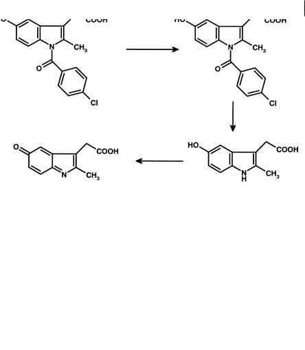
8.3 Quinone Imines 107
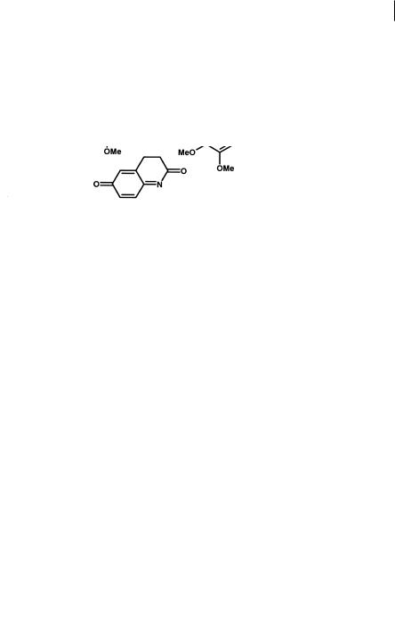
Fig. 8.12 Metabolism of indomethacin to a reactive iminoquinone metabolite.
Fig. 8.13 Structures of practolol and atenolol.
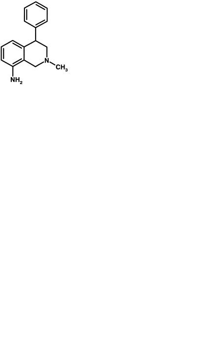
teins. That the toxic functionality is the acetanilide is confirmed by the safety of a fol- low-on drug atenolol. Atenolol (Figure 8.13) has identical physicochemical properties and a very similar structure except that the acetyl-amino function has been replaced with an amide grouping. This structure cannot give rise to similar aromatic amine reactive metabolites. The withdrawal of practolol from the market is obviously a severe blow to the manufacturer and to those patients who benefited from it. Although not shown to be the cause of toxicity the presence of an aromatic amine in the structure of nomifensine (Figure 8.14) has to be treated with suspicion, the compound was also withdrawn from the market 9 years after its launch due to a rising incidence of acute immune haemolytic anaemia [15].
Carbutamide was the first oral anti-diabetic, and the prototype for the sulphonamide type of agent. Carbutamide caused marked bone marrow toxicity in man, but derivatives of this, not containing the anilino function, such as tolbutamide
108 8 Toxicity
Fig. 8.14 Structure of nomifensine, an antidepressant associated with acute immune haemolytic anaemia.
Fig. 8.15 Structures of carbutamide an oral anti-diabetic, associated with bone marrow toxicity, and tolbutamide a compound without similar effects.
(Figure 8.15), were devoid of such toxicity. As for many of the agents featured in this section the structural similarity between carbutamide and tolbutamide clearly implicates the anilino function as the toxicophore.
Fig. 8.16 Structures of Na+ channel blocker antiarrythmics: lidocaine (A), procaineamide (B), tocainide (C) and flecainide (D).
8.4 Nitrenium Ions 109
A further example of the design of drugs to remove aromatic amine functionalities even when present as a amide, is illustrated by the Na+ channel class of antiarrhythmic drugs [16]. Lidocaine is very rapidly metabolized (Figure 8.16) and so is only useful as a short-term intravenous agent. Oral forms include procainamide, tocainide and flecainide (Figure 8.16). Procainamide causes fatal bone marrow aplasis in 0.2 % of patients and lupus syndrome in 25–50 %. Tocainide also causes bone marrow aplasis and pulmonary fibrosis. In contrast, flecainide, whose structure contains no aromatic amine, masked or otherwise, has adverse effects related directly to its pharmacology. Interestingly, the lupus syndrome seen with procainamide is largely absent when N-acetyl procainamide is substituted.
8.4
Nitrenium Ions
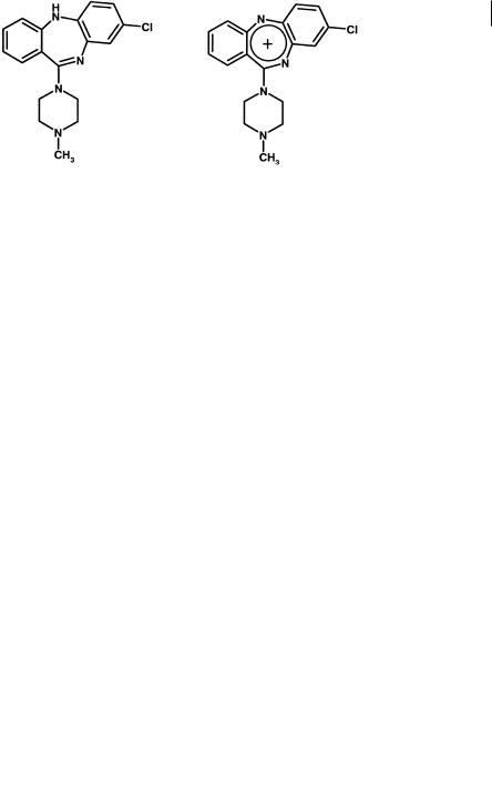
Clozapine, an antipsychotic agent, has the potential to cause agranulocytosis with an incidence of 1 %. The major oxidant in human neutrophils, HOCl, oxidizes clozapine to a nitrenium ion (Figure 8.17) in which the positive charge is highly delocalized. This metabolite is capable of binding irreversibly to the neutrophil [17]. A variety of cells including liver, neutrophils and bone marrow can form reactive clozapine metabolites which react to form glutathione thioether adducts [18].
Fig. 8.17 Structures of clozapine and its nitrenium ion, the postulated reactive metabolite generated by oxidation.
Various analogues have been investigated indicating that the nitrogen-bridge between the two aromatic rings is the target, at least for HOCl oxidation. When alter-
Fig. 8.18 Alternative structures which will not generate the nitrenium ion observed with clozapine.
110 8 Toxicity
native bridging heteroatoms are used (Figure 8.18) similar reactive metabolites are not observed [19].
8.5
Iminium Ions
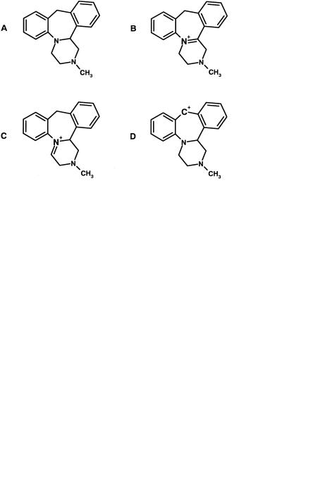
Mianserin is a tetracyclic antidepressant that causes agranulocytosis in isolated cases in patients. Detailed structural analysis [20] has indicated that oxidation by cytochrome P450 to one or more iminium ions is the likely causative first step in this toxicity, the most likely being the C4–N5 version (Figure 8.19). Evidence for this is provided by the C14β methyl and the O10 analogues which cannot form the corresponding nitrenium and carbonium ions, but are cytotoxic. Removal of the N5 nitrogen atom in analogous structures abolishes production of the reactive metabolites (Figure 8.20).
Fig. 8.19 Structures of mianserin (A) with N5 shown and its putative reactive metabolites:
N5–C14β iminium ion (B), C4–N5 iminium ion (C) and carbonium ion at C10 (D). Evidence indicates that metabolite B is the causative agent.
Fig. 8.20 Structures of mianserin analogues. A and B cannot form the N5–C14b iminium and C10 carbonium ion but are still toxic. C without the N5 nitrogen is not toxic implicating the C4–N5 iminium ion.
8.6 Hydroxylamines 111
Vesnarinone is a drug used to treat congestive heart failure and is associated with a 1 % incidence of agranulocytosis. When metabolized by activated neutrophils the major metabolite is veratrylpiperazinamide (Figure 8.21). This unusual N-dealkyla- tion (of an aromatic ring) can be rationalized by chlorination of the nitrogen to which the aromatic ring is attached by HOCl, followed by the loss of HCl to form the reactive imminium ion, which itself can react with nucleophiles. Hydrolysis of the iminium ion yields veratrylpiperazinamide and another reactive species, the quinone imine (see Section 8.3).
Fig. 8.21 Structures of vesnarinone, and its major metabolite veratrylpiperazinamide. The pathway metabolized by activated neutrophils gives rise to two highly reactive species, an iminium ion and a quinone imine.
8.6
Hydroxylamines
Sulphonamide antimicrobial agents (Figure 8.22) such as sulphamethoxazole [21] are oxidized to protein-reactive cytotoxic metabolites in the liver and also other tissues. These include hydroxylamines and further products such as nitroso-deriva- tives. Sulphonamide drugs are linked with agranulocytosis, aplastic anaemia and
Fig. 8.22 Structures of sulfamethoxazole and dapsone, drugs which form toxic hydroxylamine metabolites.

112 8 Toxicity
skin and mucous membrane hypersensitivity reactions including Stevens-Johnson syndrome, and others. Dapsone [22] is a potent anti-inflammatory and anti-parasitic compound, which is metabolized by cytochrome P450 to hydroxylamines, which in turn cause methaemoglobinemia and haemolysis.
8.7
Thiophene Rings
Thiophene rings comprise another functionality that is easily activated to electrophilic species. Thiophene itself is metabolized to the S-oxide, which is viewed as the key primary reactive intermediate. Nucleophilic groups such as thiols react at position 2 of the thiophene S-oxide via a Michael-type addition [23]. Tielinic acid (Figure 8.23) is oxidized to an S-oxide metabolite [24], creating two electron-with- drawing substituents on C2 and a resultant strongly electrophilic carbon at C5 of the thiophene ring [24]. This highly reactive metabolite covalently binds to the enzyme metabolizing it (CYP2C9), triggering an autoimmune reaction resulting in hepatitis. Rotation of the thiophene ring leads to a compound in which the sulphoxide is less reactive and [24] can create a less reactive sulphoxide metabolite which reacts primarily at C2 with nucleophiles. This metabolite is stable enough to escape the enzyme and alkylate other proteins leading to direct toxicity.
Fig. 8.23 Structure of tienilic acid (A) and an isomeric variant (B) which cause hepatotoxicity by autoimmune and direct mechanisms respectively, following conversion to sulphoxide metabolites and resultant electrophilic carbon atoms.
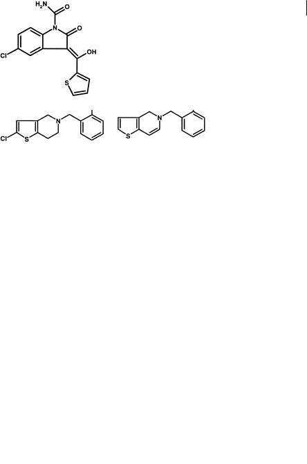
The thiophene ring has also been incorporated into a number of drugs which have diverse toxicities associated with them (Figure 8.24). Ticlopidine, a platelet function inhibitor, is associated with agranulocytosis in patients [25]. Suprofen, a non-
Fig. 8.24 Structures of suprofen and ticlopidine, compounds containing a thiophene ring and associated with diverse toxicities in patients.
8.8 Thioureas 113
steroidal anti-inflammatory agent has been withdrawn from the market due to acute renal injury [26].
The association of ticlopidine with agranulocytosis has been further investigated [27]. A general hypothesis for white blood cell toxicity is the activation of the drug to a reactive metabolite by HOCl, the principal oxidant being generated by activated neutrophils and monocytes (as in Section 8.4). Under these types of oxidation conditions ticlodipine is activated to a thiophene-S-chloride (Figure 8.25) which reacts further to form other products including a glutathione conjugate.
Fig. 8.25 Metabolism of ticlopidine by white blood cells to the reactive thiophene-S-chloride (A) and further breakdown products of this reactive metabolite.
Tenidap (Figure 8.26) is a dual cyclooxygenase (COX) and 5-lipoxygenase (5-LPO) inhibitor developed as an anti-inflammatory agent. Severe abnormalities in hepatic function were reported in Japanese clinical trials [28]. Although the thiophene is not directly implicated in these findings, the ready activation of this system to potential reactive metabolites may be suggestive of the involvement of this function.
Fig. 8.26 Structure of tenidap, a compound containing a thiophene ring and associated with changes in hepatic function.

1148 Toxicity
8.8
Thioureas
The thiourea group has been incorporated into a number of drugs. The adverse reactions of such compounds are associated with the thionocarbonyl moiety. The thionocarbonyl moiety can be metabolized by flavin-containing monooxygenases and cytchrome P450 enzymes to reactive sulphenic, sulphinic and sulphonic acids which can alkylate proteins. The prototype H2 antagonist metiamide [29] incorporated a thiourea group. This compound caused blood dyscrasias in man. Replacement of the thiourea [29] with a cyanimino grouping produced the very successful compound cimetidine (Figure 8.27).
Fig. 8.27 Structures of the thioureacontaining H2 antagonist metiamide, which caused blood dyscrasias in man, and its cyanoimino-containing analogue cimetidine which did not show similar adverse effects.
8.9
Chloroquinolines
Chloroquinolines are reactive groupings due to electron-deficient carbon to which the halogen is attached. This carbon is electron-deficient due to the combined elec- tron-withdrawing effects of the chlorine substituent and the quinoline nitrogen. The electrophilic carbon is thus able to react readily with nucleophiles present in the body. The impact of this grouping on a molecule is illustrated by 6-chloro-4-oxo-10- propyl-4H-pyrano[3,2-g]quinoline-2,8-dicarboxylate (Figure 8.28). In contrast to many related compounds (chromone-carboxylates) lacking the chloroquinoline, 6- chloro-4-oxo-10-propyl-4H-pyrano[3,2-g]quinoline-2,8-dicarboxylate is excreted as a
Fig. 8.28 Structure of 6-chloro-4- oxo-10-propyl-4H-pyrano[3,2-g]quino- line-2,8-dicarboxylate which, in contrast to many related compounds (chromone-carboxylates) lacking
the chloroquinoline, is excreted as a glutathione conjugate.

8.11 Toxicity Prediction - Computational Toxicology 115
glutathione conjugate [30] formed by nucleophilic attack on the halogen of the chloroquinoline function by the thiol group of glutathione and resultant halogen displacement. Other compounds are excreted as unchanged drug with no evidence of any metabolic breakdown. Moreover the reactivity of the chloroquinoline is illustrated by the observation that the reaction with glutathione occurs without enzyme (glu- tathione-S-transferase) present in vitro, albeit at a slower rate.
8.10
Stratification of Toxicity
Table 8.2 gives a summary of the various toxicities and the stages at which they can occur. Also summarized are the causes for the specificity of the effect. With mutagenicity, certain pre-clinical toxicity, carcinogenicity and late clinical toxicology, the actual structure of the molecule is important and care should be taken to avoid the incorporation of toxicophores into compounds, as outlined about. Direct toxicity is addressed by ensuring the daily dose size is low, the intrinsic selectivity high and the physicochemical properties within reasonable boundaries.
Tab. 8.2 Various expressions of toxicity and their cause relating to the stage of drug development.
Mutagenicity |
Metabolites react with nucleic acid |
Metabolites sufficiently stable |
|
|
to cross nuclear membrane |
Pre-clinical and |
Toxicity directly related to dose size |
Overstimulation of receptor |
early clinical |
and intrinsic selectivity. Metabolites |
and others in superfamily. |
|
react with protein and cause cell death |
|
|
|
Metabolites not detoxified |
|
|
by glutathione etc. |
Carcinogenicity |
Metabolites react with nucleic acid |
|
or hormonal effects |
Metabolites not detoxified by glutathione etc. Effects on thyroxine etc. leads to pituitary tumours
Late clinical |
Metabolites react with protein |
Protein adduct acts as |
|
|
immunogen; immune |
|
|
response involved in toxicity. |
|
|
|
8.11
Toxicity Prediction - Computational Toxicology
With increasing toxicity data of various kinds, more reliable predictions based on structure–toxicity relationships of toxic endpoints can be attempted [31–36]. Even the Internet can be used as a source for toxicity data, albeit with caution [37]. A number of predictive methods have been compared from a regulatory perspective [35]. Often traditional QSAR approaches using multiple linear regression are used [38]. Newer approaches include the use of neural networks in structure–toxicity relationships

1168 Toxicity
[39]. An expert system such as DEREK is yet another approach. Attempts have been made to integrate physiologically-based pharmacokinetic/pharmacodynamic (PBPK/PD) and quantitative structure–activity relationship (QSAR) modelling into toxicity prediction [40]. Most methods need further development [41]. Currently these approaches can serve to give a first alert, rather than being truly predictive. In addition, animal models are not perfect or fully predictive. By comparing animal models to the human data obtained for 131 pharmaceutical agents, an accurate prediction rate for human toxicity of 69 % was observed [42].
New technologies are based on advances in understanding and analysing the effects of chemicals on gene expression or protein expression and processing [43, 44]. More developments can be expected in the coming years.
8.12
Toxicogenomics
A new subdispline derived from a combination of the fields of toxicology and genomics is termed toxicogenomics [45]. Using genomic resources, its aim is to study the potential toxic effects of drugs and environmental toxicants. DNA microarrays or “chips” are now available to monitor the expression levels of thousands of genes simultaneously as a marker for toxicity. The complexity of the microarrays yields almost too much data. For instance initial comparisons of the expression patterns for 100 toxic compounds using all the genes on a DNA microarray, failed to discriminate between cytotoxic anti-inflammatory drugs and DNA-damaging agents [46]. A major obstacle encountered in these studies was the lack of reproducible gene responses, presumably due to biological variability and technological limitations. Thus multiple replicate observations for the prototypical DNA-damaging agent, cisplatin, and the non-steroidal anti-inflammatory drugs (NSAIDs), diflunisal and flufenamic acid, were made and a subset of genes yielding reproducible inductions/repressions was selected for comparison. Many of the “fingerprint genes” identified in these studies were consistent with previous observations reported in the literature (e. g. the wellcharacterized induction by cisplatin of p53-regulated transcripts such as p21waf1/ cip1 and PCNA (proliferating cell nuclear antigen)). These gene subsets not only discriminated among the three compounds in the learning set, but also showed predictive value for the rest of the database (approximately 100 compounds with various toxic mechanisms). Further refinement of the clustering strategy, yielded even better results and demonstrated that genes which ultimately best discriminated between DNA damage and NSAIDs were involved in such diverse processes as DNA repair, xenobiotic metabolism, transcriptional activation, structural maintenance, cell cycle control, signal transduction, and apoptosis. The genes span the cycle a cell will go through from initial insult to repair response, and a more detailed understanding of these cycles will not only lead to the development of very sensitive assays for toxicants, but also to a greater understanding of the molecular events of toxicity.
An even more focused direction of this work is to explore the pathway resulting from oxidative stress. Exposure of cells to toxic chemicals can result in reduced glu-
