

Pharmacokinetics and Metabolism in Drug Design Edited by D. A. Smith, H. van de Waterbeemd, D. K. Walker, R. Mannhold, H. Kubinyi, H.Timmerman
.pdf86 7 Metabolic (Hepatic) Clearance
Fig. 7.16 Steps in the discovery of cromakalim: initial structure (A), more potent pyrrolidine analogue (B) and active metabolite (cromakalim C).
The discovery of the potassium channel opener cromakalim is another example of metabolism providing novel active molecules [11]. The programme was designed to find agents that were antihypertensive without having β-adrenoceptor blocking activity. This was due to the belief that β-adrenoceptor blockade was not solely responsible for the antihypertensive effects of β-adrenoceptor blockers. Early compounds were synthesized as cyclized derivatives of β-adrenoceptor blockers. The initial lead is illustrated in Figure 7.16. The gem-dimethyl group and an electron-withdrawing group on the aromatic ring were essential. Cyclic amino groups were preferred to the original isopropylamine, leading to the pyrrolidine derivative. The eventual candidate cromakalim was produced by investigating the metabolites of the pyrrolidine derivative, an oxidation to amines to produce amides being a common metabolic step in cyclic amide systems.
7.4
Non-Specific Esterases
7.4.1
Function of Esterases
Non-specific esterases are distributed widely throughout the body. The activity of these enzymes varies markedly within different tissues. In mammals the highest levels are found in liver and kidney. Numerous isoenzymes exist which have broad substrate overlap. A loose categorization divides the two enzyme types likely to be involved in drug hydrolysis into arylesterases and aliesterases. Aliesterases have a wide substrate range, arylesterases require a phenolic ester. Since most of the major tissues contain a mixture this division is not of great importance. Where esters are of great benefit to drug design is in the design of rapidly cleared molecules, either to an inactive or active form. The most rapid clearance is by blood metabolism. An important point in the screening of compounds designed for rapid metabolism is that the erythrocyte surface has a high esterase content and whole blood is therefore the medium of choice. Moreover rodent blood has very high esterase levels, and may
7.4 Non-Specific Esterases 87
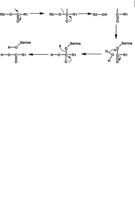
Fig. 7.17 Proposed mechanism for non-specific esterase catalysis involving a serine residue.
give a misleading view of stability if this species is used in isolation. It is highly likely that many of these enzymes are serine esterases and a suggested mechanism is proposed in Figure 7.17.
Ester functions present in molecules tend to be considered labile although steric effects etc. may be utilized to produce drugs without inherent chemical or metabolic problems due to ester lability. For instance a series of antimuscarinic compounds which had selectivity for the M3 receptor (Figure 7.18) were stabilized by the incorporation of a hydroxyethyl side chain or a cyclic ring system at positions surrounding the ester function. Presumably the proximity of these groups to the ester function (carbonyl) prevents close approach of the “attacking” nucleophile, in this case probably a serine hydroxyl.
Fig. 7.18 Stabilization of antimuscarinic compounds to esterase activity by steric effects. Stability was achieved when groupings corresponding to those illustrated were incorporated.
887 Metabolic (Hepatic) Clearance
7.4.2
Ester Drugs as Intravenous and Topical Agents
Lability can be used to advantage to create drugs that are designed for topical or intravenous infusion administration. For topical administration compounds may benefit from rapid systemic clearance to overcome possible side-effects. Thus the compound is stable at its topical site of action (skin, eye etc.) but rapidly degraded by the esterases present in blood, liver and kidneys to its inactive metabolites. This approach renders the compound selective.
The aim of intravenous infusion is often to achieve a steady state plasma concentration as rapidly as possible and to ensure that the concentration of the drug declines as rapidly as possible once the infusion is stopped. This gives the clinician complete control and the opportunity to react quickly to the patient’s needs. Fig-
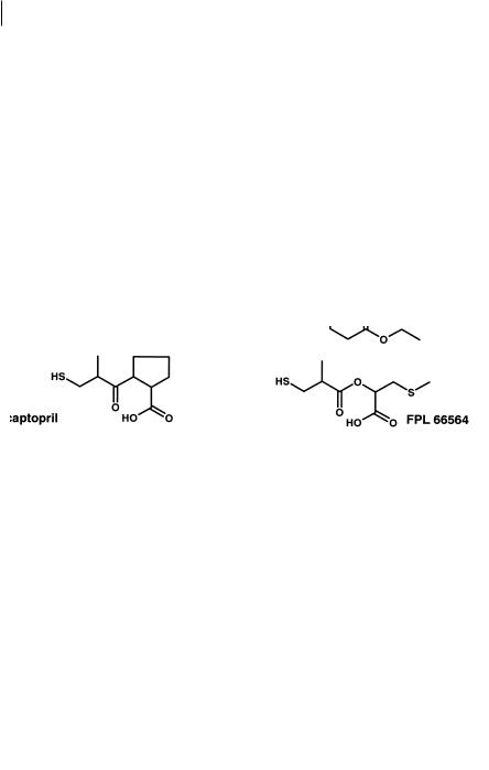
ure 7.19 shows how existing drugs such as the anaesthetic/analgesic sulfentanil, the β-blocker propranolol and the ACE inhibitor captopril, have been used as the starting
point for the design of short-acting infusion agents. Essential to this design is the
Fig. 7.19 Design of rapidly cleared ester analogues of sulfentanil propranolol and captopril. The compounds remifentanil, esmolol and FPL 66564 are all cleared to inactive metabolites.
7.5 Pro-drugs to Aid Membrane Transfer 89
need for the acid metabolites produced from ester hydrolysis to be devoid of activity. In the case of remifentanil two sites of hydrolysis were incorporated into the molecule to provide sufficient metabolic lability [12, 13].
Topical agents can also be produced by the “soft-drug” approach. Bodor [14] has produced “soft” analogues of methatropine and methscopolamine. These are potent anticholinergics with a short duration of mydriatic action. Moreover the compounds show no systemic side-effects. They thus have a highly selective local action with a much decreased potential for systemic side-effects. Again the design of these drugs depends on knowing that the acidic metabolites produced are inactive.
7.5
Pro-drugs to Aid Membrane Transfer
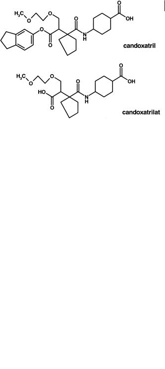
Esterification of a carboxylic acid function in a molecule has the immediate effect of a reduction in H-bonding potential and an increase in lipophilicity. Such parameters are important in the oral absorption of compounds as described earlier. Candoxatrilat (Figure 7.20), an inhibitor of neutral endopeptidase (NEP), has poor oral bioavailability [15]. The compound has a log D of – 2. The indanyl ester analogue candoxatril (Figure 7.20) has a log D of 1.5, and a reduced Raevsky score [15]. As such the compound is well within the requirements expected for an oral agent. The pro-drug is well absorbed, rapidly hydrolyzed but complete conversion is not achieved. The proportion of candoxatrilat liberated depends on competing clearance processes for candoxatril clearance e. g. hepatic uptake/ biliary clearance. For candoxatrilat the values of systemic availability after oral administration to the mouse, rat, dog and man are 88, 53, 17 and 32 % respectively and depend on the esterase activity (which in rodents is the highest) and the competing processes (which in man are probably the lowest).
Fig. 7.20 Structures of candoxatril, the orally absorbed indanyl ester pro-drug of candoxatrilat.

907 Metabolic (Hepatic) Clearance
7.6
Enzymes Catalysing Drug Conjugation
7.6.1
Glucuronyland Sulpho-Transferases
One of the most important phase II conjugation reactions is that catalyzed by the glucuronyl transferases. A number of functional groups have the potential to be glucuronidated as shown in Table 7.3, but phenol and carboxylic acid functions are of prime importance to the medicinal chemist.
Tab. 7.3 Reactions performed by the glucuronyl transferases.
Function |
Typical example |
|
|
Aliphatic hydroxyl |
Tiaramide |
Phenol |
Morphine |
Aromatic carboxyl |
Furosemide |
Aromatic tetrozole |
Losartan |
Aliphatic carboxyl |
Benoxaprofen |
Immidazole |
Tioconazole |
Aromatic amine |
Dapsone |
Tertiary amine |
Chlorpromazine |
Triazine |
Lamotrigine |
|
|
Glucuronidation involves the transfer of D-glucuronic acid from UDP-α-glu- curonic acid to an acceptor compound. The family of enzymes which catalyze this reaction are the UDP-glucuronyl transferases [16]. The reaction proceeds by nucleophilic SN2 substitution of the C-1 carbon of glucuronic acid, the product undergoing inversion of configuration. The mechanism is illustrated schematically in Figure 7.21.
Fig. 7.21 Schematic showing mechanism of glucuronidation reactions.
7.6 Enzymes Catalysing Drug Conjugation 91
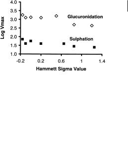
Similar mechanisms apply to sulphate transferases in which the donor is 3’-phos- phoadenosine-5-phosphosulphate (PAPS). The accepting groups in the molecule are phenols, alcohols and hydroxylamines. The sulphotransferases are relatively nonspecific, however, phenol-sulphotransferase is probably the most relevant to the medicinal chemist. The similarity in mechanism [17, 18] is shown by comparing the Vmax values for glucuronyl transferase and sulphotransferase for a series of power substituted phenols. Figure 7.22 shows the log Vmax for these series plotted against the Hammett sigma value.
Fig. 7.22 Relationship between sigma value and enzyme rate for glucuronyl and sulphotransferases indicating the role of nucleophilicity.
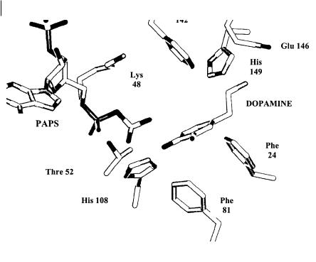
The negative slope of both curves indicates the greater the nucleophilicity (elec- tron-donating ability) of the phenolate anion the faster the rate of the reaction. The initial deprotonation of the phenol is apparently not rate limiting but must occur rapidly so that those compounds with high pKa values can be deprotonated. Recently X-ray crystallography data [19, 20] has been obtained for various sulphotransferases. The active site comprises a hydrophobic pocket formed by phenylalanine residues (see Figure 7.23). In the case of the catecholtransferase SULT1A3, a glutamic acid residue provides an ion pair interaction with the basic nitrogen of many of its natural substrates such as dopamine. A critical residue in the catalytic process is a lysine which stabilizes the transition state and via a hydrogen bond interaction with the bridge oxygen, between the 5’-phosphate group and the sulphate group of PAPS, acts as a catalytic acid to enhance the dissociative nature of the sulphuryl transfer mechanism. The other critical residue is an histidine, which acts as the base which deprotonates the phenol (or other group) to a phenoxide. The resultant nucleophile can then attack the sulphur atom of the transferring sulphuryl group.
The glucuronide and sulphotransferases are present in the gut as well as the liver and catalyze the metabolism of many phenolor catechol-containing drugs (morphine, isoprenaline etc.) during their passage through the gut. The ready conjugation of phenolic functions by both glucuronyl and sulphotransferase systems means that drugs such as morphine are cleared as both glucuronide and sulphate metabolites.
92 7 Metabolic (Hepatic) Clearance
Fig. 7.23 Schematic of the active site of catechol sulphotransferase (SULT1A3). with dopamine in the active site. Phe 142, 81 and 24 form a hydrophobic pocket whilst Glu 146 provides an ion pair interaction with the sub-
7.6.2
strate. Leu 48 stabilizes the transition state and His 108 deprotonates one of the catechol hydroxyls to form the phenoxide nucleophile to allow the reaction to proceed.
Methyl Transferases
Structural data is available on a third member of the transferases, catechol methyl transferase. This crystal structure data gives further clues on how the transferases metabolize their substrates, particularly with regard to the deprotonation step [21]. This enzyme catalyzes the transfer of the methyl group from S-adenosyl-L-methion- ine (SAM) to one of the hydroxyl group of catechols. Catechols are occasionally present in drug candidates (e. g. felodopam) but are frequently encountered as metabolites of drugs (e. g. methylene dioxy-containing compounds such as zamifenacin and paroxetine). The active site of COMT includes the co-enzyme-binding motif and the catalytic site situated in the vicinity of the Mg2+ ion. The methyl transfer from SAM to the catechol substrate catalyzed by COMT is a direct bimolecular transfer of the methyl group from the sulphur of α-Me-DOPA to the oxygen of the catechol hydroxyl in an SN2-like transition state. The exact juxtaposition of the substrate to the methyl group is possible because of the binding of the two hydroxyl groups to a Mg2+

7.7 Stability to Conjugation Processes 93
ion. One hydroxyl of the substrate is surrounded by three positively charged groups inducing it to release its proton to become a negatively charged phenolate ion. These moieties are the Mg2+, the methyl group of SAM and Lys 144. The Mg2+ ion in particular probably lowers the pKa of the hydroxyl group significantly. In contrast, the proton of the other hydroxyl is stabilized by the negatively charged carboxyl group of Glu 199. The ionized hydroxyl makes a direct nucleophilic attack on the electron-de- ficient methyl of α-Me-DOPA.
7.6.3
Glutathione-S-Transferases
Glutathione-S-transferases (GSTs) are the most important family of enzymes involved in the metabolism of alkylating compounds and their metabolites. They are a major defence system in deactivating toxic materials within the body. The cytosolic GSTs function as dimeric proteins that are assembled from identical or non-identi- cal subunits. Catalytic diversity for the cytosolic isoenzymes originates from the multiplicity of different homo-and hetero-dimeric forms that collectively metabolize a very broad range of structurally diverse electrophilic substrates, although all are highly specific for the thiol-containing substrate glutathione. Understanding the mechanism is considerably helped by the availability of X-ray crystallography data [22].
Each subunit has an active site that appears as a cleft along the domain interface. Each site can be separated into two distinct functional regions: a hydrophilic G-site for binding the physiological substrate glutathione, and an adjacent hydrophobic H- site for binding structurally diverse electrophilic substrates. Although the active sites of glutathione S-transferases are catalytically independent, the full active site is formed by structural elements from both subunits of the dimer. Residues contributing to binding glutathione at the G-site form a network of specific polar interactions. Of key importance is a hydrogen bond between a conserved G-site tyrosine residue and the glutathione thiol group. This hydrogen bond stabilizes the thiolate anion of the active site-bound glutathione. Estimated pKa values for the bound glutathione are at least two pKa units below the pKa value for glutathione free in aqueous solution. The H-site is formed of clusters of non-polar amino acid side chains which provide a highly hydrophobic surface, which, in the absence of a drug substrate is open to bulk solvent. Binding of substrates to this site has been shown to relate to increased lipophilicity for substrates (4-hydroxyalkenes). The actual conjugation reaction, with the thiolate anion acting as a nucleophile proceeds via an SN2-type mechanism yielding the “deactivated” product.
7.7
Stability to Conjugation Processes
Conjugation with glucuronic acid or sulphate requires a nucleophilic substituent to be present in the molecule, normally an hydroxyl function. In the case of the glu-
94 7 Metabolic (Hepatic) Clearance
curonyl transferases this can be phenol, primary, secondary or even tertiary alcohol or carboxylate or in the case of sulphotransferases, normally phenol. In some cases primary alcohols can also form sulphate conjugates. The most “reactive” grouping is the phenol and a simple rule is to eliminate such groupings unless essential for activity. In some cases bioisosteres can be introduced to retain pharmacological activity and overcome conjugation. To act as agonists of the dopamine receptor an H-bond donor group is essential in the correct position on a phenyl ring whose centre is situated 5.1 Å from a protonated nitrogen atom [23]. Figure 7.24 illustrates 7-hydroxy- (amino) tetralin analogues which are potent agonists.
Fig. 7.24 Substitution of a pyrrolo for a phenolic function to act at as a H-bond donor for receptor interactions but is resistant to glucuronidation.
These compounds have very low bioavailability and short duration due to extremely rapid glucuronidation [24]. Substitution of the phenolic hydroxy group with a pyrrolo ring (Figure 7.20) gives a series of compounds with a suitable H-bond donor in the correct position (also the geometry matches that of the phenolic hydroxyl), but resistant to glucuronidation [23]. It is not sufficient to replace functionality with
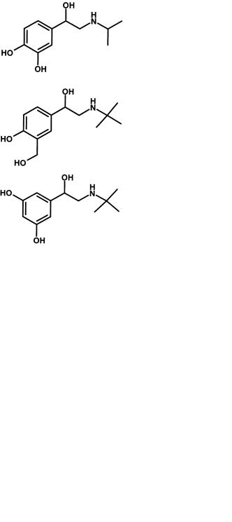
Fig. 7.25 Design of beta-2 selective adrenoceptor agonists resistant to catechol O-methyl transferase (COMT).
7.8 Pharmacodynamics and Conjugation 95
groupings with similar chemical properties. For instance tetrazolyl is almost an exact mimic of the carboxyl group and readily undergoes glucuronidation.
Catechol methyl transferases require the catechol function to be present to bind to the Mg2+ ion. In the search for β2-adrenoceptor selectivity to produce potent bronchodilators with low cardiovascular effects, changing the 3,4-hydroxy grouping of the catechol to 3,5- or 3-hydroxyl, 4-methyl-hydroxy, proved to be important (Figure 7.25). These compounds now have much improved bioavailability and pharmacokinetics due to their resistance to catechol methyl transferases.
7.8
Pharmacodynamics and Conjugation
In a number of cases the transferase enzymes metabolize compounds to active species. Morphine is a highly potent opioid analgesic (Figure 7.26) and is metabolized by glucuronidation of both its hydroxyl functions in both the gastrointestinal tract and the liver. Glucuronidation of the 6-position to form morphine-6-glu- curonide gives a compound that is also active [25]. Given systemically, the metabolite is twice as potent as morphine itself. When administered intrathecally the compound is approximately 100-fold more potent than the parent morphine.
Fig. 7.26 Structure of morphine, which is metabolized to a more active opioid analgesic by glucuronidation at the 6 position.
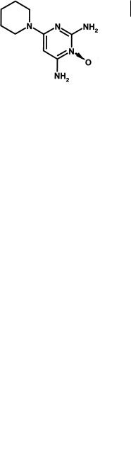
Unlike morphine, minoxidil [26] is not active itself but is metabolized by hepatic sulphotransferases to minoxidil N-O sulphate (Figure 7.27).
Fig. 7.27 Structure of minoxidil a compound metabolized by sulphotransferases to a potassium channel activator.
Minoxidil sulphate is a potent activator of the ATP-modulated potassium channel and thereby relaxes vascular smooth muscle to give a resultant antihypertensive effect. The actual sulphate metabolite is a relatively minor metabolite, the principal