

Pharmacokinetics and Metabolism in Drug Design Edited by D. A. Smith, H. van de Waterbeemd, D. K. Walker, R. Mannhold, H. Kubinyi, H.Timmerman
.pdf96 7 Metabolic (Hepatic) Clearance
metabolite being the N-O glucuronide. The discovery of minoxidil illustrates the caution which should be applied to screening compounds solely in vitro. Occasionally in vivo experiments will provide significant advances as a result of the metabolism of novel active agents.

97
References
1Smith DA, Jones BC, Biochem. Pharmacol. 1992, 44, 2089–2104.
2Gillis AM, Kates RE, Clin. Pharmacokinet. 1984, 9, 375–403.
3Lee JT, Kroemer HK, Silberstein DJ, Funck-Brentano C, Lineberry MD, Wood AJ, Roden DM, Woosley RL, New Engl. J. Med. 1990, 322, 1764–1768.
4 Manoury PM, Binet JL, Rousseau J, Le- ferre-Borg FM, Cavero IG, J. Med. Chem. 1987, 30, 1003–1011.
5Marchetti P, Natalesi R, Clin. Pharmacokinet. 1989, 16, 100–1286.
6Verbeck RK, Blackburn JL, Loewen GR,
Clin. Pharmacokinet. 1983, 8, 297–331.
7Dugar S, Yumibe N, Clader JW, Vizziano M, Huie K, Heek MV, Compton DS, Davis HR, Biorg. Med. Chem. Lett. 1996, 6, 1271–1274.
8Penning TD, Talley JJ, Bertenshaw SR, Carter JS, Collins PW, Docter S, Graneto MJ, Lee LF, Malecha JW, Miyashiro JM, Rogers RS, Rogier DJ, Yu SS, Anderson GD, Burton EG, Cogburn EG, Gregory SA, Koboldt CM, Perkins WE, Seibert K, Veenhuizen AW, Zhang AW, Isaakson PC, J. Med. Chem. 1997, 40, 1347–1365.
9Floyd DM, Dimball SD, Drapcho J, Das J, Turk CF, Moquin RV, Lago MW, Duff KJ, Lee VG, White RE, Ridgewell RE, Moreland S, Brittain RJ, Normandin DE, Hedberg SA, Cucinotta GC, J. Med. Chem. 1992, 35, 756–772.
10Rosenblum SB, Huynh T, Afonso A, Davis HR, Yumibe N, Clader JW, Burnett DA, J. Med. Chem. 1998, 41, 973–980.
11Evams ME, Stemp G, Chem. Brit. 1991, 27, 439–442.
12Hoke F, Cunningham F, James MK, Muir KT, Hoffman WE, J. Pharmacol. Exp. Ther. 1997, 281, 226–232.
13Baxter AJ, Carr RD, Eyley SC, FraserRae L, Hallam C, Harper ST, Hurved PA, King SJ, Menchani P, J. Med.
Chem. 1992, 35, 3718–3720.
14Kumar GN, Bodor N, Curr. Med. Chem. 1996, 3, 23–36.
15Kaye B, Brearley CJ, Cussans NJ, Herron M, Humphrey MJ, Mollatt AR,
Xenobiotica 1997, 27, 1091–1102.
16Burchell B, In: Handbook of Drug Metabolism. (Ed. Woolf TF), pp. 153– 173. Marcel Dekker, New York, 1999.
17Yin H, Bennett G , Jones JP, ChemicoBiol. Int. 1994, 90, 47–58.
18Duffel MW, Jacoby WB, J. Biol. Chem. 1981, 256, 11123–11127.
19Dajani R, Cleasby A, Neu M, Wonacott AJ, Jhoti H, Hood AM, Modi S, Hersey A, Taskinen J, Cooke RM, Manchee GR, Coughtrie MWH, J. Biol. Chem. 1999, 274, 37862–37868.
20Kakuta Y, Petrotchenko EV, Pedersen LC, Negishi M, J. Biol. Chem. 1998, 273, 27324–27330.
21Vidgren J, Svensson LA, Liijas A, Nature 1994, 368, 354–358.
22Dirr H, Reinemer P, Huber R, Eur. J. Biochem. 1994, 220, 645–661.
23Asselin AA, Humber LG, Roith K, Metcalf G, J. Med. Chem. 1986, 29, 648–654.
24Stjernlof P, Gullme M, Elebring T, Anderson B, Wikstrom H, Lagerquist S, Svensson K, Ekman A, Carlsson A, Sundell S, J. Med. Chem. 1993, 36, 2059–2065.
25Paul D, Standifer KM, Inturrisi CE, Pasternack GW, J. Pharmacol. Exp. Ther. 1989, 251, 477–483.
26McCall JM, Aiken JW, Chidester CG, DuCharme DW, Wendling MG,
J. Med. Chem. 1983, 26, 1791–1793.

Pharmacokinetics and Metabolism in Drug Design 99
Edited by D. A. Smith, H. van de Waterbeemd, D. K. Walker, R. Mannhold, H. Kubinyi, H. Timmerman Copyright © 2001 Wiley-VCH Verlag GmbH ISBNs: 3-527-30197-6 (Hardcover); 3-527-60021-3 (Electronic)
8
Toxicity
Abbreviations
ANF |
Atrial natriuretic factor (also ANP: atrial natriuretic peptide) |
COX |
Cyclooxygenase |
ENCC |
Electroneutral Na–Cl co-transporter |
hFGF |
Human fibroblast growth factor |
GSH |
Glutathione |
HMG-CoA |
3-Hydroxy-3-methylglutaryl coenzyme A |
LH |
Luteinizing hormone |
5-LPO |
5-Lipoxygenase |
NK |
Neurokinin |
NKCC |
Old name for ENCC |
PBPK/PD |
Physiologically-based pharmacokinetic/pharmacodymanic |
|
(modelling) |
PCNA |
Proliferating cell nuclear antigen |
PPAR-γ |
Peroxisome proliferator-activated receptor γ |
TA2 |
Thromboxane |
VEGF |
Vascular endothelial growth factor |
8.1
Toxicity Findings
A complex series of in vitro tests, animal test and then human exposure can at any stage reveal adverse findings that can be termed toxicity. Broadly, toxicity findings can be broken down into the following three sub-divisions.
8.1.1
Pharmacophore-induced Toxicity
This involves findings relating to the pharmacology of the compound. Within this category the adverse effects are either a direct or an indirect extension of the pharmacology. With the indirect extension the original selectivity of the compound for a target is lost at elevated doses and the effects seen are triggered by effects on proteins
100 8 Toxicity
etc., which are closely related structurally to the original target. Pharmacophore-in- duced toxicity is usually seen at doses in excess of the therapeutic dose.
Pharmacophore-induced toxicity does not necessarily occur within the organ or site of intended therapy. An example of this is the toxicity of loop diuretics [1]. The target for this class of drugs are the Na-(K)-Cl co-transporters of the kidney. These cotransporters play a major role in the ion transport and fluid secretion of the utricle and semicircular canal of the ear. Perhaps not surprisingly loop diuretics are associated with ototoxicity. The selectivity and potency of various diuretics can explain their different toxicity profiles. Kidney-specific co-transporters ENCC1 and ENCC2 are expressed in the thick ascending limb and distal convoluted tubule, respectively. ENCC3 is expressed in many tissues including the cochlea. Thiazide diuretics only have activity against ENCC1 and show no toxicity. Loop diuretics inhibit NKCC2 with potencies for bumetanide < 0.2 µM and NKCC3 > 0.5 µM. The selectivity of the compound for NKCC2 is lost under the conditions which cause ototoxicity: intravenous administration of high doses.
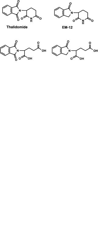
Unexpected or polypharmacology in a structure can occasionally lead to additional benefits in drugs. In the same way polypharmacology can have dramatic consequences in toxicity. Thalidomide was used as an anti-nausea drug to control morning sickness. Its use in pregnant women had terrible consequences due to the teratogenic nature of the drug.
Recently thalidomide and some of its metabolites (Figure 8.1) and related analogues have been shown to be inhibitors [2] of hFGFand VEGF-induced neovascularization (angiogenesis). Such a finding readily provides an hypothesis for the causes of teratogenicity associated with thalidomide, since limb development (the site of teratogenesis) is dependent on the formation of new blood vessels.
A problem with toxicity produced by an extension of the pharmacology of a compound, is that the conventional use of no-effect doses based on pre-clinical animal studies may not apply. Moreover pre-clinical studies may be complicated by the often understated ranges of response seen across species due to species differences in the receptors, enzymes and ion channels that comprise drug targets. Table 8.1 lists some of these known variations and the consequences range from an exaggerated response, to an absence of a response.
Fig. 8.1 Structures of thalidomide, EM-12 and their metabolites which are all angiogenesis inhibitors and teratogens or potential teratogens.
8.1 Toxicity Findings 101
Tab. 8.1 Receptors, ion channels and enzymes which are drug targets and show species differences.
Receptors |
|
|
|
Adenosine |
A1, A2 |
Luteinizing |
|
|
|
hormone |
LH |
Adrenoceptors |
α1B, α1C, α2A |
Muscarinic |
M2 |
Atrial nutriuretic factor |
ANF-R1 |
Neurokinin |
NK1, NK3 |
Bradykinin |
B2 |
Purinoceptors |
P2 |
Cholecystokinin |
CCK |
Thromboxane |
TA2 |
Dopamine |
D1 |
Vanilloid |
|
Endothelin |
ETB |
Vasopressin |
V1 |
Serotonin |
5-HT1A, B, D |
|
|
|
5-HT2, 5-HT3, |
|
|
|
5-HT4 |
|
|
|
|
|
|
Ion Channels
Rapidly activating delayed rectifier K+ channel
Enzymes |
|
Carboxypeptidase B |
Renin |
Na+/K+ ATPase |
HMG-CoA |
|
reductase |
|
|
8.1.2
Structure-related Toxicity
Findings related to the structure of the compound but not related to the pharmacology can provide another possible source of toxicity. This category is distinguished by the adverse events or effects being triggered by structural features or physicochemical properties etc. which allow the compound or metabolites to interact at sites distinct from the intended target or related proteins, etc. This type of toxicity can occur at any dose level including the therapeutic dose.
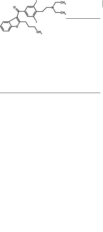
Amiodarone (Figure 8.2) is an efficacious drug that causes a number of side-ef- fects. The presence of iodine in the molecule is unusual and hypoand hyperthyroidism have been reported in patients. Although the loss of iodine is relatively slow the relatively large daily dose size and long half-life of the drug and its de-ethylated metabolite suggest that the presence of iodine in the molecule is responsible for its toxicity [3].
Fig. 8.2 Structure of the highly lipophilic antiarrythmic, amiodarone.
102 8 Toxicity
The drug is also a highly lipophilic base and accumulates in a number of tissues including the lung. This combination of extreme physicochemical properties can result in more specific interactions such as the condition of phospholipidosis (increase in total lung phospholipids) caused by inhibition of phospholipid breakdown [4]. The medicinal chemist has to decide if extreme lipophilicity and the presence of iodine are essential for activity and, in the case of amiodarone, proven clinical efficacy or whether alternative structures are possible.
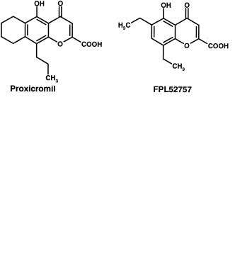
Proxicromil and FPL 52757 [5] were oral anti-allergy agents that utilized the strongly acidic “chromone” skeleton as a starting point (Figure 8.3). This skeleton contained the pharmacophore. To achieve oral absorption substantial lipophilicity was added and these changes resulted in surface active (detergent) molecules. The hepatobiliary route of excretion and resultant high concentrations of the compounds at the biliary cannaliculus resulted in hepatotoxicity [5].
Fig. 8.3 Structures of proxicromil and FPL 52757, two compounds which exhibited toxicity due to their physicochemical properties.
8.1.3
Metabolism-induced Toxicity
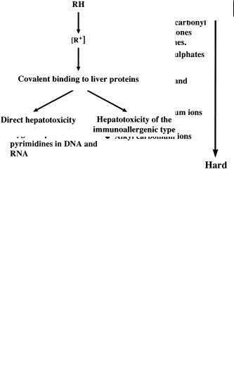
Metabolism-induced toxicity results when a key function or grouping is altered by oxidation, reduction or conjugation to become reactive, normally an electrophile [6]. The electrophilic group is then capable of reacting with nucleophiles in the body. Nucleophilic functions are present in proteins, nucleic acids and small peptides such as glutathione (see Section 8.1.2). Reactions with these targets can lead to organ toxicity including carcinogenesis or simply excretion from the body (glutathione conjugates). Some indication of the possible targets [7] is indicated by the nature of the electrophile produced (soft–hard), as indicated in Figure 8.4.
Compounds that react with amino acids or proteins can trigger toxicity by two mechanisms. The direct mechanism involves reaction with specific proteins thus altering their function such that cell death and necrosis occurs. Such toxicities are often seen in a large number of subjects and are dose related. They are also often predicted from animal studies. Alternative mechanisms of toxicity involve an immune component, whereby the protein–metabolite conjugate triggers an immune response. Such toxicity is termed idiosyncratic, occurring in only a subset of the patients receiving the drug. This type of toxicity is not predicted normally by animal studies. These two types of toxicity are illustrated in the schematic shown in Figure 8.5. Glutathione and the glutathione transferase enzymes protect the body from
8.2 Epoxides 103
Fig. 8.4 Schematic showing relative softness and hardness of nucleophiles and electrophiles as an indicator of sites of reaction of electrophilic metabolites.
the reactive metabolites. This system is saturable, so that a threshold dose, or other factors leading to glutathione depletion, is needed to trigger toxicity.
Fig. 8.5 Schematic showing the generation of a reactive metabolite, its reaction with a protein target, and toxicity resulting from a direct mecha-
nism (protein essential to cell function) or one involving the immune system.
8.2
Epoxides
Epoxide metabolites can be generated from a variety of aromatic systems. Anticonvulsants are a class of drug whose side-effects, such as hepatic necrosis and aplastic anaemia, are thought to be mediated by chemically reactive epoxide metabolites formed by cytochrome P450 oxidation. For instance phenytoin (Figure 8.6) toxicity is correlated with oxidation and the inhibition of epoxide hydrolase [8].
Carbamazepine exerts its anticonvulsant activity through its own action on voltage sensitive sodium channels and those of its relatively stable 10-11-epoxide. The compound shows a number of potential toxicities including skin rash, hepatic necrosis and teratogenicity. It is possible the 10-11-epoxide is the causative agent, but struc-
104 8 Toxicity
Fig. 8.6 Structure of phenytoin, a drug believed to assert its toxicity through reactive epoxide metabolites.
tural studies [9] suggest other epoxide metabolites of the aromatic ring may be responsible in part. Oxcarbazepine (Figure 8.7) is a related drug that cannot form the 10-11-epoxide and owes part of its activity to its hydroxyl metabolite. Oxcarbazepine is much less teratogenic in animal models and shows a lower preponderance of skin rash [8,10].
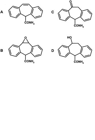
Fig. 8.7 Structures of carbamezepine (A), its 10-11-epoxide metabolite (B), and oxcarbazepine (C) and its hydroxyl metabolite (D).
8.3
Quinone Imines
Phenacetin is a classical example of a quinone imine, with oxidation of the compound by cytochrome P450 leading to a benzoquinone intermediate (Figure 8.8). The benzoquinone reacts with various cytosolic proteins to trigger direct hepatotoxicity [6].
Toxicity by metabolism is not confined to the liver since oxidative systems occur in many organs and cells. Amodiaquine is a 4-aminoquinoline antimalarial that has been associated with hepatitis and agranulocytosis. Both side-effects are probably triggered by reactive metabolites produced in the liver or in other sites of the body. For instance polymorphonuclear leucocytes can oxidize amodiaquine. It appears that amodiaquine is metabolized to a quinone imine by the same pathway as that seen in

8.3 Quinone Imines 105
Fig. 8.8 Oxidation of phenacetin to a benzoquinone intermediate.
the case of acetaminophen [11] (Figure 8.9), suggesting that such structural features in a molecule should be avoided.
Fig. 8.9 Structures of amodiaquine and its quinone imine metabolite.
Such reactions can occur in other molecules containing aromatic amine functions without a para oxygen substituent. For instance diclofenac can be oxidized to a minor metabolite (5-OH) diclofenac which can be further oxidized [12] to the benzoquinine imine metabolite (Figure 8.10). Again, the reactivity of this intermediate has been implicated in the hepatotoxicity of the compound.
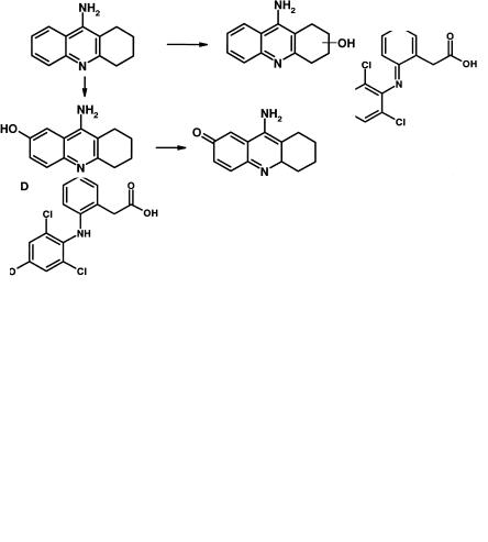
Another drug with a high incidence of hepatotoxicity is the acetylcholinesterase inhibitor tacrine. Binding of reactive metabolites to liver tissue correlated with the formation of a 7-hydroxy metabolite [13], highly suggestive of a quinone imine metabolite as the reactive species. Such a metabolite would be formed by further oxidation of 7-hydroxy tacrine (Figure 8.11).
Indomethacin is associated, in the clinic, with a relatively high incidence of agranulocytosis. Although indomethacin itself is not oxidized to reactive metabolites, one of its metabolites, dsemethyldeschlorobenzoylindomethacin (DMBI) forms an iminoquinone [14]. Formation of the iminoquinone from DMBI is catalysed by
106 8 Toxicity
Fig. 8.10 Scheme showing metabolism of diclofenac by oxidation (A) to benzoquinone imine (C) metabolites.
Fig. 8.11 Metabolism of tacrine to hydroxyl metabolites, the 5-hydroxy derivative of which can be further oxidized to the reactive quinone imine.
myeloperoxidase (the major oxidizing enzyme in neutrophils) and HOCl (the major oxidant produced by activated neutrophils). The pathway for formation of the iminoquinone is illustrated in Figure 8.12.
Practolol (Figure 8.13) was the prototype cardioselective β-adrenoceptor blocking agent. Selectivity was achieved by substitution in the para position with an acetyl anilino function. The similarity of this drug with those outlined above is obvious. Practolol caused severe skin and eye lesions in some patients which led to its withdrawal from the market [6]. These lesions manifested as a rash, hyperkeratosis, scarring, even perforation of the cornea and development of a fibrovascular mass in the conjunctiva, and sclerosing peritonitis. Some evidence is available that the drug is oxidatively metabolized to a reactive product that binds irreversibly to tissue pro-