

Pharmacokinetics and Metabolism in Drug Design Edited by D. A. Smith, H. van de Waterbeemd, D. K. Walker, R. Mannhold, H. Kubinyi, H.Timmerman
.pdf767 Metabolic (Hepatic) Clearance
Tab. 7.1 Division of enzymes into phase I and phase II. Phase I enzymes are normally oxidative and phase II conjugative.
Cytochrome P450 monooxygenase
Azo and nitro group reductase
Aldehyde dehydrogenase
Alcohol dehydrogenase Phase I
Epoxide hydrolase
Monoamine oxidase
Flavin monoamine oxidase
Non-specific esterases
Non-specific N- and O-methyltransferases
D–glucuronic acid transferase
Catechol O-methyltransferase Phase II
Glutathione transferase
Sulphate transferase
7.2
Cytochrome P450
The cytochrome P450 system can carry out a variety of oxidation reactions as listed in Table 7.2.
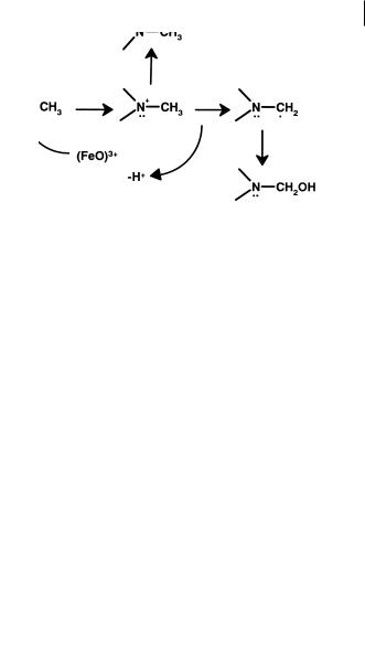
The overall scheme for these reactions is the insertion of a single oxygen atom into the drug molecule. Current views indicate that the chemistry of cytochrome P450 is radical by nature.
The mechanism of cytochrome P450 catalysis is probably constant across the system. It is determined by the ability of a high valent formal (FeO)3+ species to carry out one-electron oxidations through the abstraction of hydrogen atoms or electrons. The resultant substrate radical can then recombine with the newly created hydroxyl radical (oxygen rebound) to form the oxidized metabolite. Where a heteroatom is the (rich) source of the electron more than one product is possible. There can be direct recombination to yield the heteroatom oxide or radical relocalization within the
Tab. 7.2 Reactions performed by the cytochrome P450 system.
Reaction |
Product |
Typical example |
|
|
|
Aromatic hydroxylation |
Phenyl to phenol |
Phenytoin |
Aliphatic hydroxylation |
Methyl to carbinol |
Ibuprofen |
N-dealkylation |
Tertiary to secondary amine |
Lidocaine |
O-dealkylation |
Ether to alcohol |
Naproxen |
S-dealkylation |
Thioether to thiol |
6-methylthiopurine |
N-oxidation |
Pyridine to pyridine N-oxide |
Voriconazole |
S-oxidation |
Sulphoxide to sulphone |
Omeprazole |
Alcohol oxidation |
Alcohol to carboxylic acid |
Losartan |
|
|
|
7.2 Cytochrome P450 77
Fig. 7.1 Heteroatom oxidation of drugs by cytochrome P450 leading to heteroatom oxides or dealkylation products.
substrate to a carbon and oxidation of this function to form the unstable carbinol and ultimately heteroatom dealkylation. A possible reaction sequence is illustrated as Figure 7.1.
Many of the investigations into the enzymology of cytochrome P450 over the previous 20 years have focused on the pathway that generates this reactive species as illustrated in Figure 7.2 particularly the donation of electrons and protons to yield the (FeO)3+ substrate complex which is the oxidizing species. As part of the cycle substrate binds to the enzyme as an initial step before the addition of electrons and molecular oxygen. The final stage of the cycle is the actual attack of the (FeO)3+ species on the substrate.
Fig. 7.2 Cytochrome P450 cycle showing the key stages of substrate interaction.
The critical points of the cycle involving substrate–enzyme interactions are illustrated in Figure 7.2 and explored below:
a)The initial binding of the substrate to the CYP which causes a change in the spin state of the haem iron eventually resulting in the formation of the (FeO)3+-sub-
strate complex. This is obviously a key substrate–protein interaction and depends on the actual 3D-structure of the substrate and the topography of the active site.
78 7 Metabolic (Hepatic) Clearance
However, it cannot be assumed that this initial binding is the same as the final conformation that the protein and substrate adopt during actual substrate attack. b)The final stages of the cycle, when the geometry and chemical reactivity of this
complex determine the structure of the metabolite produced.
Analysis of the literature indicates that three major forms of CYPs are involved in the metabolism of pharmaceuticals in man: CYP2D6, CYP2C9 and CYP3A4, CYP1A2, CYP2C19 and CYP2E1 are also involved, but this involvement is much less extensive. The catalytic selectivity of the major CYPs has been reviewed [1].
7.2.1
Catalytic Selectivity of CYP2D6
Substrates for CYP2D6 include tricyclic antidepressants, β-blockers, class 1 antiarrhythmics. In brief, the structural similarities of many of the substrates and inhibitors in terms of position of hydroxylation, overall structure (aryl-alkylamine) and physicochemistry (ionized nitrogen at physiological pH), have allowed template models such as that illustrated as Figure 7.3 to be constructed.
Fig. 7.3 Template model for CYP2D6 with Y the site of oxidation, a is the distance from Y to a heteroatom which is positively charged (normally
5–7 Å).
All the template models produced have the same common features of a basic nitrogen atom at a distance of 5–7 Å from the site of metabolism which is in general on or near a planar aromatic system. It is currently believed that aspartic acid residue 301 provides the carboxylate residues which binds the basic nitrogen of the substrates.
With CYP2D6 therefore the catalytic selectivity relies heavily on a substrate–pro- tein interaction. The relative strength of the proposed ion pair association between the basic nitrogen and the active site of aspartic acid means that the affinity for substrates will be high. This is borne out by the enzyme having lower Km and Ki values than other CYPs. Thus CYP2D6 is often a major enzyme in drug oxidation despite its low abundance in human liver. This statement is particularly true for low concentrations or doses of drugs, the low Km values rendering the enzyme easily saturable
Fig. 7.4 Structure of propafenone.
7.2 Cytochrome P450 79
(see Section 2.12). An example of this is the antiarrythmic compound propafenone (Figure 7.4) [2] which is converted to 5-hydroxy propafenone by CYP2D6.
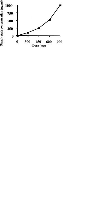
The dependence on CYP2D6 metabolism and the relatively high clinical dose (see Figure 7.5) mean that the metabolism is readily saturable over a narrow clinical dose range, so that small increases in dose can lead to disproportionate increases in plasma concentration, and a resultant steep dose–response curve.
Fig. 7.5 Relationship between plasma concentration and dose of propafenone, a CYP2D6 substrate.
CYP2D6 is also problematic in drug therapy since the enzyme is absent in about 7 % of Caucasians due to genetic polymorphism. In these 7 % (poor metabolizers) clearance of CYP2D6 substrates such as propafenone (Figure 7.4) [3] are markedly lower and can lead to side-effects in these subsets of the population. This correlation of enhanced side-effects in poor metabolizers (lacking CYP2D6) compared to extensive metabolizers (active CYP2D6) has been made for propafenone. The example of betaxolol [4] shows how knowledge of the properties that bestow pharmacological activity can be combined with metabolism concepts to produce a molecule with improved performance. Cardioselectivity for β-adrenoceptor agents can be conferred by substitution in the para position of the phenoxy-propanolamine skeleton. The para position or methoxyethyl substituents (e.g. metoprolol) in this position are the major sites of metabolism for these compounds. This reaction is catalysed by CYP2D6, and the efficiency of the enzyme means that metoprolol shows high clearance and resultant low bioavailability and short half-life. Manoury et al. [4] designed the series of compounds leading to betaxolol on the hypothesis that bulky stable substituents in the para position (Figure 7.6) would be resistant to metabolism and also cardioselective.
Fig. 7.6 Structures of metoprolol and betaxolol an analogue designed to be more metabolically stable.
80 7 Metabolic (Hepatic) Clearance
Beside the actual steric bulk of the substituent, cyclopropyl is much more stable to hydrogen abstraction than other alkyl functions and represents an ideal terminal group. These changes make betaxolol a compound with much improved pharmacokinetics compared to its lipophilic analogues.
7.2.2
Catalytic Selectivity of CYP2C9
Substrates for CYP2C9 include many non-steroidal anti-inflammatory drugs plus a reasonably diverse set of compounds including phenytoin, (S)-warfarin and tolbutamide. All the substrates with routes of metabolism attributable to CYP2C9 have hydrogen bond donating groups a discrete distance from a lipophilic region which is the site of hydroxylation. The hydrogen bond donating groups and sites of metabolism on each of the substrates have been overlaid with those of phenytoin to produce a putative template of the active site of CYP2C9 (Figure 7.7).
Fig. 7.7 Template model of CYP2C9; Y is the site of oxidation, a is the distance from Y to a heteroatom which can act as a H-bond donor and c defines the angle of the H-bond.
The mean dimensions (± SD) for the eight compounds (a = 6.7 ± 0.8 Å, C = 133 ± 20°) illustrates the degree of overlap achieved. Like CYP2D6 the catalytic selectivity of CYP2C9 is dominated by substrate–protein interactions.
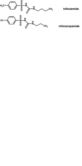
Tolbutamide (Figure 7.8) is metabolized via the benzylic methyl group by CYP2C9 as the major clearance mechanism. Chlorpropamide is a related compound incorporating a chlorine function in this position. The resultant metabolic stability gives chlorpropamide a lower clearance and a longer half-life (approximately 35 h compared to 5 h) than tolbutamide, resulting in a substantial increase in duration of action [5].
Fig. 7.8 Structures of tolbutamide and the metabolically more stable analogue chlorpropamide.
The mechanism of action of CYPs is radical rather than electrophilic and the actual substitution pattern is important: the role of chlorine is one of blocking rather than deactivation. Many non-steroidal anti-inflammatory drugs are substrates for the
7.2 Cytochrome P450 81
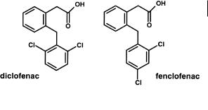
Fig. 7.9 Structures of diclofenac and fenclofenac. Fenclofenac is much more resistant to aromatic hydroxylation.
CYP2C9 enzyme and analogous structures show how metabolic stability to p-hydrox- ylation is achieved with only small changes in substitution.
Diclofenac, with ortho substitution in the aromatic ring (Figure 7.9) is metabolized principally to 4-hydroxydiclofenac by CYP2C9. In man, the drug has a short half-life of approximately 1 h due to the relatively high metabolic (oxidative) clearance. In contrast, the analogous compound, fenclofenac, is considerably more metabolically stable, due to the p-halogen substitution pattern, and exhibits a half-life of over 20 h [6].
7.2.3
Catalytic Selectivity of CYP3A4
CYP3A4 attacks lipophilic drugs in positions largely determined by their chemical lability: that is, the ease of hydrogen or electron abstraction. CYP3A4 SAR is dominated therefore by substrate–reactant interaction. Binding of substrates seems to be essentially due to lipophilic forces and results in the expulsion of water from the active site. Such an expulsion of water provides the driving force for the spin state change and hence the formation of the (FeO)3+ unit. However, the lipophilic forces holding the substrate in the active site are relatively weak (~ 1 kcal mole–1) and would allow motion of the substrate in the active site. Hence, since the substrate is able to adopt more than one orientation in the active site, the eventual product of the reaction is a product of the interaction between one of these orientations and the (FeO)3+ unit – a substrate–reactant interaction. This lack of apparent substrate structure similarity (apart from chemical reactivity) indicates a large active site that allows substrate molecules considerable mobility. The selectivity of CYP3A4 to its substrates may also be directed by the conformation they adopt within a lipophilic environment such as we are suggesting for the access channel and active site of CYP3A4. We have previously illustrated this point with cyclosporin A. In an aprotic (lipophilic) solvent cyclosporin A adopts a conformation which allows the major allylic site of CYP3A4 metabolism to extend out away from the bulk of the molecule. This is a different conformation from the one adopted in aqueous solution where the lipophilic sites are internalized and thus shielded from the solvent. As a general rule this “spreading out” of apparently sterically hindered molecules as judged by X-ray or aqueous solution structure, may help to further understand the selectivity of CYP3A4. The principle of extension of lipophilic functions normally hidden from solvent is further supported by the
82 7 Metabolic (Hepatic) Clearance
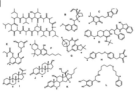
Fig. 7.10 Substrates for CYP3A4 illustrating the diversity of structure and the “selectivity” for attack at allylic and benzylic positions (major sites of metabolism indicated by asterisks). Substrates are A, cyclosporin A; B,
quinidine; C, L-696229; D, indinavir; E, lovastatin; F, D-THC; G, zatosetron H, pioglitazone; I, progesterone; J, testosterone; K, budesonide and L, salmeterol.
study of the soluble bacterial P450BM–3. In this case the substrates are fatty acids, which in aqueous solution adopt a “globular” conformation. However, upon entering the lipophilic access channel, the fatty acid opens out in an extended conformation with the lipophilic head group directed at the haem and the polar acid function directed at the solvent.
The enzyme is the principal participant in N-demethylation reactions where the substrate is a tertiary amine. The list of substrates includes erythromycin, ethylmorphine, lidocaine, diltiazem, tamoxifen, toremifene, verapamil, cocaine, amiodarone, alfentanil and terfenadine. Carbon atoms in the allylic and benzylic positions, such as those present in quinidine, steroids and cyclosporin A, are also particularly prone to oxidation by CYP3A4, a range of substrates is illustrated in Figure 7.10.
Both these routes of metabolism reflect the ease of hydrogen or electron abstraction from these functions. As with conventional radical chemistry, reactivity needs to be combined with probability. Thus, in molecules such as terfenadine the tertiary butyl group will be liable to oxidation due to its “maximum number” of equivalent primary carbons. Thus, although not a specially labile function, the site of metabolism becomes dominated by statistical probability. Terfenadine, as expected, also undergoes N-dealkylation by CYP3A4, illustrating the ability of the enzyme to produce multiple products (as for cyclosporin A, midazolam, etc.) and underlining the “flexibility” of CYP3A4 substrate binding.

7.2 Cytochrome P450 83
Overcoming metabolism by CYP3A4 is difficult due to the extreme range of substrates and the tolerance of the enzyme to variations in structure. Two strategies are available: removal of functionality and reduction of lipophilicity.
Allylic and benzylic positions are points of metabolic vulnerability. SCH48461 is a potent cholesterol absorption inhibitor [7]. Metabolic attack occurred at a number of positions including benzylic hydroxylation. Dugar and co-workers substituted oxygen for the C-3' carbon to remove this site of metabolism. This step however, produces an electron-rich phenoxy moiety in comparison to the original phenyl group and possibly makes this function more amenable to aromatic hydroxylation. Blocking of the aromatic oxidation with fluorine introduced in the para-position was required to produce the eventual more stable substitution. These steps are shown in Figure 7.11.
Fig. 7.11 Synthetic strategies to overcome benzylic hydroxylation in a series of cholesterol absorption inhibitors. Positions of metabolism are marked with an asterisk.
The lability of benzylic positions to cytochrome P450 metabolism has been exploited to decrease the unacceptably low clearance and resultant long half-life of various compounds. For example celecoxib, a selective cyclooxygenase inhibitor, has a half-life of 3.5 h in the rat. Early structural leads, represented by compounds in
Fig. 7.12 Structures of early long half-life COX2 inhibitor (A) and the candidate compound celecoxib (B) with a moderate half-life.

84 7 Metabolic (Hepatic) Clearance
which the benzylic methyl in celecoxib was substituted with a halogen (Figure 7.12), resulted in compounds with half-life values (in the male rat) of up to 220 h [8].
Diltiazem (Figure 7.13), a calcium channel blocker, is a drug that is extensively metabolized by at least five distinct pathways including N-demethylation, deacetylation, O-demethylation, ring hydroxylation and acid formation. The enzyme responsible for at least the major route (N-demethylation), has been shown to be CYP3A4 [8]. Although widely used in therapy, the compound has a relatively short duration of action. In the search for superior compounds, Floyd et al. [9] substituted the benzazepinone ring structure for the benzothiazepinone of diltiazem. Metabolic studies on this class of compound showed that the principal routes of metabolism were similar to that for diltiazem with N-demethylation, conversion to an aldehyde (precursor of an acid), deacetylation and O-demethylation all occurring. It was also noted that the N-desmethyl derivative was equipotent to the parent but much more stable metabolically. This can be rationalized as the decreased substitution on the nitrogen (secondary versus tertiary) stabilizing the nitrogen to electron abstraction (decreased radical stability). This stabilization is particularly important, since electron abstraction is the first step to both the N-desmethyl and aldehyde products (a total of 84 % of the total metabolism). The evidence of stability of secondary amines was capitalized on by synthesis of N-1 pyrrolidinyl derivatives, which were designed to achieve metabolic stability both by the decreased radical stability of secondary compounds to tertiary amines and steric hindrance afforded by β-substitution (Figure 7.11). The success of this strategy indicates how even the vulnerable alkyl substituted nitrogen grouping can be stabilized against attack.
Fig. 7.13 Structures of diltiazem and a benzozapepinone analogue resistant to metabolism.
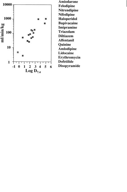
The predominant interaction of CYP3A4 is via hydrophobic forces and the overall lowering of lipophilicity can reduce metabolic lability to the enzyme. Figure 7.14 shows the relationship between unbound intrinsic clearance in man and lipophilicity for a variety of CYP3A4 substrates. The substrates are cleared by a variety of metabolic routes including N-dealkylation, aromatization and aromatic and aliphatic hydroxylation. The trend for lower metabolic lability with lower lipophilicity is maintained regardless of structure or metabolic route.
7.3 Oxidative Metabolism and Drug Design 85
Fig. 7.14 Unbound intrinsic clearance of CYP3A4 substrates and relationship with lipophilicity. The data has been calculated from various clinical studies with the drugs listed in order of decreasing lipophilicity.
7.3
Oxidative Metabolism and Drug Design
In addition to the examples indicated above the design of orally-active cholesterol absorption inhibitors combines both the concept of preventing metabolism and the serendipity of metabolites being more active than the parent drug [10]. On the basis of metabolite structure–activity relationships for SCH 48461 (Figure 7.15), SCH 58335 was designed to combine activity-enhancing oxidation and to remove or block sites of detrimental metabolic oxidation. The improvement in the pharmacodynamics of the compound is illustrated by the ED50 being reduced in the cholesterol hamster model from 2.2 to 0.04 mg kg–1 day–1.
Fig. 7.15 Structures of cholesterol absorption inhibitors SCH 48461 (A) and SCH 58235 (B). Metabolism of SCH 48461 occurs by aromatic hydroxylation (1) benzylic hydroxylation (2) and O-demethylation (3, 4). Metabolism is blocked in SCH 58235 at 1 and 4 or results in increases in potency at 2 and 3.