

Enzymes (Second Edition)
.pdf40 CHEMICAL BONDS AND REACTIONS IN BIOCHEMISTRY
The rate equation for the forward reaction must take into account the reverse reaction rate so that:
v |
d[S] |
k [S] k |
[P] |
(2.26) |
|||
dt |
|
||||||
|
|
|
|
|
|||
Integrating this equation with the usual boundary conditions, we obtain:
[S] |
[S] |
k k exp[ (k k )t] |
(2.27) |
|
k k |
||||
|
|
|
Integration of the velocity equation with respect to product formation yields:
|
|
k [S] 1 exp[ (k |
k )t] |
||
[P] |
|
|
|
|
(2.28) |
k |
k |
|
|||
|
|
|
|
||
At infinite time (i.e., when equilibrium is reached), the final concentrations of S and P are given by the following equations:
[S] |
|
k |
[S] |
|
(2.29) |
|
k |
k |
|||||
|
|
|
||||
[P] |
|
k |
[S] |
|
(2.30) |
|
k |
k |
|||||
|
|
|
||||
Hence a reaction progress curve for a reversible reaction will follow the same exponential growth of product and loss of substrate seen in Figure 2.15, but now, instead of [S] 0 and [P] [S] at infinite time, the curves will asymptotically approach the equilibrium values of the two species.
2.5.3 Measurement of Initial Velocity
The complexity of the rate equations presented in Section 2.5.2 made graphic determination of reaction rates a significant challenge prior to the widespread availability of personal computers with nonlinear curve-fitting software. For this reason researchers established the convention of measuring the initial rate or initial velocity of the reaction as a means of quantifying reaction kinetics. At the initiation of reaction no product is present, only substrate at concentration [S] . For a brief time after initiation, [P] [S], so that formation of P is quasi-linear with time. Hence, during this initial phase of the reaction one can define the velocity, d[P]/dt d[S]/dt, as the slope of a linear fit of [P] or [S] as a function of time. As a rule of thumb, this initial quasi-linear phase of the reaction usually extends over the time period between [P] 0 and [P] 0.1[S] (i.e., until about 10% of substrate has been utilized). This rule,
REFERENCES AND FURTHER READING |
41 |
however, is merely a guideline, and the time window of the linear phase of any particular reaction must be determined empirically. Initial velocity measurements are used extensively in enzyme kinetics, as we shall see in Chapters 5 and 7.
2.6 SUMMARY
In this chapter we have briefly reviewed atomic and molecular orbitals and the types of bond formed within molecules as a result of these electronic configurations. We have seen that noncovalent forces also can stabilize interatomic interactions in molecules. Most notably, hydrogen bonds, salt bridges, hydrophobic interactions, and van der Waals forces can take on important roles in protein structure and function. We have also reviewed some basic kinetics and thermodynamics as well as acid—base theories that provide a framework for describing the reactivities of protein components in enzymology. In the chapters to come we shall see how these fundamental forces of chemistry come into play in defining the structures and reactivities of enzyme.
REFERENCES AND FURTHER READING
Atkins, P. W. (1978) Physical Chemistry, Freeman, New York.
Fersht, A. (1985) Enzyme Structure and Mechanism, 2nd ed., Freeman, New York. Gray, H. B. (1973) Chemical Bonds: An Introduction to Atomic and Molecular Structure,
Benjamin/Cummings, Menlo Park, CA.
Kemp, D. S., and Vellaccio, F. (1980) Organic Chemistry, Worth, New York. Hammond, G. S. (1955) J. Am. Chem. Soc. 77, 334.
Lowry, T. H., and Richardson, K. S. (1981) Mechanism and T heory in Organic Chemistry, 2nd ed., Harper & Row, New York.
Palmer, T. (1985) Understanding Enzymes, Wiley, New York.
Pauling, L. (1960) T he Nature of the Chemical Bond, 3rd ed., Cornell University Press, Ithaca, NY.
Stryer, L. (1989) Molecular Design of L ife, Freeman, New York.

Enzymes: A Practical Introduction to Structure, Mechanism, and Data Analysis.
Robert A. Copeland Copyright 2000 by Wiley-VCH, Inc.
ISBNs: 0-471-35929-7 (Hardback); 0-471-22063-9 (Electronic)
3
STRUCTURAL
COMPONENTS
OF ENZYMES
In Chapter 2 we reviewed the forces that come to play in chemical reactions, such as those catalyzed by enzymes. In this chapter we introduce the specific molecular components of enzymes that bring these forces to bear on the reactants and products of the catalyzed reaction. Like all proteins, enzymes are composed mainly of the 20 naturally occurring amino acids. We shall discuss how these amino acids link together to form the polypeptide backbone of proteins, and how these macromolecules fold to form the three-dimensional conformations of enzymes that facilitate catalysis. Individual amino acid side chains supply chemical reactivities of different types that are exploited by the enzyme in catalyzing specific chemical transformations. In addition to the amino acids, many enzymes utilize nonprotein cofactors to add additional chemical reactivities to their repertoire. We shall describe some of the more common cofactors found in enzymes, and discuss how they are utilized in catalysis.
3.1 THE AMINO ACIDS
An amino acid is any molecule that conforms, at neural pH, to the general formula:
H N CH(R) COO
The central carbon atom in this structure is referred to as the alpha carbon (C ), and the substituent, R, is known as the amino acid side chain. Of all the possible chemical entities that could be classified as amino acids, nature has chosen to use 20 as the most common building blocks for constructing proteins and
42

THE AMINO ACIDS |
43 |
peptides. The structures of the side chains of the 20 naturally occurring amino acids are illustrated in Figure 3.1, and some of the physical properties of these molecules are summarized in Table 3.1. Since the alpha carbon is a chiral center, all the naturally occurring amino acids, except glycine, exist in two enantiomeric forms, and . All naturally occurring proteins are composed exclusively from the enantiomers of the amino acids.
As we shall see later in this chapter, most of the amino acids in a protein or peptide have their charged amino and carboxylate groups neutralized through peptide bond formation (in this situation the amino acid structure that remains is referred to as an amino acid residue of the protein or peptide). Hence, what chemically and physically distinguishes one amino acid from another in a protein is the identity of the side chain of the amino acid. As seen in Figure 3.1, these side chains vary in chemical structure from simple substituents, like a proton in the case of glycine, to complex bicyclic ring systems in the case of tryptophan. These different chemical structures of the side chains impart vastly different chemical reactivities to the amino acids of a protein. Let us review some of the chemical properties of the amino acid side chains that can participate in the interaction of proteins with other molecular and macromolecular species.
3.1.1 Properties of Amino Acid Side Chains
3.1.1.1 Hydrophobicity Scanning Figure 3.1, we note that several of the amino acids (valine, leucine, alanine, etc.) are composed entirely of hydrocarbons. One would expect that solvation of such nonpolar amino acids in a polar solvent like water would be thermodynamically costly. In general, when hydrophobic molecules are dissolved in a polar solvent, they tend to cluster together to minimize the amount of surface areas exposed to the solvent; this phenomenon is known as hydrophobic attraction. The repulsion from water of amino acids in a protein provides a strong driving force for proteins to fold into three-dimensional forms that sequester the nonpolar amino acids within the interior, or hydrophobic core, of the protein. Hydrophobic amino acids also help to stabilize the binding of nonpolar substrate molecules in the binding pockets of enzymes.
The hydrophobicity of the amino acids is measured by their tendency to partition into a polar solvent in mixed solvent systems. For example, a molecule can be dissolved in a 50 : 50 mixture of water and octanol. After mixing, the polar and nonpolar solvents are allowed to separate, and the concentration of the test molecule in each phase is measured. The equilibrium constant for transfer of the molecule from octanol to water is then given by:
[CH O]
K [C ] (3.1)

ENZYMES OF COMPONENTS STRUCTURAL 44
Figure 3.1 Side chain structures of the 20 natural amino acids. The entire proline molecule is shown.

Table 3.1 Physicochemical properties of the natural amino acids
|
|
|
Mass of |
Accessible |
|
pK of |
|
|
Van der |
|
|
|
|
Ionizable |
|
|
Waals |
||
|
Three-Letter |
One-Letter |
Residue in |
Surface Area |
Hydro- |
Side |
Occurrence in |
Relative |
Volume |
Amino Acid |
Code |
Code |
Proteins |
(Å ) |
phobicity |
Chain |
Proteins (%) |
Mutability |
(Å ) |
Alanine |
Ala |
A |
71.08 |
115 |
Arginine |
Arg |
R |
156.20 |
225 |
Asparagine |
Asn |
N |
114.11 |
160 |
Aspartate |
Asp |
D |
115.09 |
150 |
Cysteine |
Cys |
C |
103.14 |
135 |
Glutamate |
Glu |
E |
128.14 |
180 |
Glutamine |
Gln |
Q |
129.12 |
190 |
Glycine |
Gly |
G |
57.06 |
75 |
Histidine |
His |
H |
137.15 |
195 |
Isoleucine |
Ile |
I |
113.17 |
175 |
Leucine |
Leu |
L |
113.17 |
170 |
Lysine |
Lys |
K |
128.18 |
200 |
Methionine |
Met |
M |
131.21 |
185 |
Phenylalanine |
Phe |
F |
147.18 |
210 |
Proline |
Pro |
P |
97.12 |
145 |
Serine |
Ser |
S |
87.08 |
115 |
Threonine |
Thr |
T |
101.11 |
140 |
Tryptophan |
Trp |
W |
186.21 |
255 |
Tyrosine |
Tyr |
Y |
163.18 |
230 |
Valine |
Val |
V |
99.14 |
155 |
1.8 |
|
9.0 |
100 |
67 |
4.5 |
12.5 |
4.7 |
65 |
148 |
3.5 |
|
4.4 |
134 |
96 |
3.5 |
3.9 |
5.5 |
106 |
91 |
2.5 |
8.4 |
2.8 |
20 |
86 |
3.5 |
4.1 |
3.9 |
102 |
109 |
3.5 |
|
6.2 |
93 |
114 |
0.4 |
|
7.5 |
49 |
48 |
3.2 |
6.0 |
2.1 |
66 |
118 |
4.5 |
|
4.6 |
96 |
124 |
3.8 |
|
7.5 |
40 |
124 |
3.9 |
10.8 |
7.0 |
56 |
135 |
1.9 |
|
1.7 |
94 |
124 |
2.8 |
|
3.5 |
41 |
135 |
1.6 |
|
4.6 |
56 |
90 |
0.8 |
|
7.1 |
120 |
73 |
0.7 |
|
6.0 |
97 |
93 |
0.9 |
|
1.1 |
18 |
163 |
1.3 |
10.1 |
3.5 |
41 |
141 |
4.2 |
|
6.9 |
74 |
105 |
Values reflect the molecular weights of the amino acids minus that of water.
Accessible surface are for residues as part of a polypeptide chain. Data from Chothia (1975).Hydrophobicity indices from Kyte and Doolittle (1982).
Based on the frequency of occurrence for each residue in the sequence of 207 unrelated proteins. Data from Klapper (1977).Likelihood that a residue will mutate within a specified time period during evolution. Data from Dayoff et al. (1978).
45 ACIDS AMINO THE
46 STRUCTURAL COMPONENTS OF ENZYMES
where [CH O] and [C ] are the molar concentrations of the molecule in the aqueous and octanol phases, respectively. The free energy of transfer can then
be calculated from the K value using Equation 2.3. Such thermodynamic studies have been performed for the transfer of the naturally occurring amino
acids from a number of nonpolar solvent to water. To make these measurements more representative of the hydrophobicities of amino acids within proteins, workers use analogues of the amino acids in which the amino and carboxylate charges are neutralized (e.g., using N-acetyl ethyl esters of the amino acids). Combining this type of thermodynamic information for the different solvent systems, one can develop a rank order of hydrophobicities for the 20 amino acids. A popular rank order used in this regard is that developed by Kyte and Doolittle (1982); the Kyte and Doolittle hydrophobicity indices for the amino acid are listed in Table 3.1. In general, hydrophobic amino acids are found on the interior of folded proteins, where they are shielded from the repulsive forces of the polar solvent, and polar amino acids tend to be found on the solvent-exposed surfaces of folded proteins.
3.1.1.2Hydrogen Bonding Associated with the heteroatoms of the side chains of several amino acids are exchangeable protons that can serve as hydrogen donors for H-bonding. Other amino acids can participate as H-bond acceptors through the lone pair electrons on heteroatoms of their side chains. Hydrogen bonding of amino acid side chains and polypeptide backbone groups can greatly stabilize protein structures, as we shall see later in this chapter. Additionally, hydrogen bonds can be formed between amino acid side chains and ligand (substrates, products, inhibitors, etc.) atoms and can contribute to the overall binding energy for the interactions of enzymes with such molecules. Side chains that are capable of acting as H-bond donors include tyrosine ( O H), serine ( O H), threonine ( O H), tryptophan ( N H), histidine ( N H), and cysteine ( S H). At low pH the side chains of glutamic and aspartic acid can also act as H-bond donors ( COO H). Heteroatoms on the side chains of the following amino acids can serve as H-bond acceptors: tyrosine ( :O: H), glutamic and aspartic acid ( COO ), serine and threonine ( :O: H), histidine (N:), cysteine ( :S: H), and methionine (:S:). Several of the amino acids can serve as both donors and acceptors, of H bonds, as illustrated for tyrosine in Figure 3.2.
3.1.1.3Salt Bridge Formation Noncovalent electrostatic interactions can occur between electonegative and electropositive species within proteins. Figure 3.3 illustrates the formation of such an electrostatic interaction between the side chains of a lysine residue on one polypeptide chain and a glutamic acid residue on another polypeptide. Because these interactions resemble the ionic interactions associated with small molecule salt formation, they are often referred to as salt bridges. Salt bridges can occur intramolecularly, between a charged amino acid side chain and other groups within the protein, or intermolecularly, between the amino acid side chain and charged groups on a
THE AMINO ACIDS |
47 |
Figure 3.2 Tyrosine participation in hydrogen bonding as (A) a hydrogen donor and (B) a hydrogen acceptor.
ligand or other macromolecule. For example, many proteins that bind to nucleic acids derive a significant portion of their binding energy by forming electrostatic interactions between positively charged amino acid residues on their surfaces (usually lysine and arginine residues) and the negatively charged phosphate groups of the nucleic acid backbone.
Another example of the importance of these electrostatic interactions comes from the mitochondrial electron transfer cascade. Here electrons flow from the protein cytochrome c to the enzyme cytochrome oxidase, where they are used to reduce oxygen to water during cellular respiration. For the electron to jump from one protein to the other, the two must form a tight (dissociation constant 10 M) complex. When the crystal structure of cytochrome c was solved, it became obvious that the surface of this molecule contained an area with an unusually high density of positively charged lysine residues. The putative binding site for cytochrome c on the cytochrome oxidase molecule has a corresponding high density of aspartic and glutamic acid residues. It is thus believed that the tight complex formed between these two proteins is facilitated by forming a large number of salt bridges at this interface. This suggestion is supported by the ability of the complex to be dissociated by adding high concentrations of salt to the solution. As the ionic strength increases, the salt ions compete for the counterions from
Figure 3.3 Salt bridge formation between a lysine and a glutamic acid residue at neutral pH.
48 STRUCTURAL COMPONENTS OF ENZYMES
the amino acid residues that would otherwise participate in salt bridge formation.
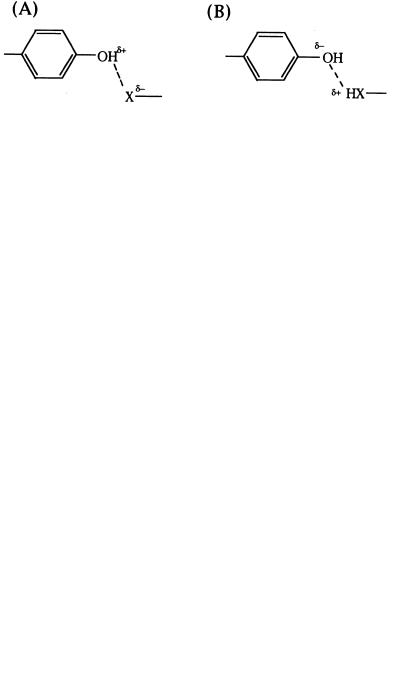
3.1.2 Amino Acids as Acids and Bases Surveying Table 3.1, we see that there are seven amino acid side chains, with titratable protons that can act as Brønsted—Lowry acids and conjugate bases. These are tyrosine, histidine, cysteine, lysine, and arginine, and aspartic and glutamic acids. The ability of these side chains to participate in acid—base chemistry provides enzymes with a mechanism for proton transfer to and from reactant and product molecules. In addition to proton transfer, side chain Lewis acids and bases can participate in nucleophilic and electrophilic reactions with the reactant molecules, leading to bond cleavage and formation. The placement of acid and base groups from amino acid side chains, at critical positions within the active site, is a common mechanism exploited by enzymes to facilitate rapid chemical reactions with the molecules that bind in the active site. For example, hydrolysis of peptide and ester bonds can occur through nucleophilic attack of the peptide by water. This reaction goes through a transition state in which the carbonyl oxygen of the peptide has a partial negative charge, and the oxygen of water has a partial positive charge:
|
O |
|
|
|
|
|
|
|
C |
C |
|
C |
¦ |
N |
|
¦ |
|
||
|
¦ |
|
|
|
¦ |
H |
|
|
O |
|
|
HH
If one could place a basic group at a fixed position close to the water molecule, it would be possible to stabilize this transition state by partial transfer of one of the water protons to the base. This stabilization of the transition state would allow the reaction to proceed rapidly:
|
O |
|
|
|
|
|
|
|
C |
C |
|
C |
¦ |
N |
|
¦ |
|
||
|
¦ |
|
|
|
¦ |
H |
|
|
O |
|
|
HH :B
THE AMINO ACIDS |
49 |
Alternatively, one could achieve the same stabilization by placing an acidic group at a fixed position in close proximity to the carbonyl oxygen, so that partial transfer of the proton from the acid to the carbonyl would stabilize the partial negative charge at this oxygen in the transition state. When one surveys the active sites of enzymes that catalyze peptide bond cleavage (a family of enzymes known as the proteases), one finds that there are usually acidic or basic amino acid side chains (or both) present at positions that are optimized for this type of transition state stabilization. We shall have more to say about stabilization of the transition states of enzyme reactions in Chapter 6.
In discussing the acid and base character of amino acid side chains, it is important to recognize that the pK values listed in Table 3.1 are for the side chain groups in aqueous solution. In proteins, however, these pK values can be greatly affected by the local environment that is experienced by the amino acid residue. For example, the pK of glutamic acid in aqueous solution is 4.1, but the pK of particular glutamic acid residues in some proteins can be as high as 6.5. Thus, while the pK values listed in Table 3.1 can provide some insights into the probable roles of certain side chains in chemical reactions, caution must be exercised to avoid making oversimplifications.
3.1.3 Cation and Metal Binding
Many enzymes incorporate divalent cations (Mg , Ca , Zn ) and transition metal ions (Fe, Cu, Ni, Co, etc.) within their structures to stabilize the folded conformation of the protein or to make possible direct participation in the chemical reactions catalyzed by the enzyme. Metals can provide a template for protein folding, as in the zinc finger domain of nucleic acid binding proteins, the calcium ions of calmodulin, and the zinc structural center of insulin. Metal ions can also serve as redox centers for catalysis; examples include heme—iron centers, copper ions, and nonheme irons. Other metal ions can serve as electrophilic reactants in catalysis, as in the case of the active site zinc ions of the metalloproteases. Most commonly metals are bound to the protein portion of the enzyme by formation of coordinate bonds with certain amino acid side chains: histidine, tyrosine, cysteine, and methionine, and aspartic and glutamic acids. Examples of metal coordination by each of these side chains can be found in the protein literature.
The side chain imidazole ring of histidine is a particularly common metal coordinator. Histidine residues are almost always found in association with transition metal binding sites on proteins and are very often associated with divalent metal ion binding as well. Figure 3.4, for example, illustrates the coordination sphere of the active site zinc of the enzyme carbonic anhydrase. Zinc typically forms four coordinate bonds in a tetrahedral arrangement about the metal ion. In carbonic anhydrase, three of the four bonds are formed by coordination to the side chains of histidine residues from the protein. The fourth coordination site is occupied by a water molecule that participates directly in catalysis. During the course of the enzyme-catalyzed reaction, the