

Enzymes (Second Edition)
.pdf180 CHEMICAL MECHANISMS IN ENZYME CATALYSIS
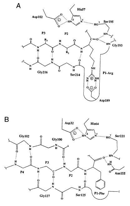
Figure 6.14 The protease subsite nomenclature of Schechter and Berger (1967): residues on the peptide substrate are labeled P1—Pn on the N-terminal side of the scissile bond, and P1 —Pn on the C-terminal side of this bond; the scissile bond is thus between residues P1 and P1 . The corresponding subsites into which these residues fit in the enzyme active site are labeled S1—Sn and S1 —Sn .
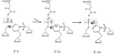
195 (the active site nucleophile) and the side chain of Asn 155. In the chymotrypsin-like enzymes, these H bonds are provided by the backbone nitrogens of the nucleophilic serine residue and an active site glycine, while in the serine carboxypeptidases these bonds are formed by the backbone nitrogens of a tyrosine and a glycine in the active site. Crystallographic data from studies of subtilisin indicate that a weak hydrogen bond exists between Asn 155 and the substrate in the ES complex, and that this H-bonding is significantly strengthened in the transition state (an example of conformational alterations in the enzyme active site that facilitate transition state stabilization). Mutation of Asn 155 to any of a variety of non-hydrogen-bonding amino acids significantly decreases the rate of the enzymatic reaction, as would be expected from our discussion of enzymatic rate enhancement by transition state stabilization.
The transition state then decays as a proton is donated from the active site histidine to the amine group of P1 , followed by dissociation of the first product of the reaction, the peptide starting at P1 , and simultaneous formation of a covalent intermediate with an acyl group (from P1) bound to the active site serine. The enzyme is then deacylated by nucleophilic attack by a water molecule that enters the enzyme active site from the cavity resulting from the departure of the first product peptide. The deacylation reaction proceeds with formation of another tetrahedral transition state, very similar to that formed during the acylation reaction, and engaging the same stabilizing H bonds with
THE SERINE PROTEASES: AN ILLUSTRATIVE EXAMPLE |
181 |
Figure 6.15 Schematic representation of the general acyl transfer mechanism of serine proteases. [Reprinted with permission from Nature (Carter and Wells (1988) 332, 564), copyright (1988) Macmillan Magazines Limited.]
the enzyme. This transition state decays with proton transfer to the active site histidine and release of the second peptide product having the P1 group at its amino terminus.
The active site aspartate residue is a common feature of the serine proteases and has been shown, through mutagenesis studies, to be critical for catalysis. The role of this residue in catalysis is not completely clear. Early studies suggested that together with the serine and histidine residues, this aspartate formed a catalytic ‘‘triad’’ that acts as a proton shuttle. Such a specific interaction requires a precise geometric relationship between the side chains of the aspartate and histidine residues to ensure strong H-bonding. However, the orientation of the aspartate side chain relative to the Ser-His active site residues varies considerably among the three classes of serine protease, making it unlikely that direct proton transfer occurs between the histidine and aspartate side chains in all these enzymes. In fact, some workers have suggested that the catalytic machinery of the serine proteases is most correctly viewed as two distinct catalytic dyads — one comprising the serine and histidine residue and the other comprising the histidine and aspartate residues — rather than as a single catalytic triad (Liao et al., 1992). Regardless of the molecular details, experimental data demonstrate that the presence of the carboxylate anion of the aspartate influences the reactivity of the histidine residues in a way that is critical for catalysis.
The foregoing discussion provides a good example of the interplay between substrate and enzyme active site that must accompany transition state stabilization and reaction rate enhancement. Substrate binding, however, must precede these catalytic steps, and formation of the enzyme—substrate complex is also governed by the stereochemical relationships between groups on the
182 CHEMICAL MECHANISMS IN ENZYME CATALYSIS
substrate and their counterpart subsites in the enzyme active site. For example, the differences in substrate specificity within the chymotrypsin-like and sub- tilisin-like serine proteases can be explained on the basis of their active site structures. In all these enzymes, substrate binding is facilitated by H-bonding to form -pleated sheet structures between residues in the enzyme active site and the P1—P4 residues of the substrate (Figure 6.16). These interactions provide binding affinity for the substrates but do not significantly differentiate one peptide substrate from another. The P1—S1 site interactions appear to play a major role in defining substrate specificity in these enzymes. Subtilisins generally show broad substrate specificity, with a preference for large, hydrophobic groups at the substrate P1 position. The order of preference is approximately Tyr, Phe Leu, Met, Lys His, Ala, Gln, Ser Glu, Gly (Perona and Craik, 1995). The S1 site of the subtilisins exists as a broad, shallow cleft that is formed by two strands of -sheet structure and a loop region of variable size (Figure 6.16B). In a subclass of these enzymes, in which a P1 Lys residue is accommodated, the loop contains a glutamate residue positioned to form a salt bridge with the substrate lysine residue, to neutralize the charge upon substrate binding.
In the chymotrypsin-like enzymes, the S1 pocket consists of a deep cleft into which the substrate P1 residue must fit (Figure 6.16A). The identity of the amino acid residues within this cleft will influence the types of substrate residue that are tolerated at this site. The chymotrypsin-like enzymes can be further subdivided into three subclasses on this basis. Enzymes within the trypsinlike subclass have a conserved aspartate residue at position 189, located at the bottom of the S1 well; this explains the high preference of these enzymes for substrates with arginine or lysine residues at P1. This aspartate is replaced by a serine or small hydrophobic residue in the chymotrypsin and elastase subclasses; hence both subclasses show specificity for nonpolar P1 residues. The other amino acid residues lining the S1 pocket further influence substrate specificity. The elastase subclass contains in this pocket large nonpolar groups that tend to exclude bulky substrate residues. Hence, the elastase subclass favors substrates with small hydrophobic residues at P1. In contrast, the chymotrypsin subclass has small residues in these positions (e.g., glycines), and these enzymes thus favor larger hydrophobic residues, such as tyrosine and phenylalanine at the P1 position of their substrates.
Within this overview of the serine proteases we have observed specific examples of how the active site structure of enzymes (1) engages the substrate and binds it in an appropriate orientation for catalysis (e.g., the H-bonding network developed between the P1—P4 residues of the substrate and the active site residues), (2) stabilizes the transition state to accelerate the reaction rate (e.g., the stabilization of the tetrahedral oxyanionic intermediate through H-bonding interactions), and (3) differentiates between potential substrates on the basis of stereochemical relationships between the substrate and active site subsites. While the molecular details differ from one enzyme to another, the
THE SERINE PROTEASES: AN ILLUSTRATIVE EXAMPLE |
183 |
Figure 6.16 Substrate—active site interactions in the serine proteases. (A) Interactions within the trypsinlike class of serine proteases. (B) Interactions within the subtilisin-like class of serine proteases. [Reprinted with the permission of Cambridge University Press from Perona and
Craik (1995).]

184 CHEMICAL MECHANISMS IN ENZYME CATALYSIS
general types of interaction illustrated with the serine proteases also govern substrate binding and chemical transformations in all the enzymes nature has devised.
6.5 ENZYMATIC REACTION NOMENCLATURE
The hydrolytic activity illustrated by the serine proteases is but one of a wide variety of bond cleavage and bond formation reactions catalyzed by enzymes. From the earliest studies of these proteins, scientists have attempted to categorize them by the nature of the reactions they provide. Group names have been assigned to enzymes that share common reactivities. For example, ‘‘protease’’ and ‘‘proteinase’’ are used to collectively refer to enzymes that hydrolyze peptide bonds. Common names for particular enzymes are not always universally used, however, and their application in individual cases can lead to confusion. For example, there is a metalloproteinase known by the common names stromelysin, MMP-3 (for matrix metalloproteinase number 3), transin, and proteoglycanase. Some workers refer to this enzyme as stromelysin, others call it MMP-3, and still others call it transin or proteoglycanase. A newcomer to the metalloproteinase field could be quite frustrated by this confusing nomenclature. For this reason, the International Union of Pure and Applied Chemistry (IUPAC) formed the Enzyme Commission (EC) to develop a systematic numerical nomenclature for enzymes. While most workers still use common names for the enzymes they are working with, literature references should always include the IUPAC EC designations, which have been universally accepted, to let the reader know precisely what enzymes are being discussed. The EC classifications are based on the reactions that enzymes catalyze. Six general categories have been defined, as summarized in Table 6.4. Within each of these broad categories, the enzymes are further differentiated by a second number that more specifically defines the substrates on which they act. For example, 11 types of hydrolase (category 3) can be defined, as summarized in Table 6.5.
Table 6.4 The IUPAC EC classification of enzymes into six general categories according to the reactions they catalyze
First EC |
|
|
Number |
Enzyme Class |
Reaction |
|
|
|
1 |
Oxidoreductases |
Oxidation—reduction |
2 |
Transferases |
Chemical group transfers |
3 |
Hydrolases |
Hydrolytic bond cleavages |
4 |
Lyases |
Nonhydrolytic bond cleavages |
5 |
Isomerases |
Changes in arrangements of atoms in molecules |
6 |
Ligases |
Joining together of two or more molecules |
|
|
|

|
ENZYMATIC REACTION NOMENCLATURE 185 |
Table 6.5 The IUPAC EC subclassifications of the hydrolases |
|
|
|
First Two |
|
EC Numbers |
Substrates |
|
|
3.1 |
Esters, —C(O)—O- - - R, or with S or P replacing C, or |
|
—C(O)—S - - - R |
3.2 |
Glycosyl, sugar—C—O - - - R, or with N or S replacing O |
3.3 |
Ether, R—O - - - R, or with S replacing O |
3.4 |
Peptides, C - - - N |
3.5 |
Nonpeptides C - - - N |
3.6 |
Acid anhydrides, R—C(O)—O - - - C(O)—R |
3.7 |
C - - - C |
3.8 |
Halides (X), C - - - X, or with P replacing C |
3.9 |
P - - - N |
3.10 |
S - - - N |
3.11 |
C - - - P |
Hydrolyzed bonds shown as dashed lines.
Individual enzymes in each subclass are further defined by a third and a fourth number. In this way any particular enzyme can be uniquely identified. Examples of the common names for some enzymes, and their EC designations are given in Table 6.6. (The enzymes selected have served as illustrative examples in my laboratory.)
The detailed rules for assigning an EC number to a newly discovered enzyme were set forth in Volume 13 of the series Comprehensive Biochemistry (Florkin and Stotz, 1973); an updated version of the nomenclature system was published nearly 20 years later (Webb, 1992). Most of the enzymes the reader is likely to encounter or work with already have EC numbers. One can often obtain the EC designation directly from the literature pertaining to the enzyme of interest. Another useful source for this information is the Medical Subject Headings Supplementary Chemical Records, published by the National Library of Medicine (U.S. Department of Health and Human Services, Bethesda,
Table 6.6 Some examples of enzyme common names and their EC designations
Common Name(s) |
EC Designation |
|
|
Cytochrome oxidases (cytochrome c oxidase) |
EC 1.9.3.1 |
Prostaglandin G/H synthase (cyclooxygenase) |
EC 1.14.99.1 |
Stromelysin (MMP-3, proteoglycanase) |
EC 3.4.24.17 |
Dihydroorotate dehydrogenase |
ED 1.3.99.11 |
Rhodopsin kinase |
EC 2.7.1.125 |
|
|
186 CHEMICAL MECHANISMS IN ENZYME CATALYSIS
MD). This volume lists the common names of chemicals and reagents (including enzymes) that are referred to in the medical literature covered by the Index Medicus (a source book for literature searching of medically related subjects). Enzymes are listed here under their common names (with cross-references for enzymes having more than one common name) and the EC designation is provided for each. Most college and university libraries carry the Index Medicus and will have this supplement available, or one can purchase the supplement directly from the National Library of Medicine. Yet another resource for determining the EC designation of an enzyme is the Enzyme Data Bank, which can be accessed on the Internet.* This data bank provides EC numbers, recommended names, alternative names, catalytic activities, information on cofactor utilization, and associated diseases for a very large collection of enzymes. A complete description of the data bank and its uses can be found in Bairoch (1993).
6.6 SUMMARY
In this chapter we have explored the chemical nature of enzyme catalysis. We have seen that enzymes function by enhancing the rates of chemical reaction by lowering the energy barrier to attainment of the reaction transition state. The active site of enzymes provides the structural basis for this transition state stabilization through a number of chemical mechanisms, including approximation effects, covalent catalysis, general acid/base catalysis, induced strain, and solvent replacement effects. The structural architecture of the enzyme active site further dictates the substrate specificity for reaction. A structural complementarity exists between the enzyme active site and the substrate in its transition state configuration. Several models have been presented to describe this structural complementarity and the interplay of structural forces that dictate enzyme specificity and catalytic efficiency.
REFERENCES AND FURTHER READING
Bairoch, A. (1993) Nucl. Acid. Res. 21, 3155.
Bender, M. L., Bergeron, R. J., and Komiyama, M. (1984) T he Bioorganic Chemistry of Enzymatic Catalysis, Wiley, New York.
Bruice, T. C., and Lapinski, R. (1958) J. Am. Chem. Soc. 80, 2265. Cannon, W. R., and Benkovic, S. J. (1998) J. Biol. Chem. 273, 26257. Carter, P., and Wells, J. A. (1988) Nature, 332, 564.
Fersht, A. R. (1974) Proc. R. Soc. L ondon B, 187, 397.
Fersht, A. (1985) Enzyme Structure and Mechanism, Freeman, New York. Fersht, A. R., and Kirby, A. J. (1967) J. Am. Chem. Soc. 89, 4853, 4857.
*http://192.239.77.6/Dan/proteins/ec-enzyme.html.
REFERENCES AND FURTHER READING |
187 |
Fischer, E. (1894) Berichte, 27, 2985.
Florkin, M., and Stotz, E. H. (1973) Comprehensive Biochemistry, Vol. 13, Elsevier, New
York.
Goldsmith, J. O., and Kuo, L. C. (1993) J. Biol. Chem. 268, 18481.
Hammes, G. G. (1982) Enzyme Catalysis and Regulation, Academic Press, New York. Hartley, B. S., and Kilby, B. A. (1954) Biochem. J. 56, 288.
Jencks, W. P. (1969) Catalysis in Chemistry and Enzymology, McGraw-Hill, New York. Jencks, W. P. (1975) Adv. Enzymol. 43, 219.
Kirby, A. J. (1980) Effective molarity for intramolecular reactions, in Advances in Physical Organic Chemistry, Vol. 17, V. Gold and D. Bethel, Eds., Academic Press, New York, pp. 183 ff.
Koshland, D. E. (1958) Proc. Natl. Acad. Sci. USA 44, 98.
Leatherbarrow, R. J., Fersht, A. R., and Winter, G. (1985) Proc. Natl. Acad. Sci. USA, 82, 7840.
Lerner, R. A., Benkovic, S. J., and Schultz, P. G. (1991) Science, 252, 659.
Liao, D., Breddam, K., Sweet, R. M., Bullock, T., and Remington, S. J. (1992)
Biochemistry, 31, 9796.
Loewus, F., Westheimer, F., and Vennesland, B. (1953) J. Am. Chem. Soc. 75, 5018. Menger, F. M. (1992) Biochemistry, 31, 5368.
Murphy, D. J., and Benkovic, S. J. (1989) Biochemistry, 28, 3025.
Pauling, L. (1948) Nature, 161, 707.
Perona, J. J., and Craik, C. S. (1995) Protein Sci. 4, 337.
Schechter, I., and Berger, A. (1967) Biochem. Biophys. Res. Commun. 27, 157. Schowen, R. L. (1978) in Transition States of Biochemical Processes (Grandous, R. D.
and Schowen, R. L., Eds.), Chapter 2, Plenum, New York. Segal, I. H. (1975) Enzyme Kinetics, Wiley, New York.
So, O.-Y., Scarafia, L. E., Mak, A. Y., Callan, O. H., and Swinney, D. C. (1998) J. Biol. Chem. 273, 5801.
Storm, D. R., and Koshland, D. E. (1970) Proc. Natl. Acad. Sci. USA 66, 445. Walsh, C. (1979) Enzyme Reaction Mechanisms, Freeman, New York.
Webb, E. C. (1992) Enzyme Nomenclature, Academic Press, San Diego, CA. Wison, C., and Agard, D. A. (1991) Curr. Opin. Struct. Biol. 1, 617. Wolfenden, R. (1999) Bioorg. Med. Chem. 7, 647.
Yagisawa, S. (1995) Biochem. J. 308, 305.

Enzymes: A Practical Introduction to Structure, Mechanism, and Data Analysis.
Robert A. Copeland Copyright 2000 by Wiley-VCH, Inc.
ISBNs: 0-471-35929-7 (Hardback); 0-471-22063-9 (Electronic)
7
EXPERIMENTAL
MEASURES OF
ENZYME ACTIVITY
Enzyme kinetics offers a wealth of information on the mechanisms of enzyme catalysis and on the interactions of enzymes with ligands, such as substrates and inhibitors. Chapter 5 provided the basis for determining the kinetic constants k and K from initial velocity measurements taken at varying substrate concentrations during steady state catalysis. The determination of these kinetic constants rests on the ability to measure accurately the initial velocity of an enzymatic reaction under well-controlled conditions.
In this chapter we describe some of the experimental methods used to determine reaction velocities. We shall see that numerous strategies have been developed for following over time the loss of substrate or the appearance of products that result from enzyme turnover. The velocity of an enzymatic reaction is sensitive to many solution conditions, such as pH, temperature, and solvent isotopic composition; these conditions must be well controlled if meaningful data are to be obtained. Controlled changes in these solution conditions and measurement of their effects on the reaction velocity can provide useful information about the mechanism of catalysis as well. Like all proteins, enzymes are sensitive to storage conditions and can be denatured easily by mishandling. Therefore we also discuss methods for the proper handling of enzymes to ensure their maximum catalytic activity and stability.
7.1 INITIAL VELOCITY MEASUREMENTS
7.1.1 Direct, Indirect, and Coupled Assays
To measure the velocity of a reaction, it is necessary to follow a signal that reports product formation or substrate depletion over time. The type of signal that is followed varies from assay to assay but usually relies on some unique
188
INITIAL VELOCITY MEASUREMENTS |
189 |
physicochemical property of the substrate or product, and/or the analyst’s ability to separate the substrate from the product. Generally, most enzyme assays rely on one or more of the following broad classes of detection and separation methods to follow the course of the reaction:
Spectroscopy
Polarography
Radioactive decay
Electrophoretic separation
Chromatographic separation
Immunological reactivity
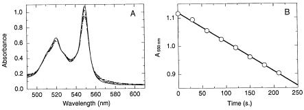
These methods can be used in direct assay: the direct measurement of the substrate or product concentration as a function of time. For example, the enzyme cytochrome c oxidase catalyzes the oxidation of the heme-containing protein cytochrome c. In its reduced (ferrous iron) form, cytochrome c displays a strong absorption band at 550 nm, which is significantly diminished in intensity when the heme iron is oxidized (ferric form) by the oxidase. One can thus measure the change in light absorption at 550 nm for a solution of ferrous cytochrome c as a function of time after addition of cytochrome c oxidase; the diminution of absorption at 550 nm that is observed is a direct measure of the loss of substrate (ferrous cytochrome c) concentration (Figure 7.1).
In some cases the substrate and product of an enzymatic reaction do not provide a distinct signal for convenient measurement of their concentrations. Often, however, product generation can be coupled to another, nonenzymatic,
Figure 7.1 (A) Absorption of ferrocytochrome c as a function of time after addition of the enzyme cytochrome c oxidase. As the cytochrome c iron is oxidized by the enzyme, the absorption feature at 550 nm decreases. (B) Plot of the absorption at 550 nm for the spectra in
(A), as a function of time. Note that in this early stage of the reaction ( 10% of the substrate has been converted), the plot yields a linear relationship between absorption and time. The reaction velocity can thus be determined from the slope of this linear function.