

Enzymes (Second Edition)
.pdf140 KINETICS OF SINGLE-SUBSTRATE ENZYME REACTIONS
Figure 5.15 Effects of positive cooperativity on the kinetics of an enzyme-catalyzed reaction:
(A) data graphed as a Michaelis—Menten (i.e., direct) plot and (B) data from (A) replotted as a Lineweaver—Burk double-reciprocal plot.
Thus, a plot of log(v/(V v)) as a function of log[S] should yield a straight
line with slope of h and a y intercept of log(K ), as illustrated in Figure 5.16. The utility of such plots is limited, however, by the need to know V a priori and because the linear relationship described by Equation 5.48 holds over only a limited range of substrate concentrations (in the region of [S] K ). Hence, whenever possible, it is best to determine V , h, and K for cooperative enzymes from direct nonlinear curve fits to Equation 5.47.
Figure 5.16 Hill plots for the data from Figure 5.15: log[v/(Vmax v)] is plotted as a function of log[S]. The slope of the best fit line provides an estimated of the Hill coefficient h, and the y intercept provides an estimate of log(K ).
TRANSIENT STATE KINETIC MEASUREMENTS |
141 |
These examples illustrate the more commonly encountered deviations from hyperbolic kinetics. A number of other causes of deviations are known, but they are less common. A more comprehensive discussion of such deviations can be found in the texts by Segel (1975) and Bell and Bell (1988).
5.10 TRANSIENT STATE KINETIC MEASUREMENTS
Much of the enzymology literature describes studies of the steady state kinetics of enzymatic reactions, and a great deal of biochemical insight can be derived from such studies. Steady state kinetics, however, does have some limitations. The steady state kinetic constants k and K are complex functions that combined rate constants from multiple steps in the overall enzymatic reaction. Hence, these constants do not provide rate information on any individual steps in the reaction pathway. For example, let us again consider a simple-single substrate enzymatic reaction (as discussed above), but this time let us define the individual rate constants for each step:
|
|
|
|
E S ES EX EP E P |
|||
|
|
|
|
In this scheme, EX represents some transient intermediate in the enzymatic reaction. This could be a distinct chemical species (e.g., an acyl—enzyme intermediate as in peptide hydrolysis by serine proteases; see Chapter 6) or a kinetically significant conformational state that is formed by a change in structure of the ES complex prior to the chemical steps of catalysis. A steady state kinetic study would define the overall reaction in terms of k and K but would not provide much information on the rates and nature of the individual steps in the reaction. Steady state kinetics also limits the mechanistic detail that one can derive. For example, in Chapter 11 we shall see how steady state kinetic measurements can define the order of substrate binding and product release for multisubstrate enzymes. These studies do not, however, give information on the specific reactions of the various enzyme species involved in catalysis, nor can they identify the rate-limiting step. The steady state kinetic constant K is often confused with the dissociation constant for the ES complex (K ), and k is often mistakenly thought of as a specific rate constant for a rate-limiting step in catalysis (i.e., k ); we have already discussed the correct interpretation of K and k .
To overcome some of these limitations, researchers turn to rapid kinetic methods that allow them to make measurements on a time scale (i.e., milliseconds) consistent with the approach to steady state (pre—steady state kinetics) and to measure the kinetics of transient species that occur after initial substrate binding. These methods are collectively referred to as transient state kinetics, and their application to enzymatic systems provides much richer kinetic detail than simple steady state measurements (Johnson, 1992).
142 KINETICS OF SINGLE-SUBSTRATE ENZYME REACTIONS
Specialized apparatus must be used to measure kinetic events on a millisecond time scale. A variety of instruments have been designed for this purpose (Fersht, 1985), but the two most commonly used methods for measuring transient kinetics are stopped-flow and rapid reaction quenching.
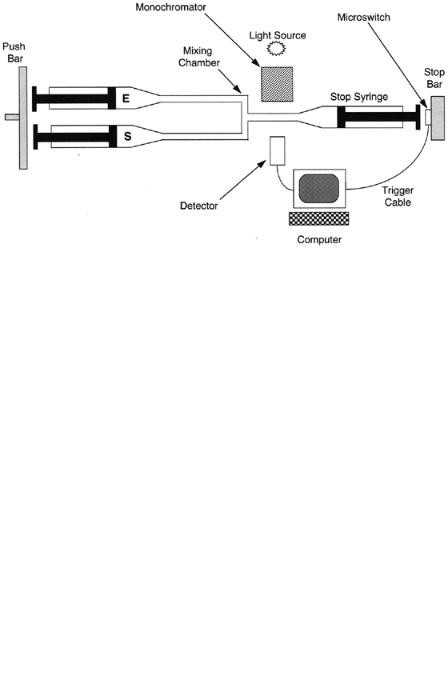
In a stopped-flow experiment, the researcher is measuring the formation of a transient species by detecting a unique optical signal (typically absorbance or fluorescence). Figure 5.17 illustrates a typical design for a stopped-flow apparatus. The instrument consists of two syringes, one holding a enzyme solution and the other holding a substrate solution. Both syringes are attached to a common drive bar that compresses the plungers of both syringes at a steady and common rate, forcing the solutions from each syringe to mix in the mixing chamber and flow through the detection tube. A third syringe is located at the end of the detection tube. As liquid is forced into this third syringe, its plunger is pushed back until it is stopped by contact with a stopping bar. The stopping bar has attached to it a microswitch, which triggers the controlling computer to initiate observation of the optical signal from the solution trapped in the detection tube. Measurements of the optical signal are then made over time as the solution ages in the detection tube.
In a rapid quench apparatus (Figure 5.18), three syringes are compressed by a common driving bar. The first two syringes contain enzyme and substrate solutions, respectively. These solutions flow into the first mixing chamber and then through a reaction aging tube to the second mixing chamber. The length of reaction time is controlled by the length of the reaction aging tube, or by the rate of flow through this tube. In the second mixing chamber, the reaction mixture is combined with a third solution containing the quenching material, which rapidly stops (or quenches) the reaction. The quenching solution must be able to instantaneously stop the reaction by denaturing the enzyme or sequestering a critical cofactor or other component of the reaction mixture. For example, strong acids are commonly used to quench enzyme reactions in this way. Also, enzymes that rely on divalent metal ions as necessary cofactors can be effectively quenched by mixing with EDTA (ethylenediaminetetraacetic acid) or other chelators. Once the reaction has been quenched, the mixed solution flows into a collection container, from which it can be retrieved by the scientist. Detection is performed off-line by any convenient method, including spectroscopy, radiometric chromatography (including thin-layer chromatography), or electrophoretic separation of substrates and products (see Chapter 7 for details).
With instruments like stopped-flow and rapid quench apparatus, one can measure the formation of products or intermediates on a millisecond time scale. A number of kinetic schemes can be studied by these rapid kinetic techniques. We describe two common situations. The first is simple, reversible association of the subtrate and enzyme to form the ES complex:
E S ES
If the experiment is performed under conditions where [S] [E], formation of
TRANSIENT STATE KINETIC MEASUREMENTS |
143 |
Figure 5.17 Schematic diagram of a typical stopped-flow instrument for rapid kinetic measurements.
the ES complex will follow pseudo-first-order kinetics and will be equivalent to the approach to equilibrium for receptor—ligand complexes, as discussed in Chapter 4. Hence, the observed rate of formation k will depend on substrate concentration as follows:
k k [S] k |
(5.49) |
A plot of k as a function of [S] will be linear with y intercept of k and slope of k , as illustrated earlier (Figure 4.2).
In the second situation, formation of an intermediate species EX is rate limiting. Here initial substrate binding comes to equilibrium on a time scale
Figure 5.18 Schematic diagram of a typical rapid quench instrument for rapid kinetic measurements.

144 KINETICS OF SINGLE-SUBSTRATE ENZYME REACTIONS
much faster than the subsequent first-order ‘‘isomerization’’ step:
|
|
E S ES EX |
|
|
|
Three forms of the enzyme appear in this reaction scheme, the free enzyme E , the ES complex, and the intermediate EX. The time dependence for each of these can be derived, yielding the following equations for the mole fraction of each species (Johnson, 1992):
|
[E] |
|
|
1 (K [S] K K [S]) |
|
|
|
||||||||
|
|
|
|
|
|
|
|
|
|
|
|
(1 |
e |
) |
(5.50) |
|
[E] |
|
|
|
|
|
|
|
|
|
|||||
|
|
|
|
|
[ES] |
K [S] |
(1 e ) |
|
|
((5.51) |
|||||
|
|
|
|
|
|
|
|
|
|
|
|||||
|
|
|
|
|
[E] |
|
|
|
|||||||
|
|
|
[EX] |
K K [S] |
|
|
|
||||||||
|
|
|
|
|
|
|
|
|
(1 e ) |
|
|
(5.52) |
|||
where |
|
|
[E] |
|
|
|
|
|
|||||||
|
|
|
|
|
|
|
|
|
|
|
|
|
|
||
|
|
1 K [S] K K [S] |
|
|
(5.53) |
||||||||||
The preexponential term in Equations 5.50—5.52 represents in each case an amplitude term that corresponds to the concentration of that enzyme species at equilibrium. The observed rate constant for formation of the EX complex,, follows a hyperbolic dependence on substrate concentration, similar to velocity in the Michaelis—Menten equation:
|
K k [S] |
|
k |
(5.54) |
|
K [S] 1 |
|||||
|
|
|
|||
Three distinctions between Equations 5.54 and 5.24 can be made. First, is a hyperbolic function of the true dissociation constant for the ES complex (i.e., K 1/K ), not of the kinetic constant K . Second, the maximal rate observed is equal to the sum of k k . Third, the y intercept is nonzero in this case, and equal the rate constant k . Thus, from fitting of the rapid kinetic data to Equation 5.54, one can simultaneously determine the values of K , k , and k .
The application of transient kinetics to the study of enzymatic reactions, and more generally to protein—ligand binding events, is widespread throughout the biochemical literature. The reader should be aware of the power of these methods for determining individual rate constants and of the value of such information for the development of detailed mechanistic models of catalytic turnover. Because of space limitations, and because these methods require specialized equipment that beginners may not have at their disposal, we shall suspend further discussion of these methods. Several noteworthy reviews on the methods of transient kinetics (Gibson, 1969; Johnson, 1992; Fierke and Hammes, 1995) are highly recommended to the reader who is interested in learning more about these techniques.
REFERENCES AND FURTHER READING |
145 |
5.11 SUMMARY
This chapter focused on steady state kinetic measurements, since these are easiest to perform in a standard laboratory. These methods provide important kinetic and mechanistic information, mainly in the form of two kinetic constants, k and K . Graphical methods for determining the values for these kinetic constants were presented. We also briefly discussed the application of rapid kinetic techniques to the study of enzymatic reactions. These methods provide even more detailed information on the individual rate constants for different steps in the reaction sequence, but they require more specialized instrumentation and analysis methods. The chapter provided references to more advanced treatments of rapid kinetic methods to aid the interested reader in learning more about these powerful techniques.
REFERENCES AND FURTHER READING
Bell, J. E., and Bell, E. T. (1988) Proteins and Enzymes, Prentice-Hall, Englewood Cliffs, NJ.
Briggs, G. E., and Haldane, J. B. S. (1925) Biochem. J. 19, 383.
Brown, A. J. (1902) J. Chem. Soc. 81, 373.
Chapman, K. T., Kopka, I. E., Durette, P. I., Esser, C. K., Lanza, T. J., IzquierdoMartin, M., Niedzwiecki, L., Chang, B., Harrison, R. K., Kuo, D. W., Lin, T.-Y., Stein, R. L., and Hagmann, W. K. (1993) J. Med. Chem. 36, 4293.
Cleland, W. W. (1967) Adv. Enzymol. 29 1—65.
Copeland, R. A. (1991) Proc. Natl. Acad. Sci. USA 88, 7281.
Cornish-Bowden, A., and Wharton, C. W. (1988) Enzyme Kinetics, IRL Press, Oxford. Eisenthal, R., and Cornish-Bowden, A. (1974) Biochem. J. 139, 715.
Fersht, A. (1985) Enzyme Structure and Mechanism, Freeman, New York. Fierke, C. A., and Hammes, G. G. (1995) Methods Enzymol. 249, 3—37. Gibson, Q. H. (1969) Methods Enzymol. 16, 187.
Henri, V. (1903) L ois Ge´ ne´ rales de l’action des diastases, Hermann, Paris. Johnson, K. A. (1992) Enzymes, XX, 1—61.
Lineweaver, H., and Burk, J. (1934) J. Am. Chem. Soc. 56, 658.
Michaelis, L., and Menten, M. L. (1913) Biochem. Z. 49 333.
Schulz, A. R. (1994) Enzyme Kinetics from Diastase to Multi-enzyme Systems, Cambridge University Press, New York.
Segel, I. H. (1975) Enzyme Kinetics, Wiley, New York.
Wahl, R. C. (1994) Anal. Biochem. 219, 383.
Wilkinson, A. J., Fersht, A. R., Blow, D. M., and Winter, G. (1983) Biochemistry, 22, 3581.

Enzymes: A Practical Introduction to Structure, Mechanism, and Data Analysis.
Robert A. Copeland Copyright 2000 by Wiley-VCH, Inc.
ISBNs: 0-471-35929-7 (Hardback); 0-471-22063-9 (Electronic)
6
CHEMICAL MECHANISMS IN ENZYME CATALYSIS
The essential role of enzymes in almost all physiological processes stems from two key features of enzymatic catalysis: (1) enzymes greatly accelerate the rates of chemical reactions; and (2) enzymes act on specific molecules, referred to as substrates, to produce specific reaction products. Together these properties of rate acceleration and substrate specificity afford enzymes the ability to perform the chemical conversions of metabolism with the efficiency and fidelity required for life. In this chapter we shall see that both substrate specificity and rate acceleration result from the precise three-dimensional structure of the substrate binding pocket within the enzyme molecule, known as the active site. Enzymes are (almost always) proteins, hence the chemically reactive groups that act upon the substrate are derived mainly from the natural amino acids. The identity and arrangement of these amino acids within the enzyme active site define the active site topology with respect to stereochemistry, hydrophobicity, and electrostatic character. Together these properties define what molecules may bind in the active site and undergo catalysis. The active site structure has evolved to bind the substrate molecule in such a way as to induce strains and perturbations that convert the substrate to its transition state structure. This transition state is greatly stabilized when bound to the enzyme; its stability under normal solution conditions is much less. Since attainment of the transition state structure is the main energetic barrier to the progress of any chemical reaction, we shall see that the stabilization of the transition state by enzymes results in significant acceleration of the reaction rate.
146
SUBSTRATE—ACTIVE SITE COMPLEMENTARITY |
147 |
6.1 SUBSTRATE--ACTIVE SITE COMPLEMENTARITY
When a protein and a ligand combine to form a binary complex, the complex must result in a net stabilization of the system relative to the free protein and ligand; otherwise binding would not be thermodynamically favorable. We discussed in Chapter 4 the main forces involved in stabilizing protein—ligand interactions: hydrogen bonding, hydrophobic forces, van der Waals interactions, electrostatic interactions, and so on. All these contribute to the overall binding energy of the complex and must more than compensate for the lose of rotational and translational entropy that accompanies binary complex formation.
These same forces are utilized by enzymes in binding their substrate molecules. It is clear today that formation of an enzyme—substrate binary complex is but the first step in the catalytic process used in enzymatic catalysis. Formation of the initial encounter complex (also referred to as the enzyme— substrate, ES, or Michaelis complex; see Chapter 5) is followed by steps leading sequentially to a stabilized enzyme—transition state complex (ES‡), an enzyme— product complex (EP), and finally dissociation to reform the free enzyme with liberation of product molecules. Initial ES complex formation is defined by the dissociation constant K , which is the quotient of the rate constants k and k (see Chapters 4 and 5). As discussed in Chapter 5, the rates of the chemical steps following ES complex formation are, for simplicity, often collectively described
by a single kinetic constant, k . As we shall see, k is most often limited by the
‡
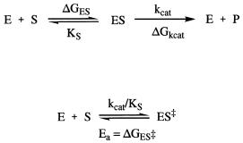
rate of attainment of the transition state species ES . Hence, a minimalist view of enzyme catalysis is captured in the scheme illustrated in Figure 6.1.
To understand the rate enhancement and specificity of enzymatic reactions, we must consider the structure of the reactive center of these molecules, the active site, and its relationship to the structures of the substrate molecule in its ground and transition states in forming the ES and the ES‡ binary complexes. While the active site of every enzyme is unique, some generalizations can be made:
1.The active site of an enzyme is small relative to the total volume of the enzyme.
2. The active site is three-dimensional — that is, amino acids and cofactors in the active site are held in a precise arrangement with respect to one another and with respect to the structure of the substrate molecule. This active site three-dimensional structure is formed as a result of the overall tertiary structure of the protein.
3.In most cases, the initial interactions between the enzyme and the substrate molecule (i.e., the binding events) are noncovalent, making use of hydrogen bonding, electrostatic, hydrophobic interactions, and van der Waals forces to effect binding.
148 CHEMICAL MECHANISMS IN ENZYME CATALYSIS
Figure 6.1 Generic scheme for an enzyme-catalyzed reaction showing the component free energy terms that contribute to the overall activation energy of reaction.
4.The active sites of enzymes usually occur in clefts and crevices in the protein. This design has the effect of excluding bulk solvent (water), which would otherwise reduce the catalytic activity of the enzyme. In other words, the substrate molecule is desolvated upon binding, and shielded from bulk solvent in the enzyme active site. Solvation by water is replaced by the protein.
5.The specificity of substrate utilization depends on the well-defined arrangement of atoms in the enzyme active site that in some way complements the structure of the substrate molecule.
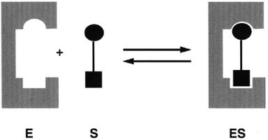
Experimental evidence for the existence of a binary ES complex rapidly accumulated during the late nineteenth and early twentieth centuries. This evidence, some of which was discussed in Chapter 5, was based generally on studies of enzyme stability, enzyme inhibition, and steady state kinetics. During this same time period, scientists began to appreciate the selective utilization of specific substrates that is characteristic of enzyme-catalyzed reactions. This cumulative information led to the general view that substrate specificity was a result of selective binding of substrate molecules by the enzyme at its active site. The selection of particular substrates reflected a structural complementarity between the substrate molecule and the enzyme active site. In the late nineteenth century Emil Fisher formulated these concepts into the lock and key model, as illustrated in Figure 6.2. In this model the enzyme active site and the substrate molecule are viewed as static structures that are stereochemically complementary. The insertion of the substrate into the static enzyme active site is analogous to a key fitting into a lock, or a jigsaw piece fitting into the rest of the puzzle: the best fits occur with the substrates that best complement the structure of the active site; hence these molecules bind most tightly.
Active site—substrate complementarity results from more than just stereochemical fitting of the substrate into the active site. The two structures must also be electrostatically complementary, ensuring that charges are
SUBSTRATE—ACTIVE SITE COMPLEMENTARITY |
149 |
Figure 6.2 Schematic illustration of the lock and key model of enzyme—substrate interactions.
counterbalanced to avoid repulsive effects. Likewise, the two structures must complement each other in the arrangement of hydrophobic and hydrogenbonding interactions to best enhance binding interactions.
Enzyme catalysis is usually stereo-, regio-, and enantiomerically selective. Hence substrate recognition must result from a minimum of three contact points of attachment between the enzyme and the substrate molecule. Consider the example of the alcohol dehydrogenases (Walsh, 1979) that catalyze the transfer of a methylene hydrogen of ethyl alcohol to the carbon at the 4-position of the NAD cofactor, forming NADH and acetaldehyde. Studies in which the methylene hydrogens of ethanol were replaced by deuterium demonstrated that alcohol dehydrogenases exclusively transferred the pro-R hydrogen to NAD (Loewus et al., 1953; Fersht, 1985). This stereospecificity implies that the alcohol bind to the enzyme active site through specific interactions of its methyl, hydroxyl, and pro-R hydrogen groups to form a three-point attachment with the reactive groups within the active site; this concept is illustrated in Figure 6.3. Having anchored down the methyl and hydroxyl groups as depicted in Figure 6.3, the enzyme is committed to the transfer of the specific pro-R hydrogen atom because of its relative proximity to the NAD cofactor. The three-point attachment hypothesis is often invoked to explain the stereospecificity commonly displayed by enzymatic reactions.
The concepts of the lock and key and three-point attachment models help to explain substrate selectivity in enzyme catalysis by invoking a structural complementarity between the enzyme active site and substrate molecule. We have not, however, indicated the form of the substrate molecule to which the enzyme active site shows structural complementarity. Early formulations of these hypotheses occurred before the development of transition state theory (Pauling, 1948), hence viewed the substrate ground state as the relevant configuration. Today, however, there is clear evidence that enzyme active sites