

Enzymes (Second Edition)
.pdf30 CHEMICAL BONDS AND REACTIONS IN BIOCHEMISTRY
Table 2.2 Examples of Bro/nsted--Lowry acids and their conjugate bases
Brønsted—Lowry Acid |
Conjugate Base |
||
|
|
||
H SO (sulfuric acid) |
HSO (bisulfate ion) |
||
|
|
|
|
HCl |
(hydrochloric acid) |
Cl (chloride ion) |
|
H O (hydronium ion) |
H O (water) |
||
|
(ammonium ion) |
|
|
NH |
|
NH (ammonia) |
|
CH COOH (acetic acid) |
CH COO (acetate ion) |
||
|
|
|
|
H O |
|
(water) |
OH (hydroxide ion) |
Table 2.2 gives some examples of Brønsted—Lowry acids and their conjugate bases. For all these pairs, we are dealing with the transfer of a hydrogen ion (proton) from the acid to some other species (often the solvent) to form the conjugate base. A convenient means of measuring the hydrogen ion concentration in aqueous solutions is the pH scale. The term ‘‘pH’’ is a shorthand notation for the negative base-10 logarithm of the hydrogen ion concentration:
pH log[H ] |
(2.8) |
Consider the dissociation of a weak Brønsted—Lowry acid (HA) into a proton (H ) and its conjugate base (A ) in aqueous solution.
HA H A
The dissociation constant for the acid, K , is given by the ratio [H ][A ]/ [HA]. Let us define the pK for this reaction as the negative base-10 logarithm of K :
pK log |
[H ][A ] |
|
[HA] |
(2.9) |
or, using our knowledge of logarithmic relationships, we can write:
pK log(HA) log(A ) log(H ) |
(2.10) |
Note that the last term in Equation 2.10 is identical to our definition of pH (Equation 2.8). Using this equality, and again using our knowledge of logarithmic relationships we obtain:
[HA] |
pH |
|
pK log [A ] |
(2.11) |

ACID--BASE CHEMISTRY |
31 |
or, rearranging (note the inversion of the logarithmic term): |
|
||
|
[A ] |
|
|
pH pK log |
|
|
(2.12) |
[HA] |
|||
Equation 2.12 is known as the Henderson—Hasselbalch equation, and it provides a convenient means of calculating the pH of a solution from the concentrations of a Brønsted—Lowry acid and its conjugate base. Note that when the concentrations of acid and conjugate base are equal, the value of [A ]/[HA] is 1.0, and thus the value of log([A ]/[HA]) is zero. At this point the pH will be exactly equal to the pK . This provides a useful working definition of pK :
The pK is the pH value at which half the Brønsted—Lowry acid is dissociated to its conjugate base and a proton.
Let us consider a simple example of this concept. Suppose that we dissolve acetic acid into water and begin titrating the acid with hydroxide ion equivalents by addition of NaOH. If we measure the pH of the solution after each addition, we will obtain a titration curve similar to that shown in Figure 2.12. Two points should be drawn from this figure. First, such a titration curve provides a convenient means of graphically determining the pK value of the species being titrated. Second, we see that at pH values near the pK , it takes a great deal of NaOH to effect a change in the pH value. This resistance to pH change in the vicinity of the pK of the acid is referred to as buffering capacity, and it is an
Figure 2.12 Hypothetical titration curve for a weak acid illustrating the graphical determination of the acid’s pK .

32 CHEMICAL BONDS AND REACTIONS IN BIOCHEMISTRY
important property to be considered in the preparation of solutions for enzyme studies. As we shall see in Chapter 7, the pH at which an enzyme reaction is performed can have a dramatic effect on the rate of reaction and on the overall stability of the protein. As a rule, therefore, specific buffering molecules, whose pK values match the pH for optimal enzyme activity, are added to enzyme solutions to maintain the solution pH near the pK of the buffer.
2.4 NONCOVALENT INTERACTIONS IN REVERSIBLE BINDING
All the properties of molecules we have discussed until now have led us to focus on the formation, stabilization, and breaking of covalent bonds between atoms of the molecule. These are important aspects of the chemical conversions that are catalyzed at the enzyme active site. Molecules can interact with one another by a number of noncovalent forces as well. These weaker attractive forces are very important in biochemical reactions because they are readily reversible. As we shall see in Chapters 4, 6, and 8, the reversible formation of binary complexes between enzymes and ligand molecules (i.e., substrates and inhibitors) is a critical aspect of both enzymatic catalysis and enzyme inhibition. Four types of noncovalent interaction are particularly important in protein structure (Chapter 3) and enzyme—ligand binding (Chapters 4, 6, and 8); these are electrostatic interactions, hydrogen bonding, hydrophobic interactions, and van der Waals forces. Here we describe these forces briefly. In subsequent chapters we shall see how each force can participate in stabilizing the protein structure of an enzyme and may also play an important role in the binding interactions between enzymes and their substrates and inhibitors.
2.4.1 Electrostatic Interactions
When two oppositely charged groups come into close proximity, they are attracted to one another through a Coulombic attractive force that is described by:
q q |
(2.13) |
F r D |
where q and q are the charges on the two atoms involved, r is the distance between them, and D is the dielectric constant of the medium in which the two atoms come together. Since D appears in the denominator, the attractive force is greatest in low dielectric solvents. Hence electrostatic forces are stronger in the hydrophobic interior of proteins than on the solvent-exposed surface. These attractive interactions are referred to as ionic bonds, salt bridges, and ion pairs.
Equation 2.13 describes the attractive force only. If two atoms, oppositely charged or not, approach each other too closely, a repulsive force between the

NONCOVALENT INTERACTIONS IN REVERSIBLE BINDING |
33 |
outer shell electrons on each atom will come into play. Other factors being constant, it turns out that the balance between these attractive and repulsive forces is such that, on average, the optimal distance between atoms for salt bridge formation is about 2.8 Å (Stryer, 1989).
2.4.2 Hydrogen Bonding
A hydrogen bond (H bond) forms when a hydrogen atom is shared by two electronegative atoms. The atom to which the hydrogen is covalently bonded is referred to as the hydrogen bond donor, and the other atom is referred to as the hydrogen bond acceptor:
N H · · ·O
The donor and acceptors in H bonds are almost exclusively electronegative heteroatoms, and in proteins these are usually oxygen, nitrogen, or sometimes sulfur atoms. Hydrogen bonds are weaker than covalent bonds, varying in bond energy between 2.5 and 8 kcal/mol. The strength of a H bond depends on several factors, but mainly on the length of the bond between the hydrogen and acceptor heteroatom (Table 2.3). For example, NH O hydrogen bonds between amides occur at bond lengths of about 3 Å and are estimated to have bond energies of about 5 kcal/mol. Networks of these bonds can occur in proteins, however, collectively adding great stability to certain structural motifs. We shall see examples of this in Chapter 3 when we discuss protein secondary structure. H-bonding also contributes to the binding energy of ligands to enzyme active sites and can play an important role in the catalytic mechanism of the enzyme.
2.4.3 Hydrophobic Interactions
When a nonpolar molecule is dissolved in a polar solvent, such as water, it disturbs the H-bonding network of the solvent without providing compensat-
Table 2.3 Hydrogen bond lengths for H bonds found in proteins
Bond Type |
Typical Length (Å) |
|
|
O H---O |
2.70 |
O H---O |
2.63 |
O H---N |
2.88 |
N H---O |
3.04 |
N H---O |
2.93 |
N H---N |
3.10 |
|
|
34 CHEMICAL BONDS AND REACTIONS IN BIOCHEMISTRY
ing H-bonding opportunities. Hence there is an entropic cost to the presence of nonpolar molecules in aqueous solutions. Therefore, if such a solution is mixed with a more nonpolar solvent, such as n-octanol, there will be a thermodynamic advantage for the nonpolar molecule to partition into the more nonpolar solvent. The same hydrophobic effect is seen in proteins. For example, amino acids with nonpolar side chains are most commonly found in the core of the folded protein molecule, where they are shielded from the polar solvent. Conversely, amino acids with polar side chains are most commonly found on the exterior surface of the folded protein molecule (see Chapter 3 for further details). Likewise, in the active sites of enzymes hydrophobic regions of the protein tend to stabilize the binding of hydrophobic molecules. The partitioning of hydrophobic molecules from solution to the enzyme active site can be a strong component of the overall binding energy. We shall discuss this further in Chapters 4, 6 and 8 in our examination of enzyme—substrate interactions and reversible enzyme inhibitors.
2.4.4 Van der Waals Forces
The distribution of electrons around an atom is not fixed; rather, the character of the so-called electron cloud fluctuates with time. Through these fluctuations, a transient asymmetry of electron distribution, or dipole moment, can be established. When atoms are close enough together, this asymmetry on one atom can influence the electronic distribution of neighboring atoms. The result is a similar redistribution of electron density in the neighbors, hence an attractive force between the atoms is developed. This attractive force, referred to as a van der Waals bond, is much weaker than either salt bridges or H bonds. Typically a van der Waals bond is worth only about 1 kcal/mol in bond energy. When conditions permit large numbers of van der Waals bonds to simultaneously form, however, their collective attractive forces can provide a significant stabilizing energy to protein—protein and protein—ligand interactions.
As just described, the attractive force between electron clouds increases as the two atoms approach each other but is counterbalanced by a repulsive force at very short distances. The attractive force, being dipolar, depends on the interatomic distance, R, as 1/R . The repulsive force is due to the overlapping of the electron clouds of the individual atoms that would occur at very close distances. This force wanes quickly with distance, showing a 1/R dependence. Hence, the overall potential energy of a van der Waals interaction depends on the distance between nuclei as the sum of these attractive and repulsive forces:
PE |
A |
|
|
B |
|
(2.14) |
R |
|
|||||
|
R |
|
||||
where PE is the potential energy, and A and B can be considered to be characteristic constants for the pair of nuclei involved. From Equation 2.14 we see that the optimal attraction between atoms occurs when they are separated
NONCOVALENT INTERACTIONS IN REVERSIBLE BINDING |
35 |
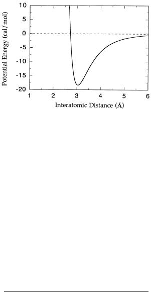
Figure 2.13 Potential energy diagram for the van der Waals attraction between two helium atoms. [Data adapted from Gray (1973) and fit to Equation 2.14.]
by a critical distance known as the van der Waals contact distance (Figure 2.13). The contact distance for a pair of atoms is determined by the individual van der Waals contact radius of each atom, which itself depends on the electronic configuration of the atom.
Table 2.4 provides the van der Waals radii for the most abundant atoms found in proteins. Imagine drawing a sphere around each atom with a radius defined by the van der Waals contact radius (Figure 2.14). These spheres, referred to as van der Waals surfaces, would define the closest contact that atoms in a molecule could make with one another, hence the possibilities for defining atom packing in a molecular structure. Because of the differences in radii, and the interplay between repulsive and attractive forces, van der Waals bonds and surfaces can play an important role in establishing the specificity of interactions between protein binding pockets and ligands. We shall have more to say about such specificity in Chapter 6, when we discuss enzyme active sites.
Table 2.4 Van der Waals radii for atoms in proteins
Atom |
Radius (Å) |
|
|
H |
1.2 |
C |
2.0 |
N |
1.5 |
O |
1.4 |
S |
1.9 |
P |
1.9 |
|
|

36 CHEMICAL BONDS AND REACTIONS IN BIOCHEMISTRY
Figure 2.14 Van der Waals radii for the atoms of the amino acid alanine. The ‘‘tubes’’ represent the bonds between atoms. Oxygen is colored red, nitrogen is blue, carbon is green, and hydrogens are gray. The white dimpled spheres around each atom represent the van der Waals radii. (Diagram courtesy of Karen Rossi, Department of Computer Aided Drug Design, The DuPont Pharmaceuticals Company.) (See Color Plates.)
2.5 RATES OF CHEMICAL REACTIONS
The study of the rates at which chemical reactions occur is termed kinetics. We shall deal with the kinetics of enzyme-catalyzed reactions under steady state conditions in Chapter 5. Here we review basic kinetic principles for simple chemical reactions.
Let us consider a very simple chemical reaction in which a molecule S,
RATES OF CHEMICAL REACTIONS |
37 |
decomposes irreversibly to a product P:
S P
The radioactive decay of tritium to helium is an example of such a chemical reactions:
H He
At the start of the reaction we have some finite amount of S, symbolized by [S] . At any time later, the amount of S remaining will be less than [S] and is symbolized by [S] . The amount of S will decline with time until there is no S left, at which point the reaction will stop. Hence, we expect that the reaction rate (also called reaction velocity) will be proportional to the amount of S present:
v |
d[S] |
k[S] |
(2.15) |
||
dt |
|
||||
|
|
|
|||
where v is the velocity and k is a constant of proportionality referred to at the rate constant. If we integrate this differential equation we obtain:
d[S] k[S]dt |
(2.16) |
Solving this integration we obtain: |
|
[S] [S] exp( kt) |
(2.17) |
Equation 2.17 indicates that substrate concentration will decay exponentially from [S] [S] at t 0 to [S] 0 at infinite time. Over this same time period, the product concentration grows exponentially. At the start of the reaction (t 0) there is no product; hence [P] 0. At infinite time, the maximum amount of product that can be produced is defined by the starting concentration of substrate, [S] ; hence at infinite time [P] [S] . At any time between 0 and infinity, we must have conservation of mass, so that:
[S] [P] [S] |
(2.18) |
or |
|
[P] [S] [S] |
(2.19) |
Using Equation 2.17, we can recast Equation 2.19 as follows: |
|
[P] [S] [S] e |
(2.20) |
or: |
|
[P] [S] (1 e ) |
(2.21) |
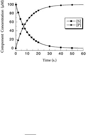
Hence, from Equations 2.17 and 2.21 we expect the concentrations of S and P to respectively decrease and increase exponentially, as illustrated in Figure 2.15.
38 CHEMICAL BONDS AND REACTIONS IN BIOCHEMISTRY
Figure 2.15 Progress curves of product development (circles) and substrate loss (squares) for a first-order reaction.
From Equation 2.17 we could ask the question, How much time is required to reduce the concentration of S to half its original value? To answer this we first rearrange Equation 2.17 as follows:
|
|
|
|
|
|
[S] |
|
|
e |
|
(2.22) |
||||
|
|
|
|
|
[S] |
|
|
||||||||
|
|
|
|
|
|
|
|
|
|
|
|
|
|||
when [S] |
|
is half of [S] |
|
the ratio [S] |
/[S] |
|
is obviously |
. Using this equality |
|||||||
|
|
|
|
|
|
|
|
|
|
|
|
||||
and taking the natural logarithm of both sides and then dividing both sides by |
|||||||||||||||
k, we obtain: |
|
|
|
|
|
|
|
|
|
|
|
|
|
||
|
|
|
|
ln( ) |
|
|
0.6931 |
|
|
(2.23) |
|||||
|
|
|
|
|
|
|
|
|
|
|
t |
|
|||
|
|
|
|
|
k |
|
|
k |
|
||||||
|
|
|
|
|
|
|
|
|
|
|
|
||||
The value t is referred to as the half-life of the reaction. This value is inversely related to the rate constant, but it provides a value in units of time that some people find easier to relate to. It is not uncommon, for example, for researchers to discuss radioactive decay in terms of isotope half-lives (Table 7.4 in Chapter 7 provides half-lives for four of the radioisotopes commonly used in enzyme studies).
2.5.1 Reaction Order
In the discussion above we considered the simplest of kinetic processes in which there was only one reactant and one product. From the rate equation, 2.15, we see that the velocity for this reaction depends linearly on initial reactant concentration. A reaction of this type is said to be a first-order reaction, and the rate constant, k, for the reaction is said to be a first-order rate

RATES OF CHEMICAL REACTIONS |
39 |
Table 2.5 Reaction order for a few simple chemical reactions
Order |
Reaction |
Rate Equation |
|
|
|
|
|
1 |
A P |
v k[A] |
|
2 |
2A |
P |
v k[A] |
2 |
A B |
P |
v k[A][B] |
|
|
|
|
constant. Suppose that the form of our reaction was that two molecules of reactant A produced one molecule of product P:
2A P
If we now solve for the rate equation, we will find that it has the form:
v k[A] |
(2.24) |
A reaction of this type would be said to be a second-order reaction. Generally, the order of a chemical reaction is the sum of the exponent terms to which reactant concentrations are raised in the velocity equation. A few examples of this are given in Table 2.5. A more comprehensive discussion of chemical reaction order and rate equations can be found in any good physical chemistry text (e.g., Atkins, 1978).
As we have just seen, reactions involving two reactants, such as A B P, are strictly speaking always second order. Often, however, the reaction can be made to appear to be first order in one reactant when the second reactant is held at a constant, excess concentration. Under such conditions the reaction is said to be pseudo—first order with respect to the nonsaturating reactant. Such reactions appear kinetically to be first order and can be well described by a first-order rate equation. As we shall see in Chapters 4 and 5, under most experimental conditions the rate of ligand binding to receptors and the rates of enzyme-catalyzed reactions are most often pseudo—first order.
2.5.2 Reversible Chemical Reactions
Suppose that our simple chemical reaction S P is reversible so that there is some rate of the forward reaction of S going to P, defined by rate constant k , and also some rate of the reverse reaction of P going to S defined by the rate constant k . Because of the reverse reaction, the reactant S is never completely converted to product. Instead, an equilibrium concentration of both S and P is established after sufficient time. The equilibrium constant for the forward reaction is given by:
K |
[P] |
|
k |
(2.25) |
|||
|
|
|
|
||||
[S] |
|
k |
|||||
|
|
|
|||||