Gastrointestinal Tract Pathology 1 7
ESOPHAGUS
1. Congenital and mechanical disorders
a.Tracheoesophageal fistula is a congenital connection between the esophagus and trachea that is often associated with esophageal atresia. It is often discovered soon after birth because of aspiration.
b.Esophageal webs are web-like protrusions of the esophageal mucosa into the lumen which typically present with dysphagia. Plummer-Vinson syndrome is a disease of middle-aged women characterized by esopha geal webs, iron deficiency anemia, and increased risk of carcinoma. Schatzki rings are web-like narrowings at the gastroesophageal junction.
c.Achalasia is a failure of the lower esophageal sphincter (LES) to relax with swallowing. The etiology is unknown in most cases; in South America, achalasia may be caused by Chagas disease.
i.Clinically,the presentation is withprogressive dysphagia. The esoph agus is characteristically dilated proximal to the lower esophageal sphincter; barium swallowshows a "bird-beak" sign. Microscopically, there is a loss of ganglion cells in the myenteric plexus. Treatment is with lower esophageal sphincter balloon dilation or myotomy. There is an increased risk of esophageal carcinoma.
2.Hematemesis and esophageal bleeding
a.Mallory-Weiss syndrome has linear lacerations at the gastroesophageal junction that are produced by severe prolonged vomiting; the most common cause is acute alcohol ingestion and/or chronic alcoholism. Mallory-Weiss syndrome presents with hematemesis; it can be rarely complicated by esophageal rupture (Boerhaave syndrome).
b.Esophageal varices are dilated submucosal veins in the lower third of the esophagus, usually secondary to portal hypertension. The most common cause is cirrhosis. Clinically, the presentation is asymptomat ic, though there is massive hematemesis when the varices are ruptured. Complications include potentially fatal hemorrhage. Treatment is gen erally with band ligation, sclerotherapy, or balloon tamponade.
3.Esophagitis
a. Gastroesophageal reflux disease (reflux esophagitis) is esophageal irritation and inflammation due to reflux of gastric secretions into the esophagus. Reflux typically presents with heartburn and regurgitation. Complications include bleeding; stricture; bronchospasm and asthma; and Barrett esophagus.
b.Barrettesophagus is a metaplasia of the squamous esophageal mucosa to a more protective columnar type (intestinal metaplasia). It occurs because of chronic exposure to gastric secretions, usually in the setting of gastroesophageal reflux disease (GERD). The incidence of Barrett esophagus is increasing, and there is an increased risk of dysplasia and esophageal adenocarcinoma.
Clinical Correlate
The most common type of tracheoesophageal fistula:
Tracheoesophageal fistula
Clinical Correlate
Chagas disease: This tropical parasitic disease, common in South America, is caused by Trypanosoma cruzi and is transmitted by reduviid or "kissing" bugs. Chagas disease can cause:
•Romaiia's sign (swelling ofthe eyelids)
•Cardiomyopathy
•Megaesophagus and megacolon
In a Nutshell
Mallory-Weiss tears vs. esophageal varices:
Although both are associated with alcohol abuse and can presentwith hematemesis, Mallory-Weiss tears typically occur acutely as a result of retching/vomiting. Esophagealvarices result from portal hypertension and will usually present with a more significant bleeding episode.
m. Pathology.
Chapter i.7 • Gastrointestinal Tract Pathology
endocrine neoplasia (MEN) type I and Zollinger-Ellison syndromes; cirrhosis; and chronic obstructive pulmonary disease.
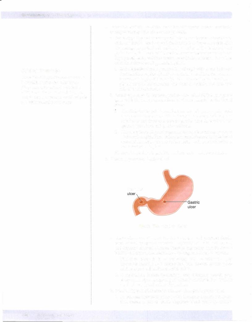
u. Clinically,most duodenal peptic ulcers are located inthe anterior wall of the proximal duodenum. The classic presentation is with burning epigastric pain 1 to 3 hours after eating, which is relieved by food.
c. Gastric peptic ulcer is associated with H. pylori (75%). The ulcers are typically located in the lesser curvature of the antrum. Grossly, these are small (<3 cm), sharply demarcated ('punched out'), solitary ulcers with round or oval shape, overhanging margins, and radiating mucosal folds. The classic presentation is with burning epigastric pain, which worsens with eating.
6. Gastric carcinoma (malignant ulcer)
a. Epidemiology. Gastric carcinoma is more common in Japan than in the United States, and has a decreasing incidence in the United States.
i.Dietaryfactors can be riskfactors,and include smoked fish and meats,
pickled vegetables, nitrosamines, benzpyrene, and decreased intake of fruits and vegetables. Other risk factors include H. pylori infection, chronicatrophicgastritis, smoking, blood type A, bacterial overgrowth in the stomach, prior subtotal gastrectomy, and Menetrier disease.
u.Clinically, gastric carcinoma is often (90%) asymptomatic until late
in the course, when it can produce weight loss and anorexia. It can also present with epigastric abdominal pain mimicking a peptic ulcer, early satiety, and occult bleeding with iron deficiency anemia.
Gastric carcinoma is most commonly located in the lesser curvature ofthe antrum, and causes a large (>3 cm), irregular ulcer with heaped-up margins and a necrotic ulcer base. It may also occur as a flat or polypoid mass. Several histological types occur.
iv.The intestinalhistologictype microscopically shows gland-forming adenocarcinoma.
v.The diffuse type of gastric carcinoma shows diffuse infiltration of stomach by poorly differentiated tumor cells, frequently numer ous signet-ring cells (whose nuclei are displaced to the periphery by intracellular mucin), and linitis plastica (thickened "leather bottle"-like stomach) gross appearance.
vi.Gastric carcinomas may specifically metastasize to the left supra clavicular lymph node (Virchow sentinel node) and to the ovary (Krukenberg tumor).
vii.Diagnosis is by endoscopy with biopsy; treatment is with gastrec tomy. The prognosis is poor, with overall 5-year survival of 20%.
SMALL AND LARGE INTESTINES
1. Mechanical obstruction
a. Volvulus is a twisting of a segment of bowel on its vascular mesentery, resulting in intestinal obstruction and infarction. Volvulus is often associated with congenital abnormalities such as intestinal malrotation. Common locations include the sigmoid colon and small bowel; compli cations include infarction and peritonitis.
b. Intussusception is the telescoping of a proximal segment of the bowel into the distal segment. It is most common in infants and children. In adults, intussusception may be associated with a mass or tumor; it presents with intestinal obstruction, abdominal pain, and "currant-jelly" stools. The intussuscepted segment can become infarcted.
MEDICAL 167
Chapter 17 • Gastrointestinal Tract Pathology
c.Whipple disease is a rare infectious disease involving many organs, including small intestines, joints, lung, heart, liver, spleen, and central nervous system. Whipple disease typicallyaffects Caucasian males aged
30 to 50.
i.The infecting organism is Tropheryma whipplei, andmicroscopically, the small bowel lamina propria is filled with macrophages stuffed with the PAS-positive, gram-positive, rod-shaped bacilli.
ii.Clinically, patients present with malabsorption, weight loss, and diarrhea. Treatment is with antibiotics.
3.Inflammatorybowel disease (IBD)
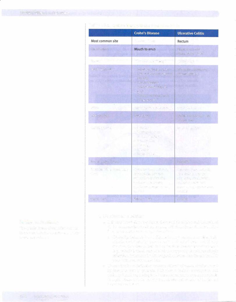
a.The three major categories of inflammatory bowel disease are Crohn's disease (CD), also called regional enteritis, ulcerative colitis (UC), and colitis ofindeterminate type.
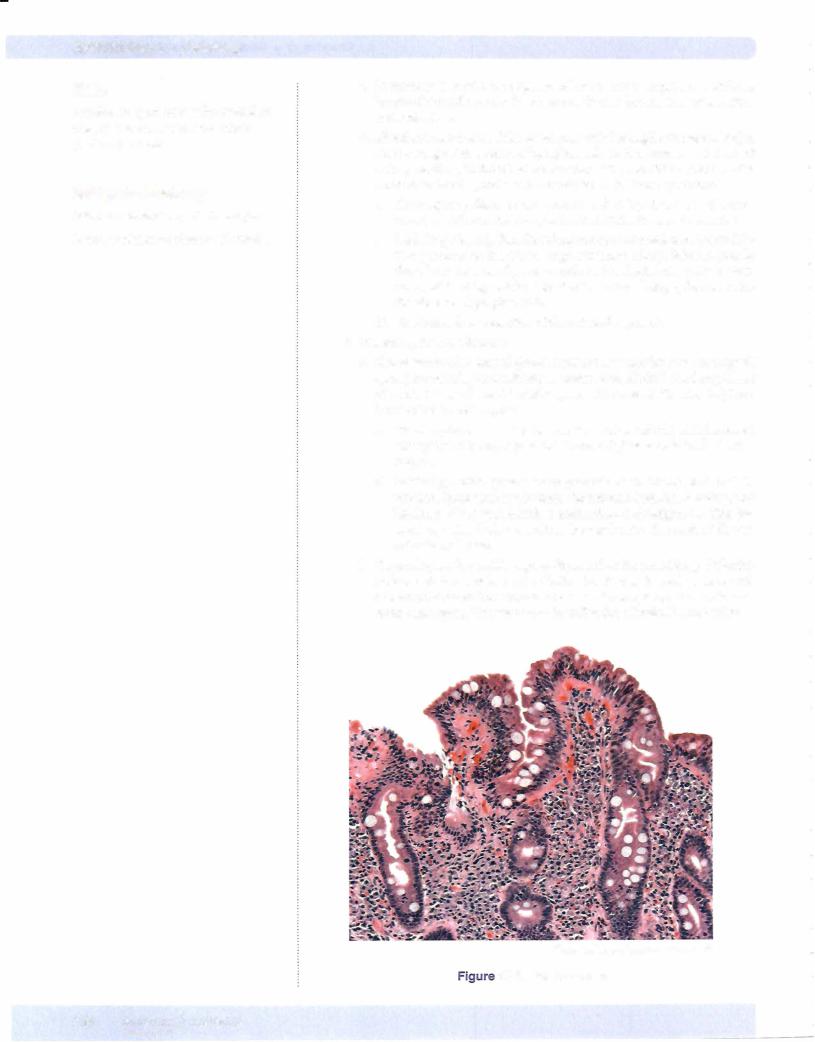
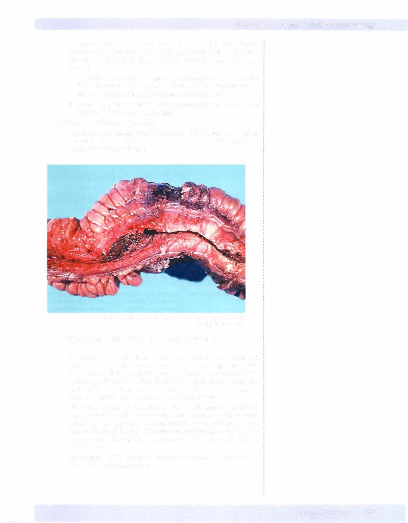
© Katsumi M. Miyai, M.D., Ph.D.; Regents of the University of California. Used with permission.
Figure 17-4. Narrowed Colon Segment in Crohn's disease
b.Epidemiology. Females develop IBD more frequently than males, and Caucasians more frequently than non-Caucasians. The age distribution varies with the disease; Crohn's disease has a bimodal distributionwith peaks at ages 10 to 30 and 50 to 70, while ulcerative colitis peaks at ages 20 to 30. The incidence of inflammatory bowel disease is increasing; ulcerative colitis is more common than Crohn's disease.
c.Clinically, the presentation of IBD can be with episodes of bloody diarrhea or stools with mucus, crarnpy lower abdominal pain, or fever. Crohn's disease may cause perianal fistulas, present with malabsorp tion, or mimic appendicitis. Extraintestinal manifestations canalso be a presentation, and are seen more commonly in ulcerative colitis than in Crohn's disease.
d.The diagnosis ofIBD is one ofexclusion,withtissuetobestudiedbeing obtainedbyendoscopicbiopsy.
Chapter 17 • Gastrointestinal Tract Pathology
c.Angiodysplasia is defined as arteriovenous malformations of the intes tines; it occurs in the cecum and right colon. It is common in individu als over age 55, presenting with multiple episodes of rectal bleeding. Angiodysplasia is associated with Osler-Weber-Rendu and CREST syndromes. Treatment is surgical resection.
d.Melanosis coli is common with laxative abuse; it causes black pig mentation of the colon due to the ingestion of the laxative pigment by macrophages in the mucosal and submucosa. Melanosis coli can mimic colitis or malignancy.
e.Pseudomembranous colitis (antibiotic-associated colitis) is an acute colitis characterized by the formation of inflammatory pseudomem branes in the intestines. It is usually due to Clostridium difficile infection (often brought on by a course of broad-spectrum antibiotics, especially clindamycin and ampicillin), but pseudomembranous colitis can also be due to ischemic bowel disease.
i.Pathology. Gross examination shows yellow-tan mucosal mem branes. Microscopic examination shows superficial colonic necrosis with an overlying pseudomembranes; the pseudomembranes are mushroom-shaped inflammatoryexudates composed ofneutrophils, mucin, fibrin, and necrotic cellular debris.
ii.Clinically, the presentation is with diarrhea, fever, and abdominal cramps, and the diagnosis is established with detection of C. difficile toxin in the stool. Treatment of clostrial pseudomembranous colitis is with vancomycin or metronidazole.
f.Appendicitis is most commonly caused by obstruction of the appendix by a fecalith. Appendicitis often starts with periumbilical pain that subse quently localizes to the right lower quadrant. Nausea, vomiting, and fever may also be present. Laboratory studies show an elevated white blood cell count. A complication is appendiceal rupture leading to peritonitis.
i.Pathology. Grossly, a fibrinopurulent exudate may be seen on the appendiceal serosa; microscopically, neutrophils are present within the mucosa and muscular wall (muscularis propria) of the appendix.
5.Diverticula
a.Meckel diverticulum is a congenital small bowel diverticulum due to persistance of a remnant of the vitelline (omphalomesenteric) duct.
i.Ruleof2s:
•2% of the normal population
•2 feet from the ileocecal valve
•2 cm in length
•2 years old or younger at the time of diagnosis
•2% of carcinoid tumors occur in a Meckel diverticulum.
ii.Clinically, most Meckel diverticula are asymptomatic, but they may contain rests of ectopic gastric mucosa and present with intestinal bleeding.
b.Colonic diverticulosis refers to acquired outpouchings of the bowel wall, characterized by herniation of the mucosa and submucosa through the muscularis propria (pseudodiverticulum). Colonic diverticulosis is extremely common in the United States, and the incidence increases with age. The major risk factor is low-fiber diet, which leads to increased intra luminal pressure; the most common location is in the sigmoid colon.
i.Clinically, the presentation varies, with many cases being asympto matic and picked up on screening colonoscopy. When symptomatic,
In a Nutshell
Osler-Weber-Rendu Syndrome
•a.k.a. Hereditary hemorrhagic telangiectasia
•Autosomal dominant
•Telangiectasias of skin and mucous membranes
•Common on lips, tongue, and fingertips
•May develop iron deficiency anemia
Note
Given that onlytwo layers ofthe bowelwall are involved, these acquired outpouchings are technically pseudodiverticula.
Chapter 17 • Gastrointestinal Tract Pathology
Chapter Summary (cont,d)
•Volvulus is twisting of a segment of bowel on its vascular mesentery, resulting in intestinal obstruction and infarction. lntussusception is telescoping of a proximal segment of bowel into the distal segment. Incarcerated hernia is a segment
ofbowel that becomes imprisoned within a hernia. Hirschsprung disease is a congenital absence ofganglion cells in the rectum and sigmoid colon resulting in intestinal obstruction.
•Celiac sprue is a hypersensitivityto gluten, resulting in loss of small bowel villi and malabsorption. Tropical sprue is a malabsorptive disease of unknown etiology affecting travelers to tropical regions, such as the Caribbean and South America. Whipple disease is a rare infectious disease involving many organs, including small intestines, joints, lung, heart, liver, spleen, and CNS.
•Inflammatory bowel disease includes Crohn disease, ulcerative colitis, and colitis of indeterminate type. Crohn disease has "skip" lesions, has transmural involvement with formation ofgranulomas, and tends to form fistulas, abscesses, and sinuses. In contrast, ulcerative colitis is confined to the rectum and colon, has inflammation limited to the mucosa and submucosa with crypt abscess, is
more likelyto have extraintestinal manifestations, and can cause toxic megacolon.
•lschemic bowel disease is the result of decreased blood flow and ischemia ofthe bowel secondary to atherosclerosis with thrombosis, thromboembolism, or reduced cardiac output from shock. Hemorrhoids are tortuous dilated submucosal veins caused by increased venous pressure. Angiodysplasia is arteriovenous malformation ofthe intestines. Melanosis
coli is a black pigmentation ofthe colon that is common with laxative abuse. Pseudomembranous colitis is characterized by formation of inflammatory pseudomembranes in the intestine following infection by Clostridium difficile, and/or ischemic bowel disease.
•Meckel diverticulum is a congenital small bowel diverticulum that is a remnant ofthe vitelline duct. Colonic diverticulosis is a common condition among
the elderly population and features acquired outpouchings ofthe bowel wall, characterized by herniation of the mucosa and submucosa through the muscularis propria.
•Adenomatous colonic polyps are benign neoplasms ofthe colonic mucosa that have the potential to progress to colonic adenocarcinoma. Familial adenomatous polyposis is a genetic condition in which patients develop thousands of colonic adenomatous polyps and have a virtually 100% chance ofdeveloping colon cancer by age 40 unless the affected colon is resected. Gardner syndrome
is a variant offamilial adenomatous polyposis with associated osteomas, fibromatosis, and epidermal inclusion cysts. Turcot syndrome is a rare variant offamilial adenomatous polyposis associated with CNS gliomas. Hereditary nonpolyposis colorectal cancer has increased risks of colon, endometrial, and ovarian cancers, but it is not associated with multiple adenomatous polyps.
Peutz-Jeghers syndrome has multiple hamartomatous polyps with increased risk of cancers ofthe lung, pancreas, breast, and uterus, but not colon.
•Colonic adenocarcinoma is the third most common cancer and a leading cause of cancer mortality in the United States. It tends to produce a polypoid mass when it involves the right side ofthe colon and a napkin ring lesion when it involves the left side. The Dukes system is used forstaging colon cancer.
•Carcinoid tumors are neuroendocrine tumors that can involve the appendix and terminal ileum and may produce carcinoid syndrome with diarrhea, flushing, bronchospasms, fibrosis, and sometimes carcinoid heart disease.