

Circulatory Pathology |
5 |
EDEMA
1.Edema is the presence ofexcess fluid in the intercellular space.
2.There are many causes of edema.
a. Increased hydrostatic pressure causes edema in congestive heart failure
(generalized edema), portal hypertension, renal retention of salt and water, and venous thrombosis (local edema).
b.Hypoalbuminemia and decreased colloid osmotic pressure cause edema in liver disease, nephrotic syndrome, and protein deficiency (e.g., kwashiorkor).
c.Lymphatic obstruction (lymphedema) causes edema in tumor, follow ing surgical removal of lymph node drainage, and in parasitic infesta tion (filariasis elephantiasis).
d.Increased endothelial permeability causes edema in inflammation, type I hypersensitivity reactions, and with some drugs (e.g., bleomycin, heroin, etc.).
e.Increased interstitial sodium causes edema when there is increased sodium intake, primary hyperaldosteronism, and renal failure.
f.Specialized forms oftissue swelling due to increased extracellular gly cosaminoglycans also occur, notablyin pretibial myxedema and exoph thalmos (Graves disease).
3.Anasarca is severe generalized edema.
4.Effusion is fluid within the body cavities.
5.Types of edema fluid:
a.Transudate is edema fluidwith low protein content; the specific gravity is less than 1.020.
b.Exudate is edemafluidwith high protein content and cells. The specific gravity is greater than 1.020. Types of exudates include purulent (pus), fibrinous, eosinophilic, and hemorrhagic.
c.Lymphedema related to lymphatic obstruction leads to accumulation ofprotein-rich fluid which produces a non-pitting edema.
d.Glycosaminoglycan-rich edema fluid shows increased hyaluronic acid and chondroitin sulfate, and causes myxedema.
6.Active hyperemia versus congestion (passive hyperemia): an excessive amount ofblood in a tissue or organ can accumulate secondary to vasodi latation (active) or diminishedvenous outflow (passive).
Note
Edema can be localized or generalized, depending on the etiology and severity.
MEDICAL 29
Chapter s • Circulatory Pathology
von Willebrand |
|
Absence or decreased |
|
expression of the GPlb |
|
factor |
|
|
|
(Bernard-Soulier syndrome) |
|
|
|
|
Deficiency |
|
t |
or dysfunction |
|
GPlb |
|
|
|
of vWF |
|
|
(von Willebrand |
|
|
disease) |
|
|
|
ADP induces |
|
|
conformational |
Inactive GPllb-GPllla |
|
change |
|
|
complex (Glanzmann |
|
|
|
|
|
|
thrombasthenia) |
|
|
t |
|
|
GPllb-llla |
|
|
complex |
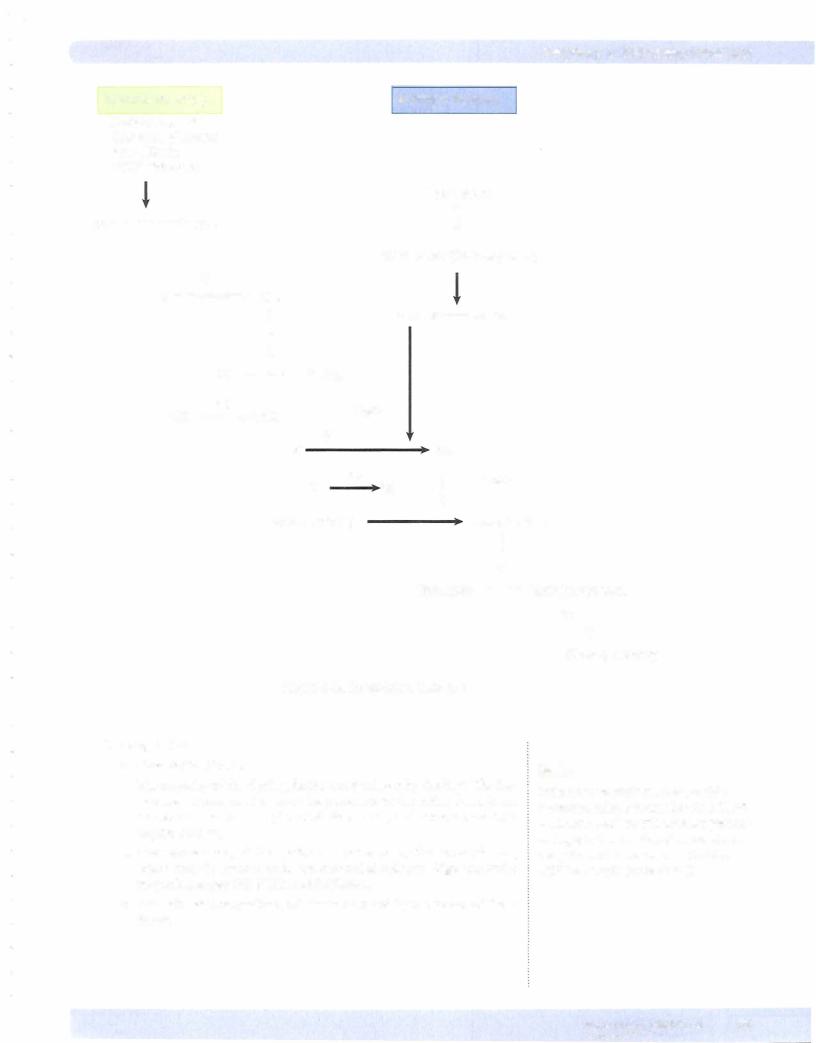
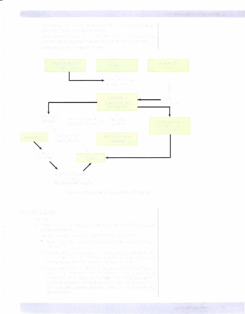
Figure 5-1. Platelet Aggregation
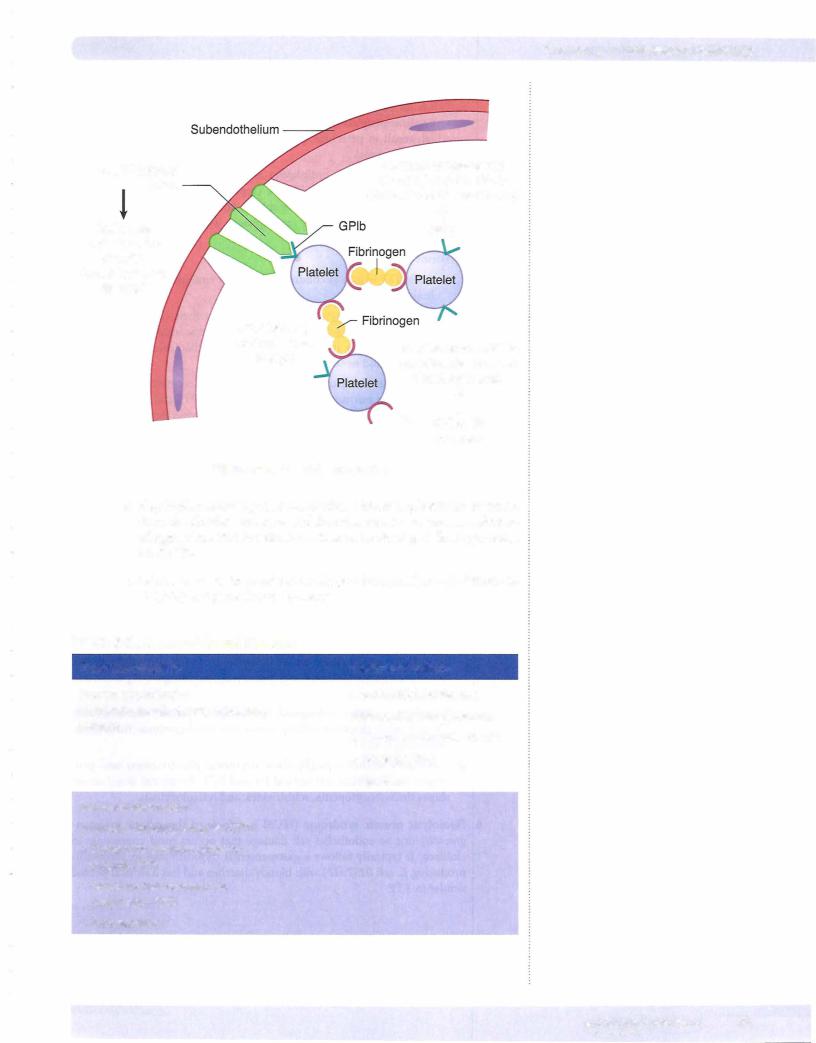
c.Step3: platelet aggregation occurs when additional platelets are recruited from the bloodstream. ADP and thromboxane A2 are potent mediators of aggregation. Platelets bind to each other by binding to fibrinogen using GPIIb-IIIa.
d.Laboratory tests for platelets include platelet count (normal: 150,000 to 400,000) and platelet aggregometry
Table 5-3. Common Platelet Disorders
Thrombocytopenia
Decreased production
•Aplastic anemia (drugs, virus, etc.)
•Tumor
Increased destruction
•Immune thrombocytopenia (ITP)
•Thrombotic thrombocytopenic purpura (TIP)
•Disseminated intravascular coagulation (DIC)
•Hypersplenism
Qualitative Defects
•von Witlebrand disease
•Bernard-Soulier syndrome
•Glanzmann thrombasthenia
•Drugs (aspirin)
•Uremia
MEDICAL 31
USMLE Step 1 • Pathology
4.Immune thrombocytopenia purpura (ITP)
a.ITP is an immune-mediated attack (usually IgG antiplatelet antibod ies) against platelets leading to decreased platelets (thrombocytopenia) which result in petechiae, purpura (bruises), and a bleeding diathesis (e.g., hematomas)
b.The etiology involves antiplatelet antibodies against platelet antigens such as GPIIb-IIIa and GPib-IX (type II hypersensitivity reaction), the antibodies are made in the spleen, and the platelets are destroyed peripherally in the spleen by macrophages, which have Fe receptors that bind IgG-coated platelets.
c.Forms ofITP
i.Acute ITP is seen in children following a viral infection and is a self limited disorder.
ii.Chronic ITP is usually seen in women in their childbearing years and may be the first manifestation of systemic lupus erythematosus (SLE). Clinically, it is characterized by petechiae, ecchymoses, men orrhagia, and nosebleeds.
d.Laboratorystudies usuallyshowdecreasedplatelet count and prolonged bleeding time but normal prothrombin time (PT) and partial throm boplastin time (PTT). Peripheral blood smear shows thrombocytopenia with enlarged immature platelets (megathrombocytes). Bone marrow biopsy shows increased numbers of megakaryocytes with immature forms.
e.The treatment can be with corticosteroids, which decrease antibody production; immunoglobulin therapy, which floods Fe receptors on splenic macrophages; and/or splenectomy, which removes the site of platelet destruction and antibody production.
5.Thrombotic thrombocytopenic purpura (TTP)
a.Definition: a rare disorder ofhemostasis where there is widespread intra vascular formation of fibrin-platelet thrombi due to a deficiency/inhibi tion of the enzyme ADAMTS13, which is responsible for cleaving large multimers ofvon Willebrand factor.
b.Clinically, TTP most often affects adult women. The characteristic pentad of signs includes fever, thrombocytopenia, microangiopathic hemolytic anemia (intravascular hemolysis), neurologic symptoms, and renal failure.
c.Pathology includes widespread formation of platelet thrombi with fibrin (hyaline thrombi) leading to intravascular hemolysis (thrombotic microangiopathy).
d.Laboratory studies typically show decreased platelet count and pro longed bleeding time but normal PT and PTT. Peripheral blood smear shows thrombocytopenia, schistocytes, and reticulocytosis.
6.Hemolytic uremic syndrome (HUS) is a form of thrombotic rnicroan giopathy due to endothelial cell damage that occurs most commonly in children. It typically follows a gastroenteritis (typically due to verotoxin producing E. coli 0157:H7) with bloody diarrhea and has a clinical pentad similar to TTP.
32 MEDICAL

USMLE Step 1 • Pathology
d.Laboratory tests for coagulation
i.Prothrombin time (PT)
PT tests the extrinsic and common coagulation pathways; more spe cifically, it tests factors VII, X, V, prothrombin, and fibrinogen. The international normalized ratio (INR) standardizes the PT test so that results throughout the world can be compared.
11.Partial thromboplastin time (PTT)
PTT tests the intrinsic and common coagulation pathways; more specifically, it tests factors XII, XI, IX, VIII, X, V, prothrombin, and fibrinogen.
iii. Thrombin time (TT) tests for adequate fibrinogen levels.
iv.Fibrin degradation products (FDP) tests the fibrinolytic system (in creased with DIC).
8.HemophiliaA (classic hemophilia) is an X-linked recessive condition that is due to a deficiency of factor VIII.
a.Clinically, hemophilia A predominately affects males. Symptoms are variable dependent on the degree of deficiency. Newborns may develop bleeding at the time of circumcision. Other problems include sponta neous hemorrhage into joints (hemarthrosis), easy bruising and hema toma formation after minor trauma, and severe prolonged bleeding after surgery or lacerations.
b.Laboratory studies typically show normal platelet count and normal bleeding time, normal PT and prolonged PTT.
c.Treatment: factor VIII concentrate
9. Hemophilia B (Christmas disease) is an X-linked recessive condition due to deficiency of factor IX that is clinically identical to hemophilia A.
10. Acquired coagulopathies include vitamin K deficiency (decreased synthe sis offactors II, VII, IX, X, and protein C & S) and liver disease (decreased synthesis ofvirtually all clotting factors).
1 1. Von Willebrand disease is an inherited bleeding disorder characterized by either a deficiency or qualitative defect in van Willebrand factor.
a. vWF is normally produced by endothelial cells and megakaryocytes.
b.Clinical features include spontaneous bleeding from mucous mem branes, prolonged bleeding from wounds, and menorrhagia in young females, though bleeding into joints is uncommon.
c.Laboratory studies show normal platelet count, a prolonged bleeding time, normal PT and often aprolongedPTT. Abnormalplatelet response to ristocetin (adhesion defect) is an important diagnostic test.
d.Treatment: treat mild cases (type I) with desmopressin (an antidiuretic hormone [ADH] analog), which releases vWF from Weibel-Palade bod ies of endothelial cells.
12.Disseminated intravascular coagulation (DIC)
a.DIC is always secondary to another disorder.
b.Causes are diverse. Obstetric complications can cause DIC because placental tissue factor activates clotting; Gram-negative sepsis can cause DIC because tumor necrosis factor [TNF] activates clotting. Other causes include microorganisms (especially meningococcus and rick ettsiae), AML M3 (cytoplasmic granules in neoplastic promyelocytes activate clotting), and adenocarcinomas (mucin activates clotting).
34 MEDICAL

USMLE Step 1 • Pathology
Bridge to Anatomy
The dual blood supply to the lungs is from the pulmonary artery and the bronchial arteries.
Clinical Correlate
The classic presentation of pulmonary embolism, which occurs in fewer than 20% of patients, includes hemoptysis, dyspnea, and chest pain.
Table 5-4. Comparison ofa Thrombus with a Blood Clot
|
Thrombus |
Blood Clot |
Location |
lntravascular |
Extravascular or intravascular |
|
|
(postmortem) |
Composition |
Platelets |
Lacks platelets |
|
Fibrin |
Rbrin |
|
RBCs and WBCs |
RBCs and WBCs |
Lines ofZahn |
Present |
Absent |
Shape |
Has shape |
Lacks shape |
c.Common locations ofthrombus formation include coronary and cere bral arteries; heart chambers in atrial fibrillation or post-MI (mural thrombus); aortic aneurysms; heart valves (vegetations); and deep leg veins (DVTs).
d.Outcomes of thrombosis include vascular occlusion and infarctions; embolism; thrombolysis; and organization and recanalization.
EMBOLISM
1. An embolism is any intravascular mass that has been carried down the bloodstream from its site of origin, resulting in the occlusion of a vessel.
2.Composition of emboli
There are manytypes ofemboli. Thromboemboli are most common (98%). Other types include atheromatous emboli (severe atherosclerosis), fat em boli (bone fractures and soft-tissue trauma), and bone marrow emboli (bone fractures and cardiopulmonary resuscitation [CPR]). Gas emboli cause decompression sickness ("the bends" and Caisson disease) when rapid ascent results in nitrogen gas bubbles in the blood vessels. Amniotic fluid emboli are a complication oflabor thatmay result in DIC. Fetal squa mous cells are seen in the maternal pulmonaryvessels. A fewmore types of emboli include tumor emboli (metastasis), talc emboli (intravenous drug abuse), and bacterial/septic emboli (infectious endocarditis).
3.Pulmonaryemboli (PE)
a.Pulmonary emboli are often clinically silent and are the most common ly missed diagnosis in hospitalized patients. They are found in almost half of all hospital autopsies.
b.Most (95%) pulmonary emboli arise from deep leg vein thrombosis (DVT) in the leg; other sources include the right side ofthe heart and the pelvic venous plexuses ofthe prostate and uterus.
c.Diagnosis of a pulmonary embolus can be established when V/Q lung shows a scan V/Q mismatch. Doppler ultrasound of the leg veins can be used to detect a DVT. Additionally, plasma D-dimer ELISA test is elevated.
d.Potential outcomes of PEs
i.No sequelae (75%) is the most common outcome and PEs can be asymptomatic or have transient dyspnea/tachypnea. No infarction (dual blood supply) is seen and there is complete resolution in such cases.
36 MEDICAL
Chapter 5 • Circulatory Pathology
ii.Infarction (15%) is more common in patients with cardiopulmonary compromise. Symptoms include shortness ofbreath (SOB), hemop tysis, pleuritic chest pain, and pleural effusion. On gross examina tion, there is typically a hemorrhagic wedge-shaped infarct. The infarction heals by regeneration or scar formation.
iii.Sudden death (5%) can occur when large emboli lodge in the bifur cation (saddle embolus) or large pulmonary artery branches and obstruct more than 50% ofthe pulmonary circulation.
iv.Chronic secondary pulmonary hypertension (3%) is caused by recur rent PEs, which increase pulmonary resistance and lead to secondary pulmonaryhypertension.
4.Systemic arterial emboli
Systemic arterial embolimostlyarise in the heart. Most arterial emboli cause infarction, with common sites ofinfarction including the lower extremities, brain, intestine, kidney, and spleen. Paradoxical emboli is the term used for anyvenous embolus thatgains access to the systemic circulation by crossing over from the right to the left side ofthe heart through a septa! defect, most commonlythrough a patentforamen ovale.
INFARCTION
1. Infa rction is a localized area ofnecrosis secondary to ischemia.
a.Infarcts have multiplecauses. Most infarcts (99%) resultfrom thrombotic or embolic occlusion of an artery or vein. Less common causes include vasospasm and torsion of arteries and veins (e.g., volvulus, ovarian, and testicular torsion).
b.Factors that predict the development of an infarct include vulnerability of the tissue to hypoxia, degree of occlusion, rate of occlusion, presence of a dual blood supply or collateral circulation, and oxygen-carrying capacity of the blood (anemia, carbon monoxide poisoning, etc.).
c.Common sitesofinfarction includeheart, brain, lungs, intestines, kidneys.
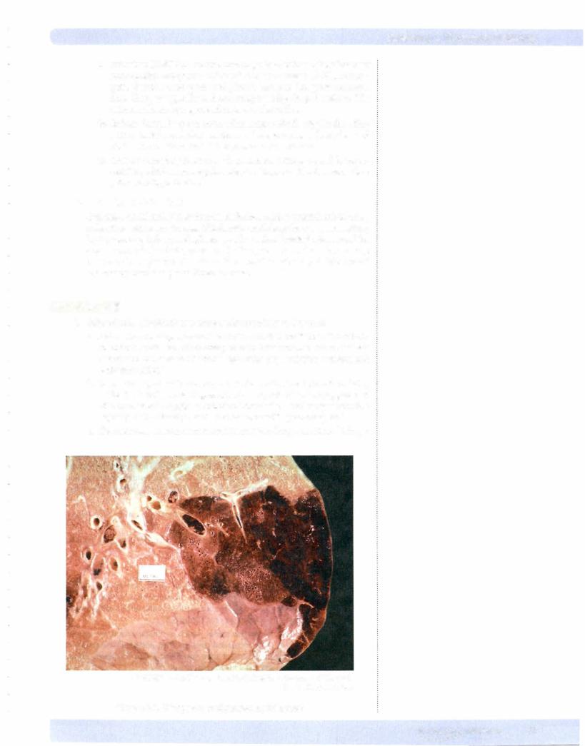
© Katsumi M. Miyai, M.D., Ph.D.; Regents of the University ofCalifornia. Used with permission.
Figure 5-4. Wedge-shaped pulmonary infarction
MEDICAL 37

USMLE Step 1 • Pathology
2.On gross examination Infarctions typically have a wedge shape, with the apex ofthe wedge tending to point to the occlusion.
a.Anemicinfarcts (pale orwhite color) occur in solid organswith a single blood supply such as the spleen, kidney, and heart.
b.Hemorrhagic infarcts (red color) occur in organs with a dual blood supply or collateral circulation, such as the lung and intestines, and can also occur with venous occlusion (e.g., testicular torsion).
3.Microscopicpathology of infarction can show either coagulative necrosis (most organs) or liquifactive necrosis (brain). The general sequence oftis sue changes after infarction is:
ischemia -7 coagulative necrosis -7 inflammation -7 granulation tissue -7 fibrous scar
SHOCK
1. General
Shock is characterized by vascular collapse and widespread hypoperfusion ofcells and tissue due to reduced bloodvolume, cardiac output, or vascular tone. The cellular injury is initially reversible; if the hypoxia persists, the cellular injury becomes irreversible, leading to the death of cells and the patient.
2.Major causes of shock
a.Cardiogenic shock (pump failure) can be due to myocardial infarction, cardiac arrhythmias, pulmonary embolism, and cardiac tamponade.
b.Hypovolemic shock (reduced blood volume) can be due to hemor rhage, fluid loss secondary to severe burns, and severe dehydration.
c.Septic shock (bacterial infection) is due to Gram-negative septicemia which causes the release of endotoxins (bacterial wall lipopolysaccha rides) into the circulation. High levels of endotoxin result in produc tion of cytokines TNF, IL-1, IL-6, and IL-8. These cytokines can trigger vasodilatation and hypotension, acute respiratory distress syndrome (ARDS), DIC, and multiple organ dysfunction syndrome. The mortality rate is 50%.
d.Neurogenic shock (generalized vasodilatation) can be seen with anes thesia and brain or spinal cord injury.
e.Anaphylactic shock (generalized vasodilatation) is a type I hypersensi tivityreaction.
3.Stages of shock
a.Stage I is compensation, in which perfusion to vital organs is maintained by reflex mechanisms. Compensation is characterized by increased sympathetic tone, release ofcatecholamines, and activation of the renin-angiotensin system (Figure 5-5).
b.Stage II is decompensation, which is characterized by progressive decrease in tissue perfusion; this leads to potentially reversible tissue injury with development of a metabolic (lactic) acidosis, electrolyte imbalances, and renal insufficiency.
c.Stage III is irreversible tissue injury and organ failure, ultimately result ing in death.
38 M EDICAL


USMLE Step 1 • Pathology
Chapter Summary
•Edema is the presence of excess fluid in the intercellular space. Causes of edema include increased hydrostatic pressure, increased interstitial sodium,
hypoalbuminemia and decreased colloid pressure, lymphatic obstruction, and increased endothelial permeability. Anasarca is the term used for severe generalized edema.
•Transudates have low protein content and specific gravity, while exudates have high protein content and specific gravity.
•Hyperemia is an excessive amount of blood in a tissue or organ and can be due eitherto vasodilation (active hyperemia) or diminished venous outflow (passive hyperemia or congestion).
•Hemostasis is the sequence ofevents leading to cessation of bleeding by the formation of a stable fibrin-platelet hemostatic plug. Vascular wall injury triggers transient vasoconstriction, facilitation of platelet adhesion, and activation
of both the extrinsic and intrinsic clotting pathways. Formation of a platelet thrombus occurs when platelets adhere to von Willebrand factor attached to subendothelial collagen, undergo shape change and degranulation, and then aggregate with additional platelets.
•Causes ofthrombocytopenia due to decreased platelet production include aplastic anemia and tumor. Causes ofthrombocytopenia due to increased platelet destruction include immune thrombocytopenic purpura (ITP), thrombotic thrombocytopenic purpura (TIP), disseminated intravascular coagulation (DIC), and hypersplenism. Causes of qualitative platelet defects includevon Willebrand disease, Bernard-Soulier syndrome, Glanzmann thrombasthenia, aspirin, and uremia.
•In immune thrombocytopenic purpura (ITP), antiplatelet antibodies destroy platelets, primarily in the spleen. In thrombotic thrombocytopenic purpura (TTP), there is widespread formation of platelet thrombi with fibrin but without activation ofthe coagulation system. Hemolytic uremic syndrome (HUS) can clinically resemble TTP and is triggered by E. coli strain 0157:H7.
•The intrinsic coagulation pathway is activated by contact factors and is clinically tested with the partial thromboplastin time (PTI). The extrinsic coagulation pathway is activated by the release oftissue factor, and istested with the prothrombin time (Pl), which also tests the common coagulation pathway.
•Hemophilia A is an X-linked recessive deficiency offactorVIII, which is clinically characterized by hemarthrosis, easy bruising, and severe prolonged bleeding after surgery or lacerations. Clinically, hemophilia B closely resembles hemophilia A but is due to deficiency of factor IX. Acquired coagulopathies can be due to vitamin K deficiency and liver disease. Von Willebrand disease is an inherited bleeding disorder characterized by a deficiency or qualitative defect in von Willebrand factor, which facilitates formation of platelet clots.
•Disseminated intravascular coagulation (DIC) can be triggered by a variety of severe medical conditions and results in formation of many microthrombi that consume platelets and clotting factors, leading, in turn, to a superimposed bleeding tendency.
(Continued)
40 MEDICAL
Chapter s • Circulatory Pathology
Chapter Summary (cont,dJ
•Factors involved in thrombus formation include endothelial injury, alterations in laminar blood flow, and hypercoagulability of blood. Thrombi can lead to a spectrum of outcomes, including vascular occlusion and infarction, embolism, thrombolysis, and organization and recanalization.
•The term embolism is used for any intravascular mass (solid, liquid, or gas) that has been carried downstream from its site of origin, resulting in occlusion ofa vessel. Ninety-eight percent of emboli are thromboemboli, but many other materials have also formed emboli. Pulmonary emboli are a common form of emboli that are often clinically silent but can cause infarction or sudden death. Most pulmonary emboli arise from deep vein thromboses. Systemic arterial emboli usually arise in the heart and may cause infarction in a variety of sites, depending upon where they lodge.
•Infarction is a localized area of necrosis secondary to ischemia. Ninety-nine percent of infarcts result from thrombotic occlusion ofan artery or vein . Anemic infarcts occur in organs with a single blood supply, whereas hemorrhagic infarcts occur in organs with a dual blood supply or secondary to venous occlusion.
The general sequence oftissue changes after infarction is: ischemia leads to coagulative necrosis, which leads to inflammation, which leads to granulation tissue, which leads to fibrous scar.
•Shock is characterized by vascular collapse and widespread hypoperfusion of cells and tissues due to reduced blood volume, cardiac output, orvascular tone. Major forms of shock include cardiogenic shock, hypovolemic shock, septic shock, and neurogenic shock. Shock has been clinically divided into
compensated shock (stage I), decompensated shock (stage II), and irreversible injury (stage Ill). Different organs show distinctive microscopic patterns in shock.
MEDICAL 41

DISORDERS INVOLVING CHROMOSOMAL DELETIONS
1. Cri du chat syndrome has karyotype 46 XX or XY, Spand is due to dele tion of the short arm of chromosome 5.
a.Clinical findings include a characteristic high-pitched catlike cry; men tal retardation; congenital heart disease; and microcephaly.
2.Microdeletions of which you should be aware include 13q14 (the retino blastoma gene) and 1 1pl3 (WAGR complex [Wilms tumor, Aniridia, Geni tourinary anomalies, and mental Retardation]).
DISORDERS INVOLVING SEX CHROMOSOMES
The following conditions involving sex chromosomes are now called disorders of sexual development (differentiation).
1.Klinefelter syndrome has karyotype 47 XXY; is caused by meiotic nondis junction; and is a common cause of male hypogonadism.
a.Laboratory studies show elevated FSH and LH with low levels of tes tosterone.
b.Clinical findings include testicular atrophy, infertility due to azoosper mia, eunuchoid body habitus, high-pitched voice; female distribution of hair; and gynecomastia.
2.Turner syndrome has karyotype 45 XO and is a common cause of female hypogonadism. The second X chromosome is necessary for oogenesis and normal development of the ovary. No Barr body is present on histological examination of cells.
a.Clinically, Turner syndrome patients fail to develop secondary sex characteristics and have short stature with widely spaced nipples. Other features related to the abnormal sexuality include gonadal dysgenesis with atrophic "streaked" ovaries; primary amenorrhea; and infertility. Clinical features involving other organ systems include cystic hygroma and webbing of the neck; hypothyroidism; congenital heart disease (preductal coarctation of the aorta and bicuspid aortic valve); and hydrops fetalis. Females with 45,X; 46,XY mosaicism are at risk for gonadoblastoma.
Chapter 6 • Genetic Disorders
Note
The presence of a Y chromosome determines male phenotype due to the presence ofthe testes-determining factor gene (also called the sex-determining region Y [SRY]) on the Y chromosome.
In a Nutshell
Lyon's Hypothesis of
X-lnactivation
•Only one X is genetically active.
•The otherX chromosome is inactivated (becomes the Barr body) in the blastocyst stage of development.
•Females are mosaics.
•Either the maternal or paternal X chromosome is inactivated at random.
MEDICAL 45

USMLE Step 1 • Pathology
Bridge to Biochemistry
The majority of cystic fibrosis cases resultfrom deletion of phenylalanine at position 508 (Delta F508), which interferes with proper protein folding and the post-translational processing of oligosaccharide side chains. The abnormal chloride channel protein is degraded by the cytosolic proteasome complex rather than translocated to the cell membrane.
Bridge to Biochemistry
Phenylalanine hydroxylase converts phenylalanine into tyrosine.
AUTOSOMAL RECESSIVE DISORDERS
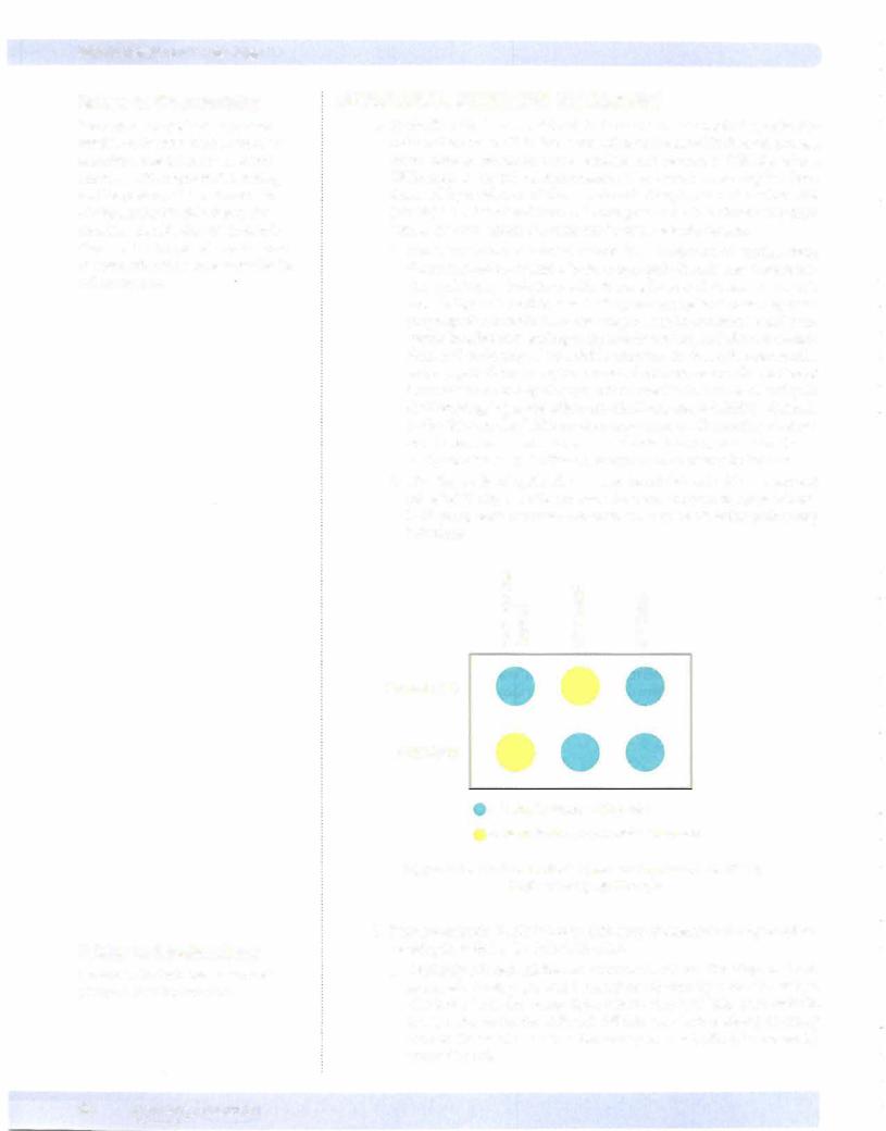
1.Cystic fibrosis (mucoviscidosis) is the most common lethal genetic dis order in Caucasians. It is due to mutation of the chloride channel protein, cystic fibrosis transmembrane conductance regulator (CFTR), whose CFTR gene is located on chromosome 7 and most commonly has been damaged by a deletion of the amino acid phenylalanine at position 508 ( F508). The defective chloride channel protein leads to abnormally thick viscous mucous, which obstructs the ducts of exocrine organs.
a.The distribution of disease reflects the distribution of eccrine sweat
glands and exocrine glands. In the lungs, cystic fibrosis may cause recur rent pulmonary infections with P. aeruginosa and S. aureus; chronic bronchitis; and bronchiectasis. In the pancreas; cystic fibrosis may cause plugging of pancreatic ducts resulting in atrophy and fibrosis; and pan creatic insufficiency leading to fat malabsorption, malodorous steator rhea, and deficiency of fat-soluble vitamins. In the male reproductive system, cystic fibrosis may be associated with absence or obstruction of the vas deferens and epididymis, which often leads to male infertility. In the liver, plugging ofthe biliary canaliculi may result in biliary cirrhosis. In the GI tract, the thick secretions may cause small intestinal obstruc tion (meconium ileus). The salivary glands do not appear to be signifi cantly affected in cystic fibrosis, except for more mucus in the ducts.
b.The diagnosis of cystic fibrosis can be established with a sweat test (elevated NaCl) or DNA probes. The mean survival in cystic fibrosis is 30 years, with the most common cause of death being pulmonary infections.
|
C/l |
|
|
|
|
::J |
"E |
|
|
|
0 |
(i) |
||
|
Cl |
|||
|
>. |
Q) |
·;:: |
|
|
( i j |
|
Cii |
|
|
Cl.. |
(.) |
|
|
|
E E |
LL. |
LL. |
|
|
0 0 |
|||
|
: C Z |
(.) |
(.) |
|
Normal ASO |
0 0 0 |
|
||
F508 ASO |
0 0 0 |
|
||
|
O = Sample reacts with probe |
|
||
|
O= Sample does not react with probe |
|||
Figure 6-5. The Use of Allele-Specific Oligonucleotide (ASO)
Probes for Cystic Fibrosis
2.Phenylketonuria (PKU) is due to deficiency of phenylalanine hydroxylase, resulting in toxic levels of phenylalanine.
a.Clinically, affected children are normal at birth but, if undiagnosed and untreated, develop profound mental retardation by 6 months of age. The lack of tyrosine causes light-colored skin and hair, since melanin is a tyrosine derivative. Affected children may have a mousy or musty odor to the sweat and urine (secondary to metabolite [phenylacetate] accumulation).
48 MEDICAL
b.Screening for phenylketonuria is done at birth and treatment is with dietary restriction of phenylalanine, including avoidance of the artificial sweetener aspartame.
c.A genetic variant, benign hyperphenylalaninemia, has partial enzyme deficiency with mildly increased levels of phenylalanine which are insufficient to cause mental retardation.
3.Alkaptonuria (ochronosis) occurs when deficiency of homogentisic acid oxidaseresults in the accumulation ofhomogentisic acid. The homogentisic acid has an affinity for connective tissues (especially cartilage), resulting in a black discoloration (as a consequence of oxidation of homogentisic acid).
a.Clinical features include urine that is initially pale yellow, but which turns black upon standing, and black-stained cartilage, which causes discoloration of the nose and ears, and also predisposes for early onset of degenerative arthritis.
4.Albinism is a due to a lack of the enzyme tyrosinase that is needed for melanin production. Affected individuals show deficiency of melanin pig mentation in the skin, hair follicles, and eyes (oculocutaneous albinism), with resulting increased risk of basal cell and squamous cell carcinomas.
5.The glycogen storage diseases are a group of rare diseases that have in common a deficiency in an enzyme necessary for the metabolism of glyco gen, which results in the accumulation of glycogen in the liver, heart, and skeletal muscle.
a.Type I (von Gierke disease) is due to a deficiency ofglucose-6-phospha tase, and is characterized clinically by hepatomegaly and hypoglycemia.
b.Type II (Pompe disease) is due to a deficiency of lysosomal a-1,4- glucosidase (acid maltase), and is characterized clinically by hepato megaly, skeletal muscle hypotonia, cardiomegaly, and death from cardiac failure by age 2 years.
c.Type V (McArdle syndrome) is due to a deficiency of muscle glycogen phosphorylase, and is characterized clinically by exercise-induced muscle cramps.
6.Tay-Sachs disease is due to a deficiency ofhexosaminidase A (due to muta tion of HEXA gene on chromosome 15), which leads to the accumulation of GM2 ganglioside in the lysosomes of the CNS and retina. Tay-Sachs disease is common in Ashkenazi Jews (1 in 30 carrier rate).
a.The distribution of disease involves the retina (cherry-red spot due to accentuation of the macula) and central nervous system (dilated neurons with cytoplasmic vacuoles). Affected children are normal at birth, but by 6 months show onset ofsymptoms (progressive mental deterioration and motor incoordination) that progress to death by age 2-3 years.
b.Electron microscopy shows distended lysosomes with whorled mem branes; the diagnosis can also be established with enzyme assays and DNA probes.
Chapter 6 • Genetic Disorders
Note
Lysosomal Storage Diseases
Defined as a deficiency of a lysosomal enzyme (acid hydrolase), which leads to the accumulation of a complex substrate within the lysosome leading to enlarged cells that become dysfunctional
•Tay-Sachs
•Niemann-Pick
•Gaucher
•Mucopolysaccharidosis
•Fabry
•Metachromatic leukodystrophy
MEDICAL 49


Chapter 6 • Genetic Disorders
AUTOSOMAL DOMINANT DISORDERS
1. Familial hypercholesterolemia is the most common inherited disorder (with incidence of 1 in 500) and is due to a mutation in the low density lipoprotein (LDL) receptor gene on chromosome 19.
a.There are five major classes ofmutations.
Class I has no LDL receptor synthesis.
Class II has a defect in transport out of the endoplasmic reticulum.
-Class III has a defect in LDL receptor binding.
-Class IV has a defect in ability to internalize bound LDL.
-Class V has a defect in the recycling of the LDL receptor. The muta- tions in the LDL receptor cause increased levels ofcirculating choles terol, loss of feedback inhibition of HMG-coenzyme A (HMG-CoA) reductase, and increased phagocytosis of LDL by macrophages.
b.Clinical features include elevated serum cholesterol (heterozygotes have elevations of 2 to 3 times the normal level and homozygotes have elevations of 5 to 6 times the normal level), skin xanthomas (collections of lipid-laden macrophages), xanthelasma around the eyes, and prema ture atherosclerosis (homozygotes often develop myocardial infarctions in late teens and twenties).
2.Marfan syndrome is due to a mutation of the fibrillin gene (FBNl) on chromosome 15q21. Fibrillin is a glycoprotein that functions as a scaffold for the alignment of elastic fibers.
a.Clinical features include skeletal changes (tall, thin build with long extremities, hyperextensible joints, pectus excavatum [inwardly depressed sternum], and pectus carinatum [pigeon breast] ) and abnormal eyes (ectopia lentis, characterized by bilateral subluxation of the lens). The cardiovascular system is also particularly vulnerable; it may show cystic medial degeneration of the media of elastic arteries with a loss of elastic fibers and smooth muscle cells with increased risk of dissecting aortic aneurysm (a major cause of death), dilatation of the aortic ring poten tially leading to aortic valve insufficiency, and/or mitral valve prolapse.
3.Ehlers-Danlos syndrome (EDS) is a group of inherited connective tissue diseases that have in common a defect in collagen structure or synthesis. Clinically, the disease causes hyperextensible skin that is easily traumatized and hyperextensible joints secondary to effects on the joints and adjacent ligaments.
a.There are 10 variants with different modes of inheritance, of which the most important are:
EDS Type 3 (autosomal dominant with unknown genetic defect; most common type)
-EDS Type 4 (autosomal dominant defect in the type III collagen gene)
-EDS Type 6 (autosomal dominant defect in the lysyl hydroxylase gene, which is the enzyme responsible for hydroxylation oflysine residues)
-EDS Type 9 (X-linked recessive mutation of copper-binding protein on X chromosome, leading to low levels of ceruloplasmin and serum copper; and to decreased activity of lysyl oxidase, which is copper dependent and necessary for cross-linking of collagen fibers).
b.Complications of Ehlers-Danlos include poor wound healing, joint dis locations; diaphragmatic hernias (EDS Type 1), retinal detachment and kyphoscoliosis (EDS Type 6), and arterial or colonic rupture (EDS Type 4).
Bridge to Biochemistry
HMG-CoA reductase is the rate-limiting enzyme in the synthesis of cholesterol. Normally, cholesterol represses the expression of the HMG-CoA reductase gene (negative feedback).
Note
Disorders of collagen biosynthesis include scurvy, osteogenesis imperfecta, Ehlers Danlos syndrome, Alport syndrome, and Menkes disease.
Clinical Correlate
Menkes disease is an X-linked recessive condition that is caused by mutations in the gene encoding a Cu2+ efflux
protein. Cells from an affected individual accumulate high concentrations of Cu2+ that cannot be released.
MEDICAL 51
USMLE Step 1 • Pathology
4.Neurofibromatosis
a.Type 1 (von Recklinghausen disease) neurofibromatosis accounts for 90% of cases of neurofibromatosis, with an incidence of 1 in 3,000. The condition is due to a mutation of the tumor suppressor gene NF- 1 located on chromosome 17 (17ql l .2). The normal gene product (neurofibromin) inhibits p21 ras oncoprotein. Type 1 neurofibroma tosis characteristically has multiple neurofibromas, benign tumors of peripheral nerves that are often numerous and may be disfiguring. The plexiform variant of the neurofibromas are diagnostic. Rarely (3%), malignant transformation of a neurofibroma may occur. Other clinical features include pigmented skin lesions (6 or more "cafe-au-lait spots" that are light brown macules usually located over nerves); pigmented iris hamartomas (Lisch nodule); and increased risk of meningiomas and pheochromocytoma (dark/dusky-colored tumor).
© Richard P. Usatine, M.D. Used with permission.
Figure 6-6. Multiple Subcutaneous Neural Tumors of Neurofibromatosis
b.Type 2 (bilateralacoustic) neurofibromatosis accounts for 10% ofcases of neurofibromatosis, with an incidence of 1 in 45,000. In this form of neurofibromatosis, the tumor suppressor gene: is NF-2 (22ql2.2) located on chromosome 22. The normal gene product (merlin) is a critical regulator of contact-dependent inhibition of proliferation. Clinical features include bilateral acoustic neuromas; neurofibromas
52 MEDICAL

Chapter 6 • Genetic Disorders
and cafe-au-lait spots (usually both in smaller numbers than in type 1), and increased risk ofmeningioma and ependymomas.
5.von Hippel-Llndau disease is due to a mutation of the tumor suppressor gene VHL, which is located on chromosome 3p (3p26-p25). The normal gene product's main action is to tag proteins (e.g., hypoxia inducible factor la [a transcription factor that induces the expression ofangiogenesis factors]) with ubiquitin for degradation.
a.Clinical manifestations can include retinal hemangioblastoma (von Hippel tumor); hemangioblastomas of the cerebellum, brain stem, and spinal cord (Lindau tumor); cysts ofthe liver, pancreas, and kidneys; and multiple bilateral renal cell carcinomas.
X-LINKED RECESSIVE CONDITIONS
1. In X-linked recessive conditions, males with a mutant recessive gene on the X chromosome have the condition while daughters of affected males are obligate carriers, who in many situations are asymptomatic. Sons of affected males do not carry the mutation. Daughters of carrier females may be either normal or carriers. Sons of carrier females may be affected or normal (because males are hemizygous for the X chromosome).
2. Lesch-Nyhan syndrome results from deficiency of hypoxanthine-gua nine phophoribosyltransferase (HGPRT), which impairs salvaging of the purines hypoxanthine and guanine. Clinical features include mental retar dation, hyperuricemia, and self-mutilation.
3.Testicular feminization is an androgen insensitivity that causes failure of normal masculinization of external genitalia of "X:'l males.
4.In chronic granulomatous disease, a defective NADPH oxidase enzyme complex prevents phagocytes from producing superoxide bursts to kill bacteria. Clinical features include recurrent infections, hypergammaglobu linernia, hepatosplenomegaly, and lymphadenopathy.
5.In Brutonagammaglobulinemia, defective Bruton tyrosine kinase (Btk) at band Xq21.3 causes complete failure of immunoglobulin production char acterized clinically by complete absence of antibodies in serum and recur rent bacterial infections.
X-LINKED DOMINANT CONDITIONS
1.X-linked dominant conditions are similar to X-linked recessive, but both males and females show disease. An example is Alport syndrome, which is a hereditary glomerulonephritis with nerve deafness.
TRIPLET REPEAT MUTATIONS
1. Fragile X syndrome is due to triplet nucleotide repeat mutations, so that the nucleotide sequence CGG repeats typically hundreds to thousands of times. The mutation occurs in the FMR-1 gene (familial mental retarda tion- I gene) on the X chromosome (Xq27.3), and the disease behaves as an X-linked dominant disease that causes mental retardation in all affected males and 50% of female carriers. The characteristic phenotype includes elongated face with a large jaw, large everted ears, and macroorchidism. The condition can be diagnosed with DNA probe analysis.
2.Huntington disease is due to a triplet repeat mutation (CAG) ofthe Hun tington gene that produces an abnormal protein (huntingtin), which is
Note
Most common genetic causes of mental retardation:
•Down syndrome
•Fragile X syndrome
Note
Triplet repeat expansion can occur in a coding region (Huntington and
spinobulbar muscular atrophy) or in an untranslated region ofthe gene (fragile X and myotonic dystrophy).
MEDICAL 53

USMLE Step 1 • Pathology
neurotoxic and causes atrophy of caudate nucleus. Huntington disease has an early onset (age range: 20-50 years) of progressive dementia with cho reiform movements.
GENOMIC IMPRINTING
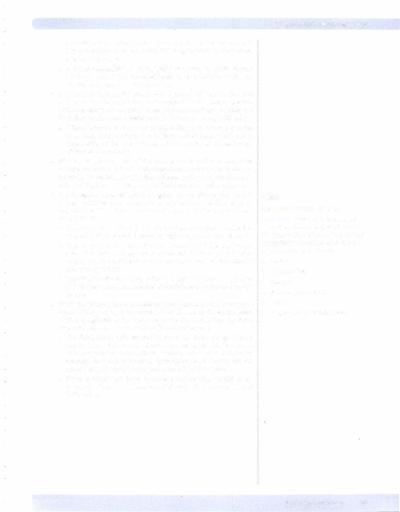
1.Genomic imprinting refers to differential expression of genes based on chromosomal inheritance from maternal versus paternal origin. In Prad er-Willi syndrome, microdeletion on paternal chromosome 15 {del(l5) (ql l;ql3)} causes mental retardation, obesity, hypogonadism, and hypoto nia. In contrast, in Angelman syndrome ("happy puppet" syndrome), mi crodeletion on maternal chromosome 15 {del(l5)(ql l;ql3)} causes mental retardation, seizures, ataxia, and inappropriate laughter.
Genomic imprinting is thought to also potentially play a role in Huntington disease, neurofibromatosis, and myotonic dystrophy.
Prader-Willi Syndrome |
|
Angelman Syndrome |
||||||
1 5 |
clt |
l |
15 |
1 5 |
|
l |
T |
15 |
Deletion |
|
|
Normal |
Normal |
|
|
Deletion |
|
Chromosome |
|
Chromosome |
Chromosome |
I :::i |
||||
|
|
|
|
|
|
|
|
Chromosome |
The inheritance ofa deletion on chromosome 15 from a male produces Prader-Willi syndrome, whereas inheritance of the same deletion from a female produces Ange/man syndrome.
Figure 6-7. Genomic Imprinting
MITOCHONDRIAL DNA DISORDERS
Mitochondrial DNA codes for mitochondrial oxidative phosphorylation enzymes; inheritance is only from mother to child, because only the ovum contributes mito chondria to the zygote. You should be aware of 2 examples of mitochondrial DNA disorders:
•Leber hereditaryopticneuropathy causes loss ofretinal cells, which leads to central vision loss.
•Myodonic epilepsyhas a pattern ofseizures with abruptjerks.
MULTIFACTORIAL INHERITANCE
1.Multifactorial inheritance refers to disease caused by a combination of multiple minor gene mutations and environmental factors. Examples include open neural tube defects and type 2 diabetes mellitus.
54 MEDICAL
Chapter 6 • Genetic Disorders
Chapter Summary
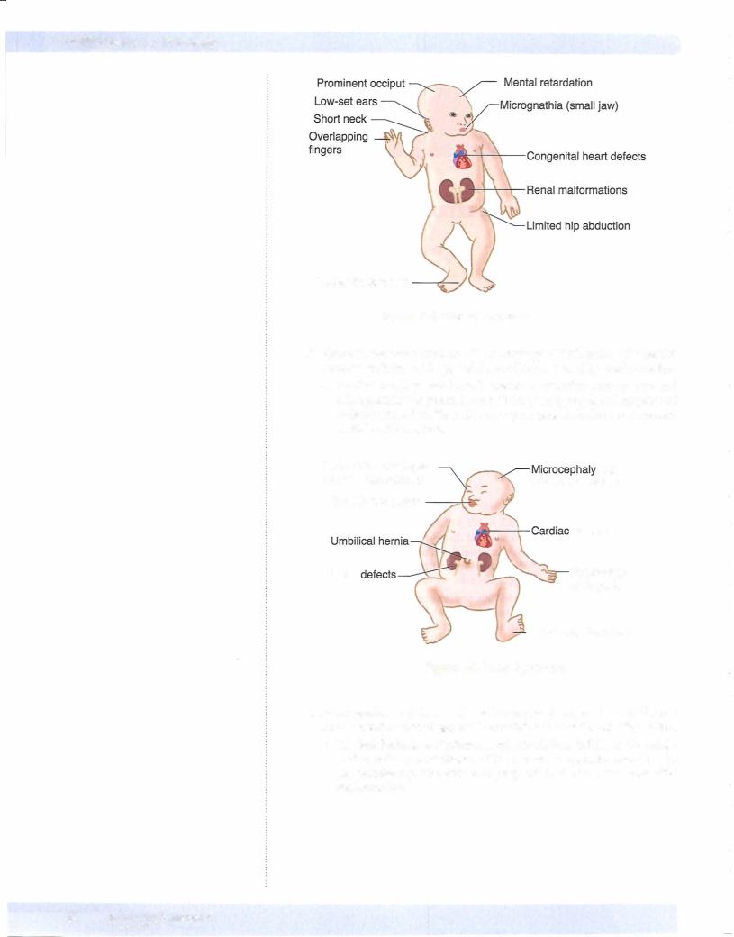
•Disorders involving an extra autosomal chromosome include Down syndrome, Edwards syndrome, and Patau syndrome. Down syndrome (trisomy 21) is the most common ofthe chromosomal disorders and is characterized by severe mental retardation, mongoloid facialfeatures, hypotonia, and palmarcreases. Serious complications of Down syndrome include congenital heart disease (endocardial cushion defects), duodenal atresia, Hirschsprung disease, acute lymphoblastic leukemia, and early onset ofAlzheimer disease. Edwards syndrome (trisomy 18) is characterized by mental retardation, low-set ears, micrognathia, congenital heart defects, overlapping flexed fingers, and rocker-bottom feet. Patau syndrome (trisomy 13) is characterized by mental retardation, cleft lip and/or palate, cardiac defects, renal abnormalities, microcephaly, and polydactyly.
•Chromosomal deletions can also cause genetic disease. Cri du chat syndrome
(Sp-) is a chromosomal deletion syndrome characterized by a high-pitched, catlike cry; mental retardation; congenital heart disease, and microcephaly. Microdeletions are associated with retinoblastoma and Wilms tumor.
•Klinefelter syndrome and Turner syndrome are important disorders ofsex chromosomes. Klinefelter syndrome (47 XXY) is a common cause of male hypogonadism and is characterized by testicular atrophy, infertility due to azoospermia, eunuchoid body habitus, high-pitched voice, female distribution of hair, and gynecomastia. Turner syndrome (45 XO) is a common cause offemale hypogonadism and is characterized by absent Barr bodies, failure to develop secondarysex characteristics, short stature, atrophic "streak" ovaries, primary amenorrhea, infertility, cystic hygroma and webbing ofthe neck, hypothyroidism, congenital heart disease (preductal coarctation ofthe aorta, bicuspid aortic valve), and hydrops fetalis.
•True hermaphrodites have both ovarian and testicular tissue and are exceptionally rare. Female pseudohermaphrodites are genetically normal females with normal female internalorgans but ambiguous orvirilized external genitalia, usually as a result of exposure to endogenous or exogenous androgens. Male pseudohermaphrodites are genetically normal males with testes and ambiguous or female external genitalia; the most common cause is testicular feminization, due to a genetically defective androgen receptor.
•Mendelian disorders are characterized by single gene mutations, which may be either point mutations or frameshift mutations. These mutations may produce autosomal dominant, autosomal recessive, orX-linked diseases.
•Cystic fibrosis is a common autosomal recessive disorder due to a defect in the chloride channel protein, the cystic fibrosis transmembrane conductance
regulator (CFTR), and can be diagnosed when elevated NaCl is identified in sweat. Cystic fibrosis now has mean survival of 30 years and is characterized clinically by recurrent severe pulmonary infections and pancreatic insufficiency.
•Phenylketonuria (PKU) is an autosomal recessive disease due to deficiency of phenylalanine hydroxylase, which can cause severe mental retardation if not identified by biochemical screening at birth.
•Alkaptonuria is an autosomal recessive disease due to deficiency of homogentisic acid, which is characterized clinically by degenerative arthritis, black discoloration of cartilage (including that in the nose and ears), and urine that turns black on standing.
(Continued)
MEDICAL 55

USMLE Step 1 • Pathology
Chapter Summary (cont,d)
•Albinism is an autosomal recessive deficiency ofmelanin pigmentation in the skin, hair follicles, and eyes that occurs secondaryto tyrosinase deficiency and is
associated with an increased risk of basal cell and squamous cell skin cancers.
•Glycogen storage diseases are rare diseases due to abnormalities of glycogen metabolism that result in accumulation ofglycogen in liver, heart, and skeletal muscle. Important subtypes include von Gierke disease, Pompe disease, and McArdle syndrome.
•Tay-Sachs disease is an autosomal recessive disease seen in Ashkenazi Jews,
which is due to deficiencyof hexosaminidase A, leading to GM2 ganglioside deposition with progressive mental deterioration, culminating in death by age 2-3.
•Niemann-Pick disease is an autosomal recessive deficiency of sphingomyelinase, leading to accumulation of sphingomyelin with hepatosplenomegaly, mental deterioration, and death by age 2.
•Gaucher disease is an autosomal recessive deficiencyofglucocerebrosidase, leading to accumulation of glucocerebroside, with hepatosplenomegaly and bone marrow involvement. Most cases present in adulthood; cases presenting at youngerages may have CNS manifestations.
•The mucopolysaccharidoses (MPS) are lysosomal storage disorders characterized by deficiencies in the lysosomal enzymes required for the degradation of mucopolysaccharides (glycosaminoglycans). Mental retardation, hepatosplenomegaly, and skeletal deformities occur in this group; Hunter syndrome (MPS II) is less severe than Hurler syndrome (MPS I).
•Familial hypercholesterolemia is a common autosomal dominant disorder with atherosclerotic manifestations (worst in homozygotes) due to genetic defects of several forms involving the low density lipoprotein (LDL) receptor gene.
•Marfan syndrome is an autosomal dominant disorder due to mutation ofthe fibrillin gene (FBNl) characterized by skeletal abnormalities (tall build with hyperextensible joints and chest abnormalities), subluxation of the lens, and cardiovascular system problems (cystic medial necrosis, dissecting aortic aneurysm, valvular insufficiency).
•Ehlers-Danlos syndrome is a group of inherited connective tissue diseases that have in common a defect in collagen structure or synthesis and are characterized clinically by hyperextensible skin and joints with complications including poor wound healing, joint dislocations, diaphragmatic hernias, retinal detachment, kyphoscoliosis, and arterial or colonic rupture.
•Von Recklinghausen disease (neurofibromatosistype 1) is an autosomal dominant defect in the tumor suppressorgene NF-1, which is characterized clinically by multiple neurofibromas, cafe-au-lait spots ofthe skin, Lisch nodules ofthe
iris, and an increased risk of meningiomas and pheochromocytomas. Bilateral acoustic neurofibromatosis (neurofibromatosis type 2) is less common than von Recklinghausen disease and due to a defect in tumor suppressorgene NF-2. It is characterized clinically by bilateral acoustic neuromas, neurofibromas, cafe-au-lait spots, and an increased risk ofmeningiomas and ependymomas.
(Continued)
56 MEDICAL
Chapter 6 • Genetic Disorders
Chapter Summary (cont'dJ
•Von Hippel-Lindau disease is due to an abnormality ofa tumor suppressor gene of chromosome 3p and is characterized clinically by hemangioblastomas in the central nervous system and retina, renal-cell carcinoma, and cysts of internal organs.
•Fragile X syndrome is an important cause offamilial mental retardation and is due to a triple nucleotide repeat mutation in the FMR-1 gene on the X chromosome. It is characterized clinically by mental retardation (affected males more severe than female carriers), elongated face, large ears, and macro-orchidism.
•Huntington disease is due to a triple repeat mutation ofthe Huntingtin gene, which clinically produces atrophy ofthe caudate nucleus with choreiform movements and progressive dementia.
•Genomic imprinting refers to differential expression of genes based on chromosomal inheritance from maternalversus paternal origin. The classic examples are the mental retardation syndromes Prader-Willi syndrome (paternal deletion of chromosome 1 5 with obesity and hypogonadism) and Angelman syndrome (maternal deletion of chromosome 15 producing ataxia and inappropriate laughter characterized as "happy puppet").
•Most X-linked disorders are recessive, with males expressing the disease and producing daughter carriers; examples include Lesch-Nyhan syndrome
(hyperuricemia, mental retardation, and self-mutilation due to impaired purine salvage), testicular feminization (androgen insensitivity leads to failure of normal masculinization), chronic granulomatous disease (impaired killing of bacteria by neutrophils leads to repeated infections), and Bruton agammaglobulinemia (lack of immunoglobulin production causes recurrent bacterial infections).
•Rare X-linked disorders are dominant, causing disease in both daughters and sons. Alport disease (hereditary glomerulonephritis with nervedeafness) is an example.
•Mitochondrial DNA disorders are transmitted from the mother, but not the father, to the offspring. These include Leber hereditary optic neuropathy and myoclonic epilepsy.
•Multifactorial inheritance occurs when disease is caused by multiple gene mutations and environmental factors; examples include open neural tube defects and type 2 diabetes mellitus.
MEDICAL 57

lmmunopathology 7
HYPERSENSITIVITY REACTIONS
1.Type I (immediate) hypersensitivity reactions (anaphylactic type) are characterized by IgE-related release of chemical mediators from mast cells and basophils. The release is triggered by exposure to an antigen and
requires prior sensitization to the antigen. The reaction mechanism requires cross-linking of IgE bound to antigen to IgE Fe receptors on the surface of mast cells and basophils. This binding triggers release from mast cells and basophils of chemical mediators that include histamine and heparin; eosin ophil chemotactic factor; leukotriene B4 and neutrophil chemotactic factor; and prostaglandin D4, platelet-activating factor (PAF), and leukotrienes C4 and D4. Influx ofeosinophils amplifies and perpetuates the reaction. Effects may be systemic (anaphylaxis, as for example due to bee stings or drugs) or localized (food allergies, atopy, and asthma).
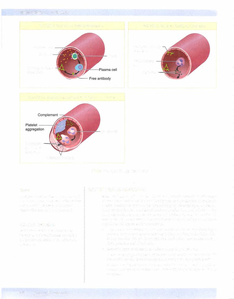
2.Type II hypersensitivity reactions (antibody-mediated) are characterized by production of an IgG or IgM antibody directed against a specific target cell or tissue. These reactions can take several forms. In complement dependent cytotoxicity, fixation of complement results in osmotic lysis or opsonization of antibody-coated cells; examples include autoimmune hemolytic anemia, transfusion reactions, and erythroblastosis fetalis.
In antibody-dependent cell-mediated cytoxicity (ADCC), cytotoxic killing of an antibody-coated cell occurs; an example is pernicious anemia.Antireceptor antibodies can activate or interfere with receptors; examples include Graves disease and myasthenia gravis.
3.Type III hypersensitivity reactions (immune complex disease) are characterized by the formation of in situ or circulating antibody-antigen immune complexes, which deposit in tissue resulting in inflammation and tissue injury. Examples include serum sickness, systemic lupus erythema tosus (SLE), and glomerulonephritis.
4.Type IV hypersensitivity reactions (cell-mediated type) are mediated by sensitized T lymphocytes. In delayed type hypersensitivity, CD4+ THl-cell lymphocytes mediate granuloma formation; examples include the PPD skin test and tuberculosis.
In cytotoxic T-cell-mediated hypersensitivity, CDS+ T-cell lympho cytes destroy antigen-containing cells; examples include viral infections, immune reaction to tumors, contact dermatitis, and graft rejection.
MEDICAL 59

Chapter 7 • lmmunopathology
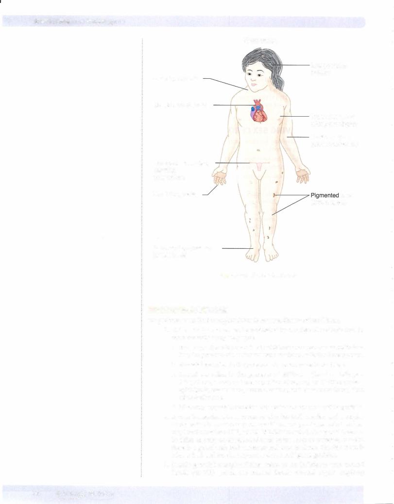
Skin (type III hypersensitivity reaction) manifestations can include a malar "butterfly" rash; maculopapular rash; and ulcerations and bullae formation.
Serosalsurfaces mayalso be affected,with resulting pericarditis, pleuritis, or pleural effusions (type III hypersensitivity reaction).
Central nervous system manifestations include focal neurologic symp toms, seizures, and psychosis (type III hype sensitivity reaction).
Cardiac manifestations include Libman-Sacks endocarditis (nonbacte rial verrucous endocarditis) (type III hypersensitivity reaction).
Of particular importance are the renal manifestations, which can be diverse. The WHO classification of kidney manifestations (type III hypersensitivity reaction) is as follows:
Class I: Normal
-Class II: Mesangial lupus nephritis
Class III: Focal proliferative glomerulonephritis
Class IV: Diffuse proliferative glomerulonephritis (most common and severe)
Class V: Membranous glomerulonephritis
d.Lupus is treated with steroids and immunosuppressive agents. It tends to have a chronic, unpredictable course with remissions and relapses. The 10-year survival is 85%, with death frequently being due to renal failure or infections.
2.Sjogren syndrome (sicca syndrome) is an autoimmune disease character ized by destruction of the lacrimal and salivary glands, resulting in the inability to produce saliva and tears. Females are affected more often than males, with typical age range of 30 to 50 years.
a.Clinical manifestations include keratoconjunctivitis sicca (dry eyes) and corneal ulcers; xerostomia (dry mouth); and Mikulicz syndrome (enlargement of the salivary and lacrimal glands). Sjogren syndrome is often associated with rheumatoid arthritis and other autoimmune dis eases. The characteristic autoantibodies are the anti-ribonucleoprotein antibodies SS-A (Ro) and SS-B (La). There is an increased risk of devel oping non-Hodgkin lymphoma.
3.Scleroderma (progressive systemic sclerosis) is an autoimmune disease characterized by fibroblast stimulation and deposition of collagen in the skin and internal organs. It affects females more than males, with typical age range of 20 to 55 years. The pathogenesis involves activation of fibro blasts by cytokines interleukin 1 (IL- 1), platelet-derived growth factor (PDGF), and/or fibroblast growth factor (FGF) with the resulting activated fibroblasts causing fibrosis.
a.Diffuse scleroderma has anti-DNA topoisomerase I antibodies (Scl-70) (70%), widespread skin involvement, and early involvement ofthevisceral organs. Organs that can be affected include the esophagus (dysphagia), GI tract (malabsorption), lungs (pulmonary fibrosis which causes dyspnea on exertion), heart (cardiac fibrosis which may manifest as arrhythmias), and kidney (fibrosis that may manifest as renal insufficiency).
b.Localized scleroderma (CREST syndrome) has anti-centromere anti bodies, skin involvement of the face and hands, late involvement of visceral organs, and a relatively benign clinical course.
4.Dermatomyositis and polymyositis are closely related conditions with immune-mediated muscle damage. Dermatomyositis has skin involvement (purple-red [heliotrope] rash on eyelids) and is due to antibody-mediated damage, while polymyositis does not have skin involvement and is due to
Butterfly Rash
Clinical Correlate
Anti-Ro antibiodies: Maternal anti-Ro (SS-A) antibodies have been implicated in the pathogenesis of congenital complete heart block.
In a Nutshell
CREST Syndrome
•Calcinosis
•Raynaud phenomenon
•Esophageal dysmotility
•Sclerodactyly
•Telangiectasia
MEDICAL 61

USMLE Step 1 • Pathology
© Richard P. Usatine, M.D. Used with permission.
Figure 7-4. Kaposi Sarcoma in an AIDS patient
Non-Hodgkin lymphomas tend to be high-grade B-cell lymphomas; extranodal CNS lymphomas are common.
Other AIDS-defining diseases include cervical cancer, HIV-wasting syndrome, AIDS nephropathy, and AIDS dementia complex.
66 MEDICAL

Chapter 7 • lmmunopathology
Chapter Summary
•Type I hypersensitivity (anaphylactic type) reactions are characterized by lgE-related release of chemical mediators from mast cells and basophils following exposure to an antigen. Examples include systemic anaphylaxis following bee stings and drugs. Localized forms of anaphylactic reaction include food allergies, atopy, and asthma.
•Type II hypersensitivity (cytotoxic type) reactions are characterized by production of an lgG or lgM antibody directed against a specific target cell or tissue. Examples include the complement-dependent cytotoxicity of autoimmune hemolytic anemia, the antibody-dependent cell-mediated cytotoxicity of pernicious anemia, and the antireceptor antibodies of Graves disease.
•Type Ill hypersensitivity (immune complex disease) reactions are characterized by the formation ofin situ or circulating antibody-antigen complexes that deposit in tissue, resulting in inflammation and tissue injury. Examples include serum sickness, systemic lupus erythematosus, and glomerulonephritis.
•Type IV hypersensitivity (cell-mediated type) reactions are mediated by sensitized T lymphocytes. Examples include the delayed hypersensitivity of PPD skin tests and tuberculosis and the cytotoxic T-cell-mediated destruction of antigen-containing cells in viral infections, immune reaction to tumors, contact dermatitis, and graft rejection.
•Systemic lupus erythematosus is a chronic systemic autoimmune disease characterized by a loss of self-tolerance and production of autoantibodies. Clinical manifestations include hemolytic anemia and other autoimmune hematologic manifestations, arthritis, skin rashes, and involvement ofthe renal, cardiovascular, and neurologic systems.
•Sjogren syndrome (sicca syndrome) is an autoimmune disease characterized by destruction of the lacrimal and salivary glands resulting in the inability to produce saliva and tears. Sjogren syndrome is associated with the antiribonucleoprotein antibodies SS-A and SS-B, and also with other autoimmune diseases such as systemic lupus erythematosus.
•Scleroderma (progressive systemic sclerosis) is an autoimmune disease characterized by fibroblast stimulation and deposition of collagen in the skin and internal organs. Scleroderma can have anti-DNA topoisomerase I antibodies (Scl-70), widespread skin involvement, and early involvement ofthe esophagus, GI tract, lung, heart, and kidney. Localized forms have a more benign course.
•X-linked agammaglobulinemia of Bruton is an inherited immunodeficiency characterized by a developmental failure to produce mature B cells and plasma cells, resulting in agammaglobulinemia with recurrent bacterial infections.
•Common variable immunodeficiency is a group of disorders characterized by a B-cell maturation defect and hypogammaglobulinemia expressed as increased susceptibility to bacterial infections, Giardia lamblia, autoimmune diseases, lymphoma, and gastric cancer.
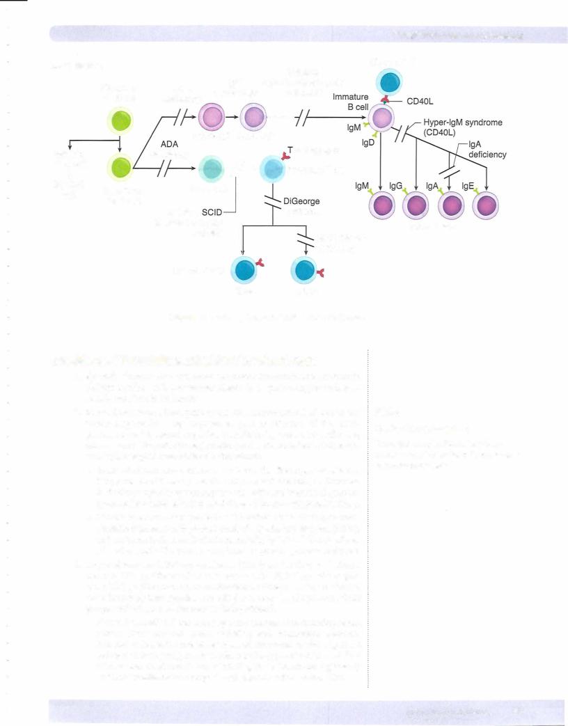
•DiGeorge syndrome is an embryologic failure to develop the third and fourth pharyngeal pouches, resulting in the absence of the parathyroid glands and thymus, leadingto hypocalcemia with tetany, T-cell deficiency, and recurrent infections with viral and fungal organisms.
(Continued)
MEDICAL 67
USMLE Step 1 • Pathology
Chapter Summary (conrd)
•Severe combined immunodeficiency (SCIO) is a combined deficiency of cell mediated and humoral immunity often caused by a stem cell defect that, without treatment, causes death by infection within 1 year. Affected infants are
susceptible to recurrent infections by bacteria, fungi, viruses, and protozoa.
• Wiskott-Aldrich syndrome is an X-linked condition characterized by recurrent infections, severe thrombocytopenia, and eczema.
• Secondary immune deficiency syndromes can be caused by systemic diseases such as diabetes mellitus, collagen vascular disease (i.e., SLE), and chronic alcoholism.
• Rejection following renal transplantation can occur in three patterns: hyperacute rejection due to preformed antibodies thattriggervascular thrombosis,
acute rejection characterized by neutrophilic vasculitis, and chronic rejection characterized by intimal fibrosis ofvessels.
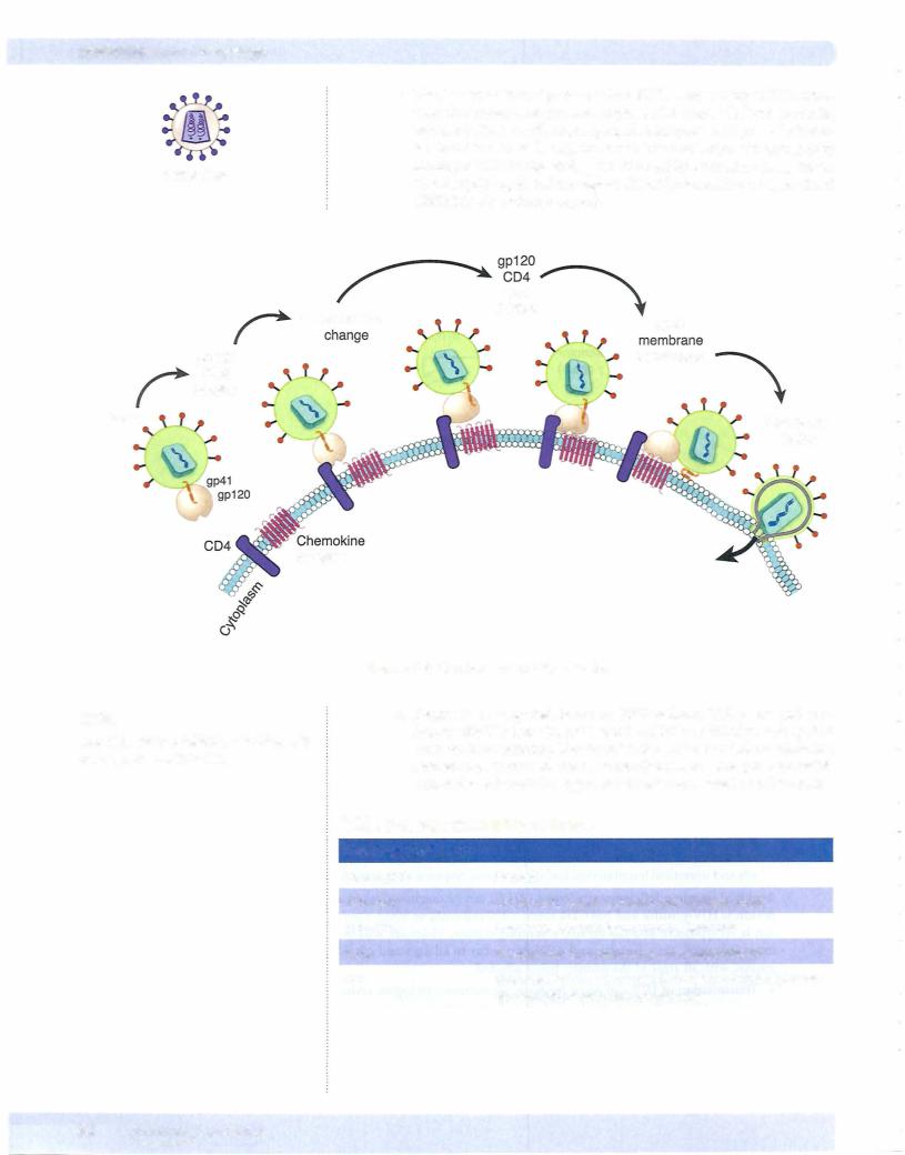
• Acquired immunodeficiency syndrome (AIDS) is said to be present when a patient is HIV positive with CD4 count less than 200 or HIV positive with an AIDS-defining disease. HIV can be spread by sexual contact, parenteral transmission, or vertical transmission. The virus is an RNA retrovirus with reverse transcriptase and a predilection for infecting CD4+ cells. Diagnosis is by HIV-antibody ELISA test followed by Western blot confirmation. A variety of drugs are now available for treatment.
• HIV infection produces a mononucleosis-like acute phase, an asymptomatic latent phase, and then progression to AIDS. Clinical AIDS is characterized by susceptibility to a wide variety of opportunistic infections. AIDS patients are also prone to develop hairy leukoplakia, Kaposi sarcoma, high-grade B-cell lymphomas, cervical cancer, a wasting syndrome, nephropathy, and dementia.
68 MEDICAL