

- •Contents
- •1. Fundamentals of Pathology
- •4. Tissue Repair
- •5. Circulatory Pathology
- •8. Amyloidosis
- •13. Vascular Pathology
- •16. Renal Pathology
- •17. Gastrointestinal Tract Pathology
- •18. Pancreatic Pathology
- •21. Central Nervous System Pathology
- •23. Female Genital Pathology
- •24. Breast Pathology
- •25. Male Pathology
- •26. Endocrine Pathology
- •27. Bone Pathology
- •28. Joint Pathology

Renal Pathology 1 6
CONGENITAL ANOMALIES OF THE KIDNEY
1 . Renal agenesis
a.Bilateral agenesis is incompatible with life. Ultrasound shows oligo hydramnios. Affected fetuses typically also have Potter facies (flattened nose, low-set ears, and recessed chin); talipes equinovarus (talus [ankle]
+pes [foot] and equino [heel] + varus [turned upward] = clubfoot); and pulmonary hypoplasia.
b.In unilateral agenesis, the remaining kidney undergoes compensatory hypertrophy. Patients often have adequate renal function, but they may develop progressive glomerular sclerosis.
2.Hypoplasia refers to failure of a kidney (usually unilateral) to develop to normal weight; the hypoplastic kidney has a decreased number of calyces and lobes.
3. Horseshoe kidney is a common congenital anomaly that is found in 1 in 750 autopsies. The kidneys show fusion, usually at the lower pole; affected individuals have normal renal function but may be predisposed to renal calculi.
4.Abnormal locations. The most common abnormal location is a pelvic kidney. The ectopic kidney usually has normal function. Tortuosity of ureters may predispose to pyelonephritis.
Clinical Correlate
The central portion of a horseshoe kidney may be found just inferiorto the inferior mesenteric artery because its embryologic ascent is arrested by the artery's presence.
CYSTIC DISEASE
1.Autosomal recessivepolycystickidneydisease (also called childhood poly cystic kidney disease) is a rare autosomal recessive disease that presents in infancywith progressive and often fatal renal failure.
a.The kidneys are bilaterally enlarged and have multiple small cysts in the cortex and medulla. The cysts occur in the collecting ducts of the nephron and are consequently oriented in a radial fashion with their long axis at right angles to the renal capsule.
b.In the liver, patients mayalso have multiplehepatic cysts and congenital hepatic fibrosis.
2.Autosomal dominantpolycystickidneydisease (also called adultpolycys tic kidney disease) is an autosomal dominant disease that affects 1 in 1,000 people. There is most frequently a mutation ofthe PKD1 gene on chromo some 16 that produces a transmembrane protein called polycystin 1 . Other mutations involve PKD2 and PKD3 genes.
a.Clinically, patients are asymptomatic with normal renal function until middle age, and then present with renal insufficiency, hematuria, and hypertension or with abdominal masses and flank pain. The diagnosis is establishedwith ultrasound and CT scans. Most patients develop end stage renal failure by their seventh decade.
Infantile
Polycystic Disease
MEDICAL 147
USMLE Step 1 • Pathology
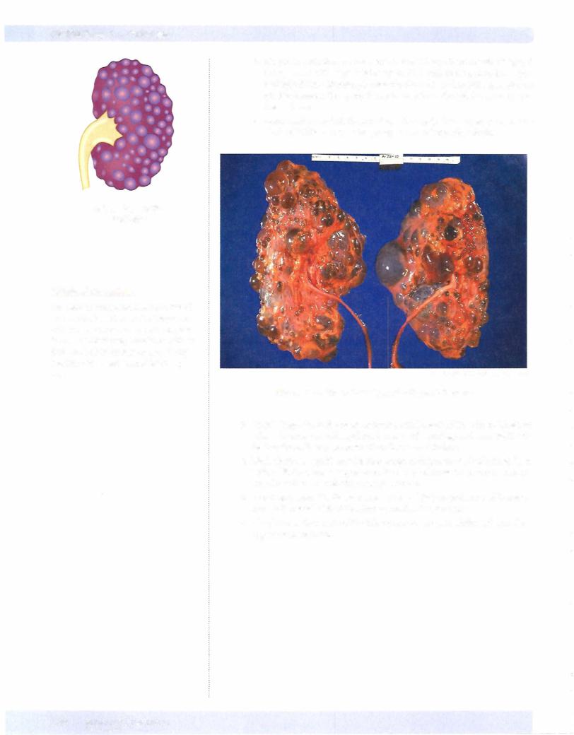
b.On gross pathologic examination, the kidneys have massive bilateral enlargement with large bulging cysts filled with serous, turbid, or hem orrhagic fluid. Microscopic examination shows functioning nephrons present between the cysts; the cysts arise from the tubular structures of the nephron.
c.Extrarenal manifestations include liver cysts, berry aneurysms of the circle ofWillis, mitralvalve prolapse, and colonic diverticula.
Adult Polycystic
Disease
Clinical Correlate
The cysts in autosomal dominant (adult) PKO involve less than 10% of nephrons, but they gradually expand and compress the rest ofthe kidney, interfering with its function. This is the reason why kidney function can remain normal for many years.
© cdc.gov. Used with permission.
Figure 16-1 . Gross Pathology of Polycystic Kidneys
3.Renal dysplasia is the most common renal cystic disease in children, in whom it causes an enlarged renal mass with cartilage and immature col lecting ducts. It mayprogress clinically to renal failure.
4.Medullary spongekidneydisease causes multiple cysts ofcollecting ducts with a "Swiss cheese" appearance. It may predispose to recurrent urinary tract infections, hematuria, and renal stones.
5.Acquired polycystic disease is seen in renal dialysis patients and is associ ated with a small risk ofdeveloping renal cell carcinoma.
6.Simpleretentioncysts ofthe kidney are common in adults and occasion ally cause hematuria.
148 MEDICAL
USMLE Step 1 • Pathology
Clinical Correlate
The classic presentation ofAPSGN is a young child with fever, malaise, periorbital edema, hypertension, smoky urine, and oliguria, beginning approximately 2 weeks aftera streptococcal throat infection.
Note
The characteristic finding in RPGN is the formation of crescents within Bowman space. The crescents are composed of fibrin, parietal epithelial cells, monocytes, and macrophages.
PRIMARY GLOMERULOPATHIES (NEPHRITIC)
1. Acute poststreptococcal glomerulonephritis (APSGN), (also called acute proliferative glomerulonephritis or postinfectious glomerulonephritis), typi cally occurs 2-4 weeks after a streptococcal infection of the throat or skin. There is a decreasing incidence in the United States; children are affected more frequently than adults.
a.The infecting organism is most commonly -hemolytic group A strep tococci, but acute proliferative glomerulonephritis can also be caused by other bacteria, viruses, and parasites, and even systemic diseases (SLE and polyarteritis nodosa).
b.Clinically, the condition presents with nephritic syndrome with elevated antistreptolysin 0 (ASO) titers (when related to streptococcal infection) and low serum complement.
c.Renal biopsy. Light microscopy shows hypercellular glomeruli with
neutrophils and monocytes; there are also red cell casts in the renal tubules. Immunofluorescence shows granular deposits of IgG, IgM, and C3 throughout the glomerulus. Electron microscopy shows subepithelial immune complex deposits (humps).
d. Clinical course. The treatment is with conservative fluid manage ment. Most children have a good prognosis, with complete recovery in >95% of cases, but severe disease (RPGN in 1% of cases and chronic glomerulonephritis in 2%) can also occur. Adults completely recover in 60% of cases, but develop either RPGN or chronic renal disease in the remaining 40%.
2. Goodpasture syndrome (anti-GBM disease) is due to the production of antibodies directed against basement membrane (anti-GBM antibodies), which results in damage of the lungs and the kidneys. The Goodpasture antigen is the noncollagenous component of type IV collagen.
a. Clinicalfeatures. The peak incidence ofGoodpasture is ages 20 to 40, and males are affected more frequentlythan females. Pulmonary involvement typically precedes the renal disease, typically presenting with pulmonary hemorrhage and recurrent hemoptysis. Most patients willalso later develop RPGN.
b. Renal biopsy findings. Light microscopy shows hypercellularity, cres cents, and fibrin depostion in glomeruli. Immunofluorescence shows a smooth and linear pattern of IgG and C3 in the glomerular basement membrane (GBM). By electron microscopy, there are no deposits, but there is glomerular basement membrane disruption.
c.Even with treatment (plasma exchange, steroids, and cytotoxic drugs), the prognosis is poor, because of risks of severe and life-threatening pulmonary hemorrhage and of RPGN leading to renal failure. Early aggressive treatment may prevent end-stage renal failure.
3.Rapidly progressive glomerulonephritis (RPGN), also called crescentic
glomerulonephritis, causes rapid progression to severe renal failure in weeks or months
a. Clinical settings. RPGN can occur following Goodpasture syndrome; following other forms of glomerulonephritis (post-streptococcal, SLE, Berger disease); in association with vasculitis (i.e., Wegner granulomato sis); or be idiopathic.
b. Renal biopsy. Light microscopy shows hypercellular glomeruli with cres cent formation in Bowman space. Immunofluorescence may show vari able results, including granular or linear deposits ofimmunoglobulin and
1 50 MEDICAL
Chapter 16 • Renal Pathology
complement. Electron microscopy may also have variable results, which may or may not include electron-dense deposits; glomerular basement membrane disruption and discontinuity are commonly seen.
c.The prognosis is poor with rapid progression to acute renal failure and end-stage renal disease.
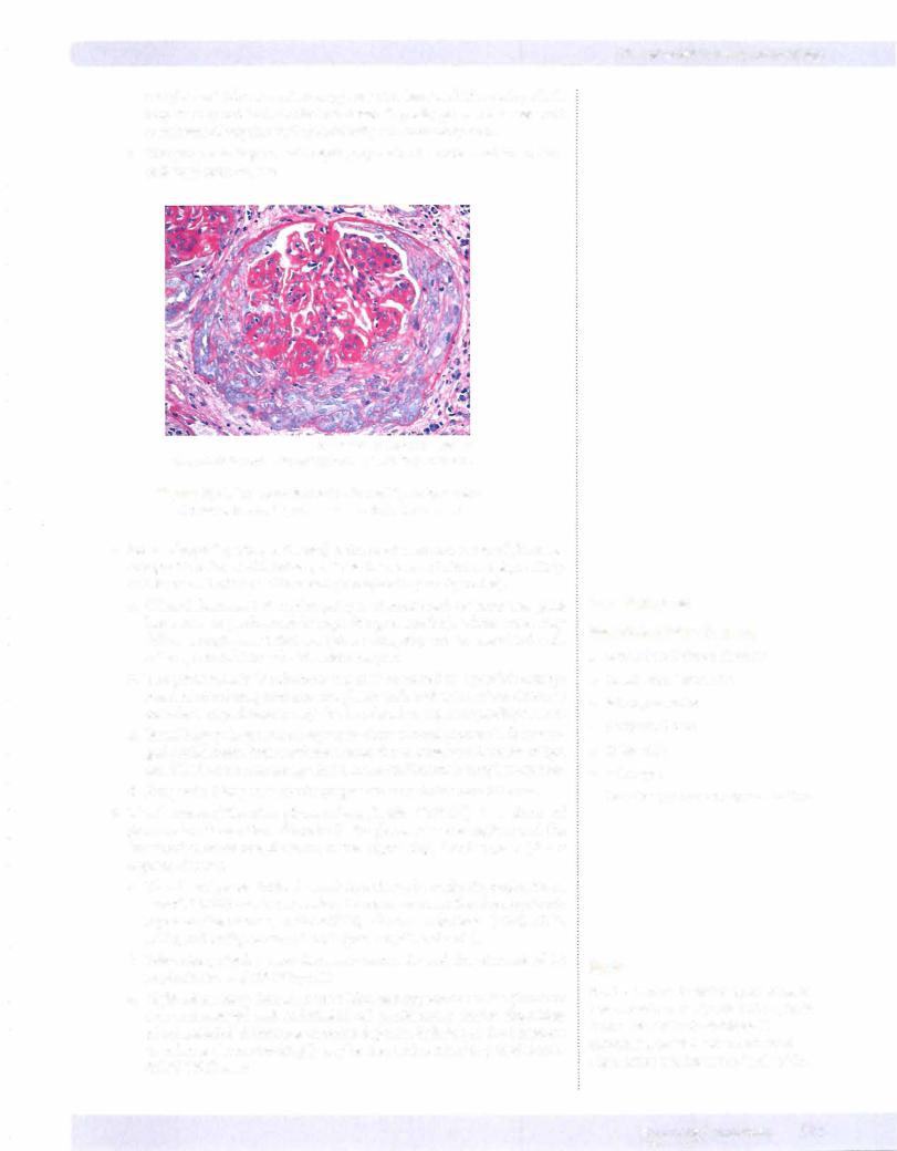
© Katsumi M. Miyai, M.D., Ph.D.; Regents ofthe University of California. Used with permission.
Figure 16-3. Crescent formation in rapidly progressive glomerulonephritis, as seen with trichrome stain
4.IgA nephropathy (Berger disease) is the most common cause of glomeru lonephritis in the world, being particularly common in France, Japan, Italy, and Austria. It affects children and young adults (mostly males).
a.Clinical features. lgA nephropathy is characterized by recurrent gross hematuria (a predominately nephritic presentation), whose onset may follow a respiratory infection. IgA nephropathy can be associated with celiac sprue and Henoch-Schonlein purpura.
b.The pathogenesis is unknown, but may be related to a possible entrap ment of circulating immune complexes with activation of the alternate complement pathway; it may also be related to a genetic predisposition.
c.Renal biopsy. Light microscopy may show normal glomeruli or mesan gial proliferation. Immunofluorescence shows mesangial deposits of IgA and C3. Electron microscopy shows mesangial immune complex deposits.
d.Prognosis. Many cases slowly progress to renal failure over 25 years.
5.Membranoproliferative glomerulonephritis (MPGN) is a form of glomerular disease that affects both the glomerular mesangium and the basement membranes; it occurs in two types, Type I and Type II (dense deposit disease).
a.The clinicalpresentation is variable, and may be nephritic, nephrotic, or mixed. MPGN may be secondary to many systemic disorders (systemic lupus erythematosus, endocarditis), chronic infections (HBV, HCV, HIV), and malignancies (chronic lymphocytic leukemia).
b.Laboratory studies show decreased serum C3 and the presence of C3 nephritic factor (MPGN type II).
c.Light microscopy demonstrates a lobulated appearance ofthe glomeruli due to mesangial and endothelial cell proliferation and/or deposition of subendothelial immune complex deposits. Splitting of the basement membrane ("tram-tracking") may be seen with a silver or periodic acid Schiff (PAS) stain.
In a Nutshell
Henoch-Schonlein Purpura
•Systemic childhood disorder
•Onset often follows URI
•lgA nephropathy
•Abdominal pain
•GI bleeding
•Arthralgia
•Palpable purpura on legs and buttock
Note
Most patients with MPGN type II disease have an autoantibodycalled C3 nephritic factor. This antibody stabilizes C3 convertase, which leads to enhanced degradation and low serum levels of C3.
MEDICAL 151
USMLE Step 1 • Pathology
Note
"Tram-Tracking"
This double contour appearance is caused by the splitting ofthe GBM by extension of the mesangial cell processes into the capillary loop.
152 MEDICAL
d.lmmunofluorescence in type I MPGN shows a granular pattern of C3 often with IgG, Clq, and C4; in type II, immunoflourescence shows a granular and linear pattern of C3.
e.Electron microscopy in type I shows subendothelial and mesangial immune complex deposits, and in type II shows dense deposits within the glomerular basement membrane.
f.Prognosis. MPGN typically has a slowly progressive course, resulting in chronic renal failure over the course of 10 years. There is a high incidence of recurrence in transplants.
6.Alport syndrome is a rare X-linked disorder caused by a defect in type IV collagen that is characterized by hereditary nephritis, hearing loss, and ocu lar abnormalities. The most common mutation causing Alport syndrome is in the COL4A5 gene coding for the alpha-5 chain of type 4 collagen.
a.Clinical features. Gross or microscopic hematuria begins in childhood. Hearing loss (leading to sensorineural deafness) and various ocular abnormalities of the lens and cornea can occur. Alport syndrome is a progressive disease that ultimately results in renal failure.
b.Electronmicroscopyshows alternating thickening and thinning of base ment membrane with splitting of the lamina densa.
PRIMARY GLOMERULOPATHIES (NEPHROTIC)
1.Membranous glomerulonephritis is a common cause of nephrotic syn drome in adults.
a.Etiology. Most cases (85%) are idiopathic. Membranous glomerulone phritis may also be caused by drugs (penicillamine), infections (hepatitis virus B and C, syphilis, etc.), and systemic diseases (SLE, diabetes mel litus, etc.). It has also been associated with malignant carcinomas of the lung and colon, and there may be a genetic predisposition.
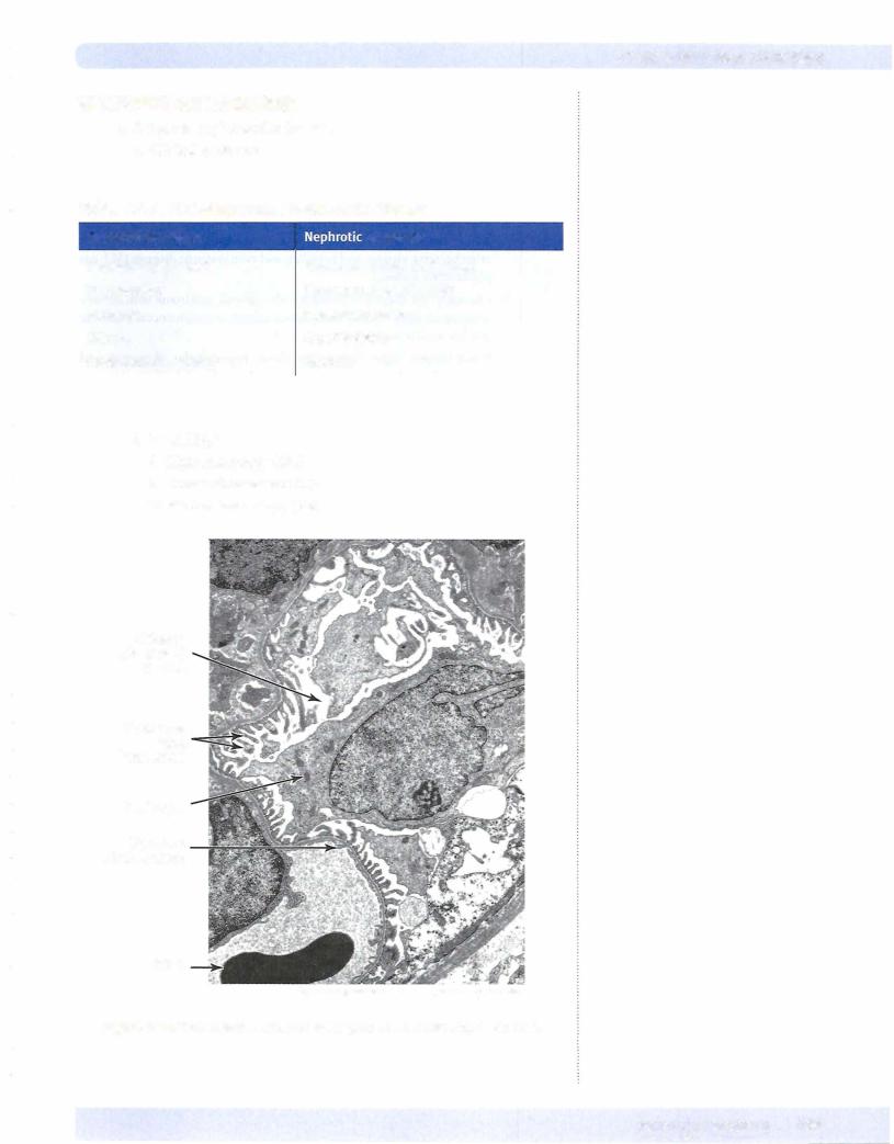
b.Renal biopsy. Light microscopy shows diffuse thickening of the capil lary walls. Basement membrane projections ("spikes") are seen on silver stains. Immunofluorescence shows a granular and linear pattern of IgG and C3. Electron microscopy shows subepithelial deposits along the basement membranes with effacement of podocyte foot processes.
c.The clinical course is variable, and may lead to spontaneous remission; persistent proteinuria; or end-stage renal disease.
2.Minimal change disease (also called lipoid nephrosis and nil disease) is the most common cause of nephrotic syndrome in children. The disease has peak incidence at ages 2 to 6.
a.The diagnosis is one of exclusion. Light microscopy shows normal glomeruli with lipid accumulation in proximal tubule cells (lipoid nephrosis). Immunofluorescence is negative, with no immune deposits being visualized. Electron microscopy shows effacement of epithe lial (podocyte) foot processes, microvillous transformation, and no immune complex deposits.
b.Clinical course. The prognosis is excellent because treatment with corticosteroids produces a dramatic response in children. The majority have a complete recovery.
3.Focal segmental glomerulosclerosis is a very common cause of nephrotic syndrome that occurs in all ages, with African Americans being affected more frequently than Caucasians.
a.Etiology.Theconditionmay beidiopathic(primary), or itmay be related to a wide variety of predisposing conditions including loss of renal tissue;
pre-existing other glomerular diseases (such as IgA nephropathy); sickle cell anemia; heroin use; AIDS; or morbid obesity.
b.Renal biopsy. Light microscopy shows focal segmental sclerosis and hyalinization of glomeruli; focal segmental glomerulosclerosis initially affects the glomeruli along the medullary border. Immunofluorescence shows IgM and C3 deposits in the sclerotic segments. Electron micros copy shows effacement of foot processes in nonsclerotic regions and increased mesangial matrix in sclerotic segments.
c.Clinical course. There is frequently a poor response to steroids,with the overallprognosis being poor (most progressing to chronic renal failure), though children do better than adults. There is a high rate ofrecurrence in renal transplants.
SECONDARY GLOMERULONEPHRITIS
1.Secondaryglomerulonephritis refers to glomerular disease that is second ary to other disease processes.
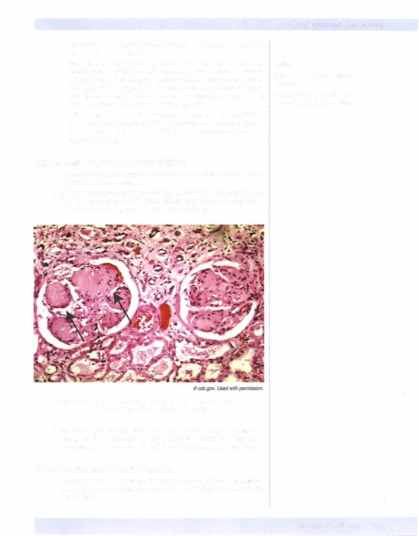
2.Diabetes causes nodularglomerulosclerosis, hyaline arteriolosclerosis, and diabetic microangiopathy. Clinically, diabetic patients may develop micro alburninuria that can progress to nephrotic syndrome.
Figure 1 6-4. Sclerotic nodules (arrows) of nodular glomerulosclerosis (Kimmelstiel-Wilson syndrome), kidney
3.Systemic lupus erythematosus can cause a wide variety of patterns of damage to the kidney with clinical features that can include hematuria, nephritic syndrome, nephrotic syndrome, hypertension, and renal failure.
CHRONIC GLOMERULONEPHRITIS
1.End-stage renal disease is the final stage of many forms of glomerular disease and is characterized by progressive renal failure, uremia, and ulti mately death.
Chapter 16 • Renal Pathology
Note
Focal: only some ofthe glomeruli are affected
Segmental: only a portion ofthe glomerulartuftexhibits sclerosis
MEDICAL 153

USMLE Step 1 • Pathology
Clinical Correlate
It may be difficult to distinguish cystitis from pyelonephritis. The presence of fever, costovertebral angle tenderness, and WBC casts in the urine are helpful clues to the diagnosis of pyelonephritis.
1 54 MEDICAL
2.Clinical features include anemia, anorexia, and malaise; and proteinuria, hypertension, and azotemia. Urinalysis shows broad, waxy casts.
3.On pathologic examination, the kidneys are grossly small and shrunken; microscopic examination shows hyalinization of glomeruli, interstitial fibrosis, atrophy oftubules, and a lymphocytic infiltrate.
4.Treatment is with dialysis and renal transplantation.
DISEASES OFTHE TUBULES AND INTERSTITIUM
1 . Acute tubular necrosis (ATN) is acute renal failure associated with poten tially reversible injury to the tubular epithelium.
a. Clinical features. ATN is the most common cause of acute renal fail ure in the United States. It is characterized by oliguria with elevation of blood urea nitrogen (BUN) and creatinine; metabolic acidosis and hyperkalemia; and dirty brown granular casts and epithelial casts on urinalysis.
b. Ischemic acute tubular necrosis is the most common cause of acute tubular necrosis. The condition is due to decreased blood flow caused by severe hemorrhage, severe renal vasoconstriction, hypotension, dehydration, or shock.
c. Nephrotoxic acute tubular necrosis has a large number of causes, including drugs (e.g., polymyxin, methicillin, gentamicin, sulfon amides); radiographic contrast agents; heavymetals (e.g., mercury, lead, gold); organic solvents (e.g., carbon tetrachloride, chloroform, methyl alcohol); ethylene glycol (antifreeze); mushroom poisoning; phenol; pesticides; and myoglobin.
d.The prognosis is excellent ifthe patient survives the disease responsible for causing the necrosis.
2.Acute and chronic pyelonephritis refer to bacterial infections involving the
renal pelvis, tubules, and interstitium. Pyelonephritis affects females much more than males.
a. Pathogenesis. Ascending infection is the most common route of infection. Causative organisms include gram-negative enteric bacilli,
Escherichia coli, Prate.us, Klebsiella, and Enterobacter.
b. Predisposingfactors include urinary obstruction, vesicoureteral reflux, pregnancy, urethral instrumentation, diabetes mellitus, benign prostatic hypertrophy, and other renal pathology.
c. Symptoms can include fever, chills, and malaise; dysuria, frequency, and urgency; and costovertebral angle tenderness. Urinalysis shows pyuria and white blood cell casts.
d. Microscopically, the kidney shows acute inflammation of the intersti tium and tubules, or chronic inflammation with fibrosis (which may progress to renal failure).
3. Tubulointerstitial nephritis is an acute or chronic inflammation of tubules and interstitum. It can be due to many causes, including medica tions, infections, acute pyelonephritis, systemic lupus erythematosus, lead poisoning, urate nephropathy, or multiple myeloma.
4. Analgesic nephropathy is the most common cause of chronic drug induced tubular interstitial nephritis. It may cause renalpapillarynecrosis, hypertension, chronic renal failure, and transitional cell carcinoma ofthe renal pelvis and bladder.
Chapter i.6 • Renal Pathology
5.Urate nephropathy is due to a deposition of urate crystals (secondary to leukemia treatment, lead poisoning, and gout) in renal tubules and inter stitium. It may produce acute renal failure.
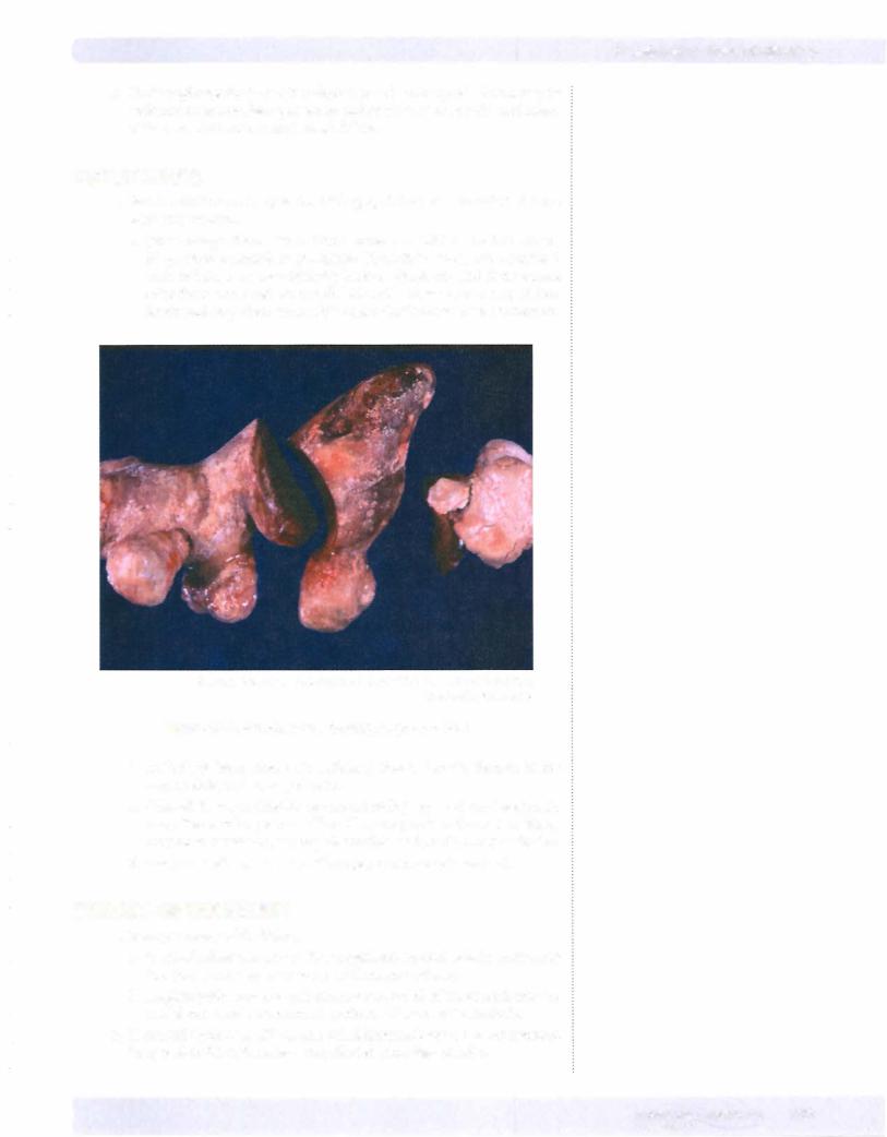
UROLITHIASIS
1. Renal calculi occur in up to 6% of the population; men are affected more often than women.
a.Stone composition. Most (75%) stones are calcium oxalate stones. Magnesium ammonium phosphate ("struvite") stones are associated with infection by urea-splitting bacteria (proteus), and these stones often form large staghorn calculi. Uric acid stones are seen in gout, leu kemia, and in patients with acidic urine. Cystine stones are uncommon.
© Katsumi M. Miyai, M.D., Ph.D.; Regents of the University of California. Used with permission.
Figure 16-5. Struvite stone forming staghorn calculi
b.Pathology. Most stones are unilateral stones that are formed in the calyx, pelvis, and urinary bladder.
c.Clinical features. Calcium stones are radiopaque and can be seen on x-ray. Renal colic may occur ifsmall stones pass into the ureters. Stones may cause hematuria, urinary obstruction, and predispose to infection.
d.Treatment of stones is with lithotripsy or endoscopic removal.
TUMORS OF THE KIDNEY
1 . Benign tumors of the kidney.
a.Cortical adenomas are small, encapsulated cortical nodules measuring less than 3 cm; they are a common finding at autopsy.
b.Angiomyolipomas are hamartomas composed of fat, smooth muscle, and blood vessels, common in patients with tuberous sclerosis.
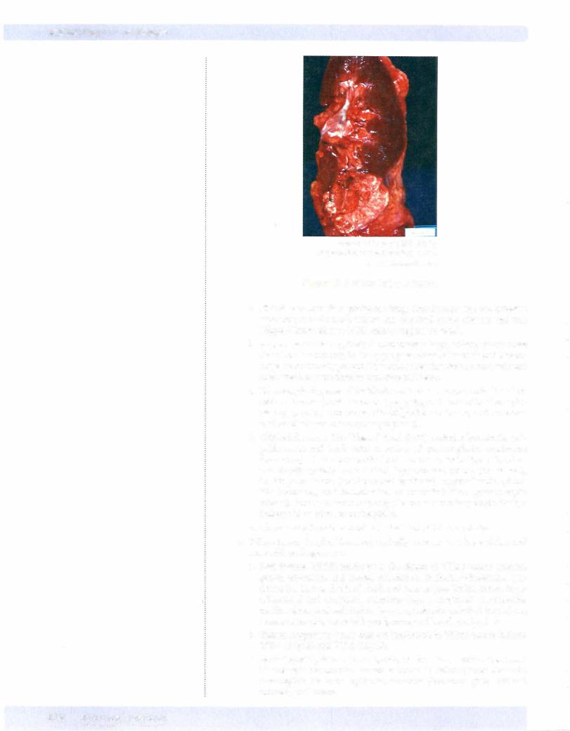
2.Renal cell carcinoma (RCC), also called hypernephroma, is most common in ages 50 to 70, with males being affected more than females.
MEDICAL 1 5 5
USMLE Step 1 • Pathology
© Katsumi M. Miyai, M.D., Ph.D.;
Regents ofthe University of California.
Used with permission.
Figure 1 6-6. Renal Cell Carcinoma
a. Risk factors include cigarette smoking; chronic analgesic use; asbestos exposure; chronic renal failure and acquired cystic disease; and von Hippel-Lindau disease (VHL tumor suppressor gene).
b. Gross examination typically demonstrates a large, solitary yellow mass found most commonly in the upper pole. Areas of necrosis and hemor rhage are commonly present. The tumor often invades the renal vein and may extend into the inferior vena cava and heart.
c. Microscopically, several histologic variants can occur, including clear cell carcinoma (most common type, polygonal cells with clear cyto plasm), papillary carcinoma, chromophobe carcinoma, and sarcoma toid renal cell carcinoma (poor prognosis).
d. Clinical features. The "classic" triad (10%) includes hematuria, pal pable mass, and flank pain. A variety of paraneoplastic syndromes from ectopic hormone production can occur, including polycythe mia (erythropoietin production), hypertension (renin production), Cushing syndrome (corticosteroid synthesis), hypercalcemia (PTH like hormone), and feminization or masculinization (gonadotropin release). Renal cell carcinoma may also cause secondary amyloidosis, a leukemoid reaction, or eosinophilia.
e. There is a high incidence of metastasis on initial presentation.
3.Wilms tumor (nephroblastoma) typically presents as a large abdominal
mass with peak age 2 to 5.
a. Risk factors. WAGR syndrome is the cluster of Wilms tumor, aniridia, genital anomalies, and mental retardation. Beckwith-Wiedemann syn drome has increased risk of childhood cancers (e.g., Wilms tumor, hepa toblastoma) and congenital anomalies (e.g., macroglossia, macrosomia, midline abdominal wall defects [e.g., omphalocele, umbilical hernia], ear creases or ear pits, neonatal hypoglycemia, and hemihypertrophy).
b. Tumor suppressor genes that are implicated in Wilms tumor include WT- 1 ( l lpl3) and WT-2 (l lplS).
c. Pathologically, Wilms tumor grossly causes a large, solitary tan mass. Microscopic exanimation reveals a tumor containing three elements: metanephric blastema, epithelial elements (immature glomeruli and tubules), and stroma.
1 56 MEDICAL
Chapter 16 • Renal Pathology
d.Treatment is with surgery, chemotherapy, and radiation; this combina tion therapyyields an excellent prognosis, with long-term survival rate of90%.
4.Transitional cell carcinomas can involve the renal pelvis as well as the urinary bladder.
CHRONIC RENAL FAILURE
1 . Chronic renal failure is the end stage ofmany different renal diseases. It is characterized pathologically by bilaterally shrunken kidneys. Clinically, it causes progressive irreversible azotemia, normocytic anemia, platelet dys function, renal osteodystrophy, and hypertension.
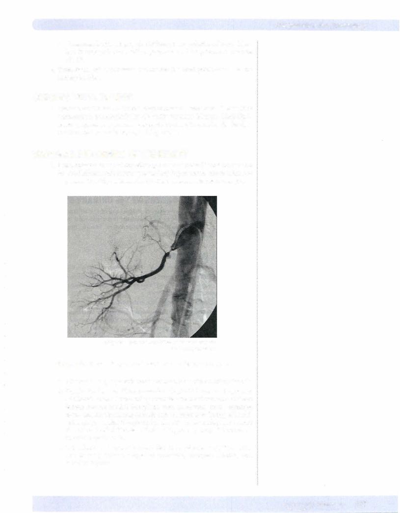
VASCULAR DISORDERS OF THE KIDNEY
1. Renal artery stenosis of any etiology causes decreased blood flow to the involved kidney, with resulting secondary hypertension that is often not responsive to antihypertensive medications; treatment is usually surgical.
© Lily Chu - National Naval Medical Center Bethesda.
Used with permission.
Figure 1 6-7. Renal artery stenosis as demonstrated by angiogram
a.Atheromatousplaque is the most commoncause ofrenal artery stenosis.
b.Dysplastic lesions ("fibromuscular dysplasia") are an important additional cause of renal artery stenosis. The most common of these lesions include medial fibroplasia with aneurysms (most common form, causing alternating stenosis and aneurysms in "string of beads" pattern), perimedial fibroplasia (involves the outer media), and medial dissection (medial fibrosis with dissecting aneurysms); all three occur in middle-aged adults.
c.Miscellaneous diseases that can affect the renal arteries (with or with out stenosis) include congenital anomalies, Takayasu arteritis, and radiation injuries.
MEDICAL 1 57
USMLE Step 1 • Pathology
2.Benign nephrosderosis is due to hypertension, and is characterized microscopically by hyaline arteriolosclerosis, tubular atrophy, interstitial fibrosis, and glomerulosclerosis. Laboratory findings include mild protein uria, hematuria, and azotemia.
3.Malignant (accelerated) hypertension can damage the kidney, causing fibrinoid necrosis of arterioles, glomerulitis, and hyperplastic arteriolo sclerosis. Clinically, malignant hypertension causes cerebral edema, papill edema, retinal hemorrhage, intracerebral hemorrhage, and oliguric acute renal failure.
4.Renalinfa rction can be due to thrombi from the left side ofthe heart, ath eroembolic disease, and vasculitis. Renal infarctions presents with sudden onset offlank pain and hematuria.
5.Sickle cellanemia can cause medullary infarctions due to blockage ofblood flow in the medullary vessels, which can result in asymptomatic hematuria, loss ofurine concentrating ability, renal papillarynecrosis, andpyelonephritis.
6.Diffuse cortical necrosis can cause anuria; the condition can occur with obstetric emergencies and disseminated intravascular coagulation.
OBSTRUCTIVE DISORDERS OF THE URINARY SYSTEM
1. Hydronephrosis is a common complication of urinary tract obstruction that is characterized by dilation of the ureter and renal pelvis. Specific causes include renal stones, retroperitoneal fibrosis, benign prostatic hyperplasia, and cervical cancer.
URETERAL DISORDERS
1 . Congenital anomalies include double ureters and congenital megaureter.
2.Ureteritiscystica is a term used when chronic inflammationcauses forma tion ofsmall mucosa! cysts in the ureter. This condition can predispose for adenocarcinoma of the ureter.
3.Renal stones commonly lodge in the ureters.
4.Retroperitoneal fibrosis is usually an idiopathic condition which causes severe fibrosis ofthe retroperitoneal area that can entrap the ureters. Some cases show sclerosing conditions in other body sites.
5.Transitional cell carcinoma is the most common ureteral carcinoma.
URINARY BLADDER PATHOLOGY
1 . Cystitis
a. The etiology of cystitis varies, with important causes including organ isms, notably from fecal flora
Enterobacter); radiation cystitis (may follow radiation therapy); and che motherapy agents such as cydophosphamide (hemorrhagic cystitis).
b.Clinically, cystitis affects females far more than males. Symptoms include frequency, urgency, dysuria, and suprapubic pain; systemic signs (e.g., fever, chills, malaise) are uncommon.
c.Predisposing factors include benign prostatic hypertrophy, bladder calculi, and cystocele.
2.Urinary bladder tumors are most commonly due to transitional cell carcinoma. There is an increasing incidence of urinary bladder tumors; males are affected more than females, and the peak incidence is between ages 40 and 60.
1 58 MEDICAL
Chapter 16 • Renal Pathology
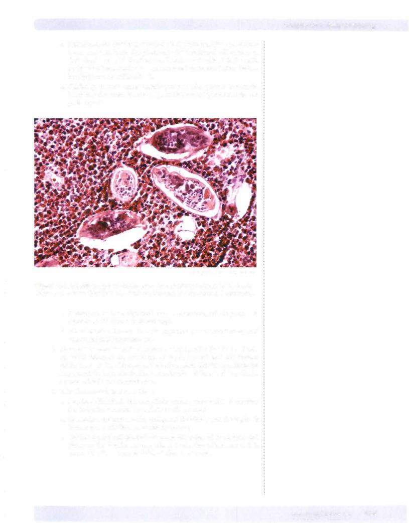
a.Riskfactors for bladder tumors include cigarette smoking and occupa tional exposure to azo dye production for transitional cell carcinoma (both due to 2-naphthylamine) aswell as chronic bladder infectionwith Schistosoma haemotobium for squamous cell carcinoma (Africa includ
ing Egypt and the Middle East).
b.Clinically, bladder cancer usually presents with painless hematuria, but it may also cause dysuria, urgency, frequency, hydronephrosis, and pyelonephritis.
@ cdc.gov. Used with permission.
Figure 16-8. Migratory eggs of Schistosoma haemotobium surrounded by dense inflammation in the bladder wall, which predisposes to squamous cell carcinoma
c.Bladder cancer has a high incidence of recurrence, and the prognosis depends on the tumor grade and stage.
d.Other bladder tumors include papillomas, adenocarcinoma, and embryonal rhabdomyosarcoma.
3.Congenital anomalies ofthe bladder. Exstrophy of the bladder is a devel opmental failure of the formation of the abdominal wall and bladder which leaves the bladder open at the body surface. Urachal cyst remnants may permit drainage ofurine from a newborn's umbilicus, and may also be a cause ofbladder adenocarcinoma.
4.Miscellaneousbladderconditions.
a.Acquired diverticuli can complicate urinary tract outlet obstruction due to benign prostatic hyperplasia or other causes.
b.Cystocele is the term used for prolapse ofthe bladder into the vagina. It is common in middle-aged to elderly women.
c.Cystitis cystica and glandularis cause formation of small cysts and glands in the bladder mucosa related to chronic inflammation. It is associated with an increased risk of adenocarcinoma.
|
|
MEDICAL |
1 59 |
USMLE Step :t • Pathology
Chapter Summary
•Renal agenesis is the failure ofone or both kidneys to develop. Bilateral renal agenesis is incompatiblewith life, but personswith unilateral agenesis may have adequate renal function. Other congenital anomalies ofthe kidney include hypoplasia, horseshoe kidney, and abnormal locations.
•Autosomal recessive polycystic kidney disease presents in infancy with progressive renal failure. Autosomal dominant polycystic kidney disease presents in adulthood with renal insufficiency, hematuria, and hypertension. The kidneys may be massively enlarged by the time of diagnosis. Renal dysplasia
is the most common renal cystic disease in children and may cause a renal mass and renal failure. Medullary sponge kidney may cause a "Swiss cheese" appearance to the kidney and predisposes for infection, hematuria, and stones. Acquired polycystic disease is seen in renal dialysis patients. Simple retention cysts are common in adult kidneys.
•Glomerular diseases can present with either nephritic syndrome or nephrotic syndrome. Nephritic syndrome is characterized by hematuria, hypertension, azotemia, oliguria, and proteinuria less than 3.5 g/day. Nephrotic syndrome is characterized by severe proteinuria greaterthan 3.5 g/day, hypoalbuminemia, generalized edema, hyperlipidemia, and lipiduria.
•Acute post-streptococcal glomerulonephritis is associated with subepithelial immune complex deposits (subepithelial humps) by electron microscopy, occurs 2-4 weeks after a streptococcal infection ofthe throat or skin, and usually causes nephritic syndrome in children.
•Goodpasture syndrome is characterized by a smooth and linear pattern of lgG and C3 by immunofluorescence. It is the result of damage by autoantibodies to the basement membranes ofthe lungs and kidneys and is characterized clinically by pulmonary hemorrhage and rapidly progressive glomerulonephritis.
•Rapidly progressive glomerulonephritis is characterized microscopically by hypercellular glomeruli with crescent formation in Bowman space. Clinically, it features rapid progression to severe renal failure in weeks or months. It can be seen idiopathically or as a complication of renal disease due to Goodpasture syndrome, other forms ofglomerulonephritis, or vasculitis.
•lgA nephropathy is characterized by mesangial deposits of lgA and C3, is the
most common cause ofglomerulonephritis worldwide, and tends to produce recurrentgross hematuria in children and young adults.
•Membranoproliferative glomerulonephritis is characterized microscopically by mesangial proliferation and basement membrane splitting and clinically may produce a nephritic pattern, a nephrotic pattern, or a mixed pattern.
•Membranous glomerulonephritis is characterized by diffuse thickening of capillary walls and basement membrane projections (spikes) visible with silver stains and is a common cause of nephrotic syndrome in adults.
•Minimal change disease is characterized by effacement of epithelial (podocyte) foot processes visible with electron microscopy and is the most common cause of nephrotic syndrome in children.
(Continued)
160 MEDICAL
Chapter :1.6 • Renal Pathology
Chapter Summary (cont'd)
•Focal segmental glomerulosclerosis is characterized by focal segmental sclerosis and hyalinization of glomeruli and is a cause of nephrotic syndrome that can occur idiopathically or secondary to other glomerular diseases, sickle cell anemia, heroin use, AIDS, and morbid obesity.
•Secondary glomerulonephritis can be caused by diabetes mellitus and systemic lupus erythematosus.
•Chronic glomerulonephritis with small, shrunken kidneys is the finalstage of many forms ofglomerular diseases and is characterized by progressive renal failure, uremia, and ultimately death.
•Acute tubular necrosis is acute renal failure associated with reversible injury to the tubular epithelium and can be due to ischemia or nephrotoxins.
•Acute and chronic pyelonephritis is a bacterial infection involving the renal pelvis, tubules, and interstitium and is most commonly due to Escherichia coli, Proteus, Klebsiella, or Enterobacter.
•Renal calculi are common and may be composed ofcalcium oxalate, struvite, uric acid, or cystine. Clinically, stones may cause renal colic, hematuria, urinary obstruction, and a predisposition forinfection.
•Benign tumors ofthe kidney include cortical adenomas and angiomyolipomas. Renal cell carcinoma tends to produce a large solitary renal mass in middle aged to older adults and may cause hematuria, palpable mass, flank pain, and paraneoplastic syndromes.
•Wilms tumor is a childhood malignancy that presentswith a large abdominal mass. It now has an excellent long-term prognosis.
•Vascular diseases ofthe kidney include renal artery stenosis (decreased blood flow to the kidney leading to secondary hypertension), benign nephrosclerosis (which develops secondaryto ordinary hypertension), hyperplastic arteriolosclerosis (which develops secondary to malignant hypertension), renal infarction (from emboli from the left side ofthe heart and secondary to sickle cell anemia), and diffuse cortical necrosis (secondary to obstetric emergencies and DIC).
•Ureteral disorders include congenital anomalies, ureteritis, cystic disease, stones lodged in a ureter, retroperitoneal fibrosis, and transitional cell carcinoma.
•Cystitis, or urinary bladder inflammation, can be due to bacterial infection, radiation, or chemotherapy; cystitis clinically produces frequency, urgency, dysuria, and suprapubic pain.
•Transitional cell carcinoma is the most common type of bladdertumor and usually presents with painless hematuria. Otherbladdertumors include papillomas, adenocarcinoma, and embryonal rhabdomyosarcoma.
•Congenital anomalies ofthe bladder include exstrophy and urachal cyst remnants.
•Other bladder conditions include acquired diverticuli, cystocele, and cystitis cystica and glandularis.
MEDICAL 161