

Fundamentals of Pathology |
1 |
DEFINITIONS OF PATHOLOGY
1.The study of the essential nature of disease, including symptoms/signs, pathogenesis, complications, and morphologic consequences including structural and functional alterations in cells, tissues, and organs.
2.The study of all aspects of the disease process focusing on the pathogenesis leading to classical structural changes (gross and histopathology) as well as molecular alterations.
OVERVIEW OF PATHOLOGY
1.The etiology (cause) of a disease may be genetic or acquired.
2.The pathogenesis of a disease defines the temporal sequence and the pat terns of cellular injury that lead to disease.
3.Morphologic changes of the disease process include both gross changes and microscopic changes.
4.The clinical significance of a disease relates to its signs and symptoms, dis ease course including complications, and prognosis.
METHODS USED IN PATHOLOGY
1. Gross examination of organs on USMLE questions has hvo major com ponents: identifying the organ and identifying the pathology. Useful gross features include size, shape, consistency, and color.
2.Microscopic examination of tissue
a.In light microscopic examination of tissue, hematoxylin and eosin (H&E) is considered the gold standard stain and is used routinely in the initial microscopic examination of pathologic specimens.
Table 1-1. Structures Stained by Hematoxylin and Eosin
Hematoxylin |
Eosin |
||
Stains blue to purple |
Stains pink to red |
||
• |
Nuclei |
• |
Cytoplasm |
• |
Nucleoli |
• |
Collagen |
• |
Bacteria |
• |
Rbrin |
• |
Calcium |
• |
RBCs |
• |
Many others |
• |
Thyroid colloid |
|
|
• |
Many others |
MEDICAL 1
USMLE Step 1 • Pathology
The common denominator of the features shown in Table 1-1 is that hematoxylin binds nucleic acids and calcium salts, while eosin stains the majority ofproteins (both extracellular and intracellular).
b. Other histochemical stains (chemical reactions): Prussian blue (stains iron), Congo red (stains amyloid), acid fast (Ziehl-Neelson, Fite) (stains acid-fast bacilli), periodic acid-Schiff (PAS, stains high carbohydrate con tent molecules), Gram stain (stains bacteria), trichrome (stains cells and connective tissue), and reticulin (stains collagen type III molecules).
© Katsumi M. Miyai, M.D., Ph.D.; Regents of the University of California. Used with permission.
Figure 1-1 . Prussian blue stain showing hemosiderin, which results from RBC breakdown within macrophages
c. Immunohistochemical (antibody) stains include cytokeratin (stains epithelial cells), vimentin (stains cells of mesenchymal origin except the 3 muscle types; stains many sarcomas), desmin (stains smooth, cardiac, and skeletal myosin), prostate specific antigen, and many others.
3. Ancillarytechniques
a. Immunofluorescence microscopy (IFM) is typically used for renal and autoimmune disease.
b.Transmission electron microscopy (EM) is typically used for renal dis ease, neoplasms, infections, and genetic disorders.
4.Molecular techniques include protein electrophoresis, Southern and
Western blots, polymerase chain reaction (PCR), and cytogenetic analysis (karyotyping, in situ hybridization studies).
2MEDICAL

Chapter 1 • Fundamentals of Pathology
Chapter Summary
•Pathology is the study of disease and concerns itself with the etiology, pathogenesis, morphologic changes, and clinical significance of different diseases.
•Gross examination of organs involves identifying pathologic lesions by evaluating abnormalities of size, shape, consistency, and color.
•Tissue sections stained with hematoxylin (nucleic acids and calcium salts) and eosin (most proteins) are used for routine light microscopic examination.
•Additional techniques that are used to clarify diagnoses in particularsettings include histochemical stains, immunohistochemical stains, immunofluorescence microscopy, transmission electron microscopy, and molecular techniques.
MEDICAL 3

USMLE Step 1 • Pathology
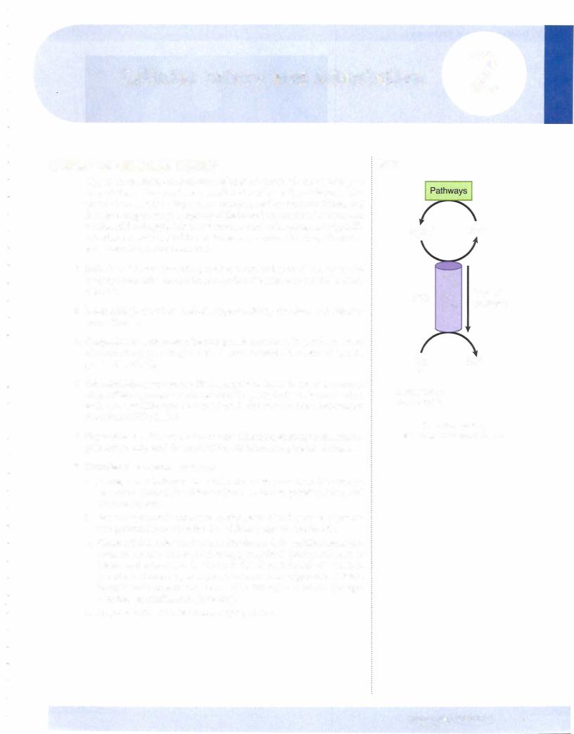
Normal
Normal cell
Injury
Note
Reversible and irreversible changes represent a spectrum. Keep in mind that any of the reversible changes can become irreversible.
Clinical Correlate
The loss of membrane integrity (cell death) allows intracellular enzymes to leak out, which can then be measured in the blood. Detection ofthese proteins in the circulation serves as a clinical marker of cell death and organ injury. Clinically important examples:
•Myocardial injury: troponin (most specific), CPK-MB, lactate dehydrogenase (LDH)
•Hepatitis: transaminases
•Pancreatitis: amylase and lipase
•Biliary tract obstruction: alkaline phosphatase
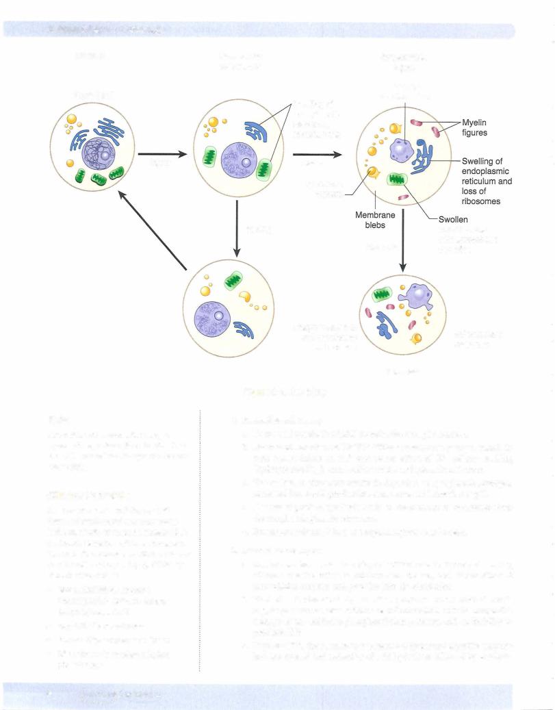
Reversible
Cell Injury
Swelling of endoplasmic reticulum, mitochondria
Death
Lysosome
rupture
Healing
Fragmentation of cell membrane and nucleus
Figure 2-4. Cell Injury
2. Reversible cellinjury
a. Decreased synthesis ofATP
Irreversible
Injury
Nuclear condensation
mitochondria
with amorphous Necrosis densities
Inflammatory response
Necrosis
by oxidative phosphorylation.
b. Decreased function of Na+K+ ATPase membrane pumps, which in turn causes influx of Na+ and water, efflux of K+, cellular swelling (hydropic swelling), and swelling of the endoplasmic reticulum.
c.The switch to glycolysis results in depletion of cytoplasmic glycogen, increased lactic acid production, and decreased intracellular pH.
d.Decreased protein synthesis leads to detachment of ribosomes from the rough endoplasmic reticulum.
e.Plasma-membrane blebs and myelin figures may be seen.
3.Irreversible cell injury
a.Severe membrane damage plays a critical role in irreversible injury, allows a massive influx of calcium into the cell, and allows efflux of intracellular enzymes and proteins into the circulation.
b.Marked mitochondrial dysfunction produces mitochondrial swell ing, large densities seen within the mitochondrial matrix, irreparable damage of the oxidative phosphorylation pathway, and an inability to produce ATP.
c.Rupture of the lysosomes causes release of lysosomal digestive enzymes into the cytosol and activation of acid hydrolases followed by autolysis.
8MEDICAL
Chapter z • Cellular Injury and Adaptation
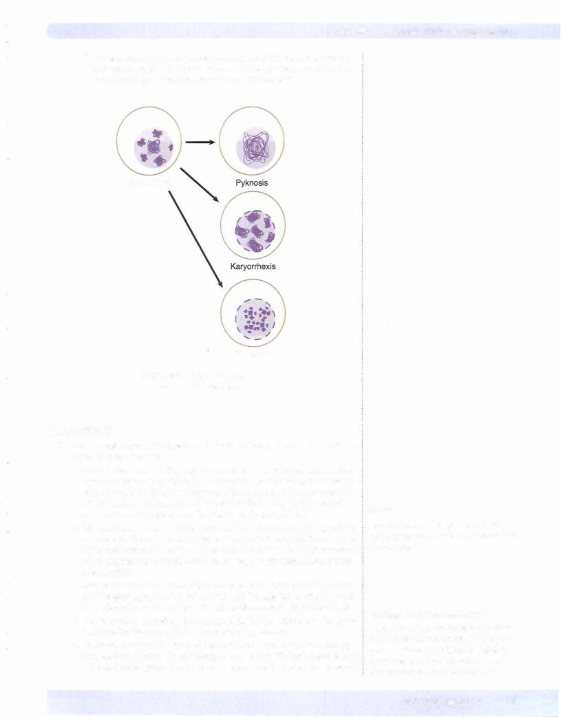
d.Nuclear changes (see figure below) can include pyknosis (degeneration and condensation of nuclear chromatin), karyorrhexis (nuclear frag mentation), and karyolysis (dissolution of the nucleus).
Normal Cell
Karyolysis
Figure 2-5. Nuclear Changes in
Irreversible Cell Injury
CELL DEATH
1.Morphologic types of necrosis (cell death in living tissue, often with an inflammatory response)
a.Coagulative necrosis, the most common form of necrosis, is most often due to ischemic injury (infarct). It is caused by the denaturing and coagula tion of proteins within the cytoplasm. Microscopic examination shows loss of the nucleus but preservation of cellular shape. Coagulative necrosis is common in most organs, including the heart, liver, and kidney.
b.Liquefaction necrosis results from cellular destruction by hydrolytic enzymes, leading to autolysis (release of proteolytic enzymes from injured cells) and heterolysis (release of proteolytic enzymes from inflammatory cells). Liquefaction necrosis occurs in abscesses, brain infarcts, and pancre atic necrosis.
c.Caseous necrosis is a combination of coagulation and liquefaction necro sis. The gross appearance is soft, friable, and "cottage cheese-like:' Caseous necrosis is characteristic of granulomatous diseases, including tuberculosis
d.Fat necrosis is caused by the action of lipases on adipocytes. On gross examination fat necrosis has a chalky white appearance.
e.Fibrinoid necrosis is a form of necrotic connective tissue that histologi cally resembles fibrin. On microscopic examination fibrinoid necrosis has an eosinophilic (pink) homogeneous appearance. It is often due to acute
Note
Liquefaction by leukocyte enzymes is called suppuration, and the resultant fluid is called pus.
Bridge to Biochemistry
Damage to fat cells releases triglycerides. The triglycerides are broken down by the action of Iipases to fatty acids. The fatty acids may associate with calcium and form calcium soaps (saponification).
MEDICAL 9
USMLE Step 1 • Pathology
Note
Necrotic tissue withinthe body evokes an inflammatory response that removes the dead tissue and is followed by healing and tissue repair. Necrotic debris may also undergo dystrophic calcification.
immunologic injury (e.g., hypersensitivity type reactions II and III) and vascular hypertensive damage.
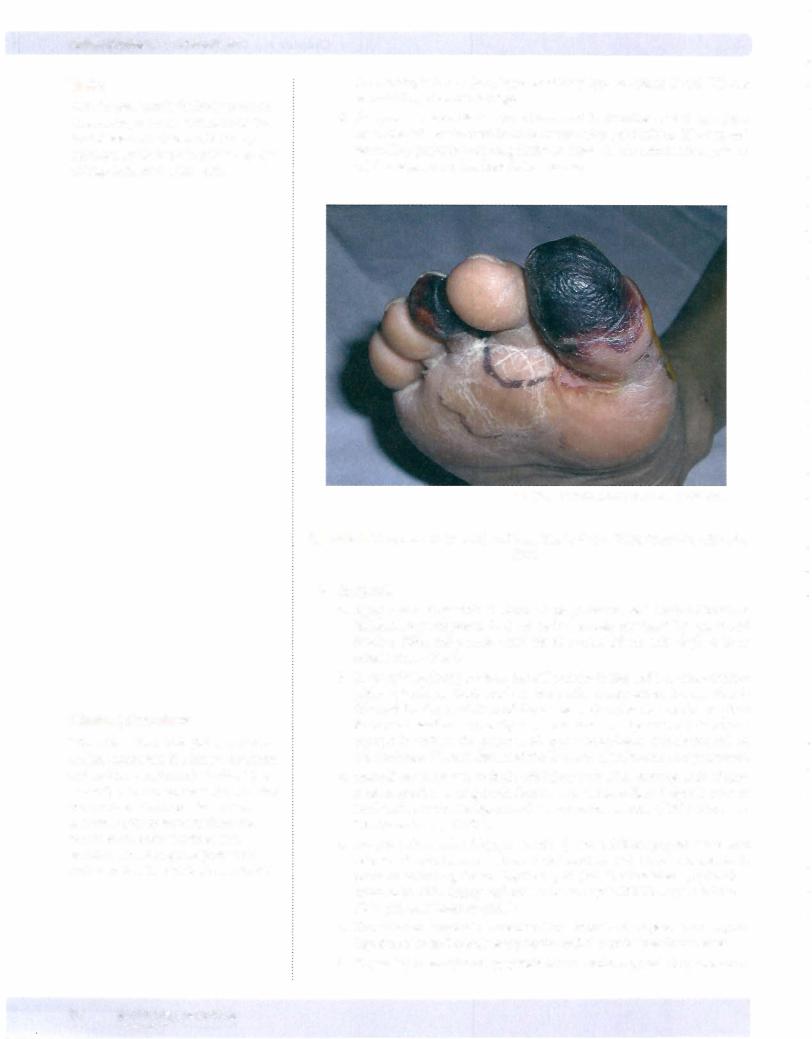
f. Gangrenous necrosis is a gross term used to describe dead tissue. Com mon sites of involvement include lower limbs, gallbladder, GI tract, and testes. Dry gangrene has coagulative necrosis for the microscopic pattern, while wet gangrene has liquefactive necrosis.
Clinical Correlate
Ifthe cells in the interdigital space fail to undergo apoptosis, the fetus will be born with webbed hands and/or webbed feet, a condition known as syndactyly. Another example is the hormone-dependent apoptosis prior to menstruation; this occurs as the body withdraws from estrogen and LH surges, signaling the endometrial cells to undergo apoptosis.
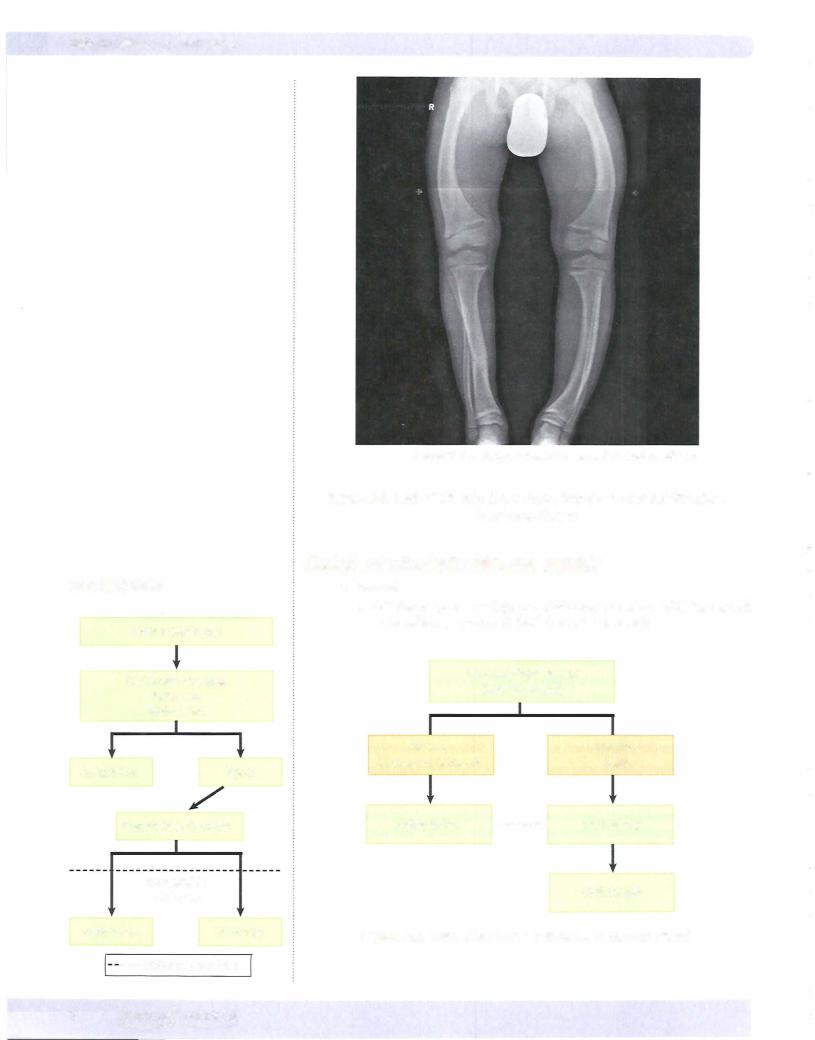
© Richard P. Usatine, M.D. Used with permission.
Figure 2-6. Gangrenous necrosis affects the first and third toes of a diabetic foot.
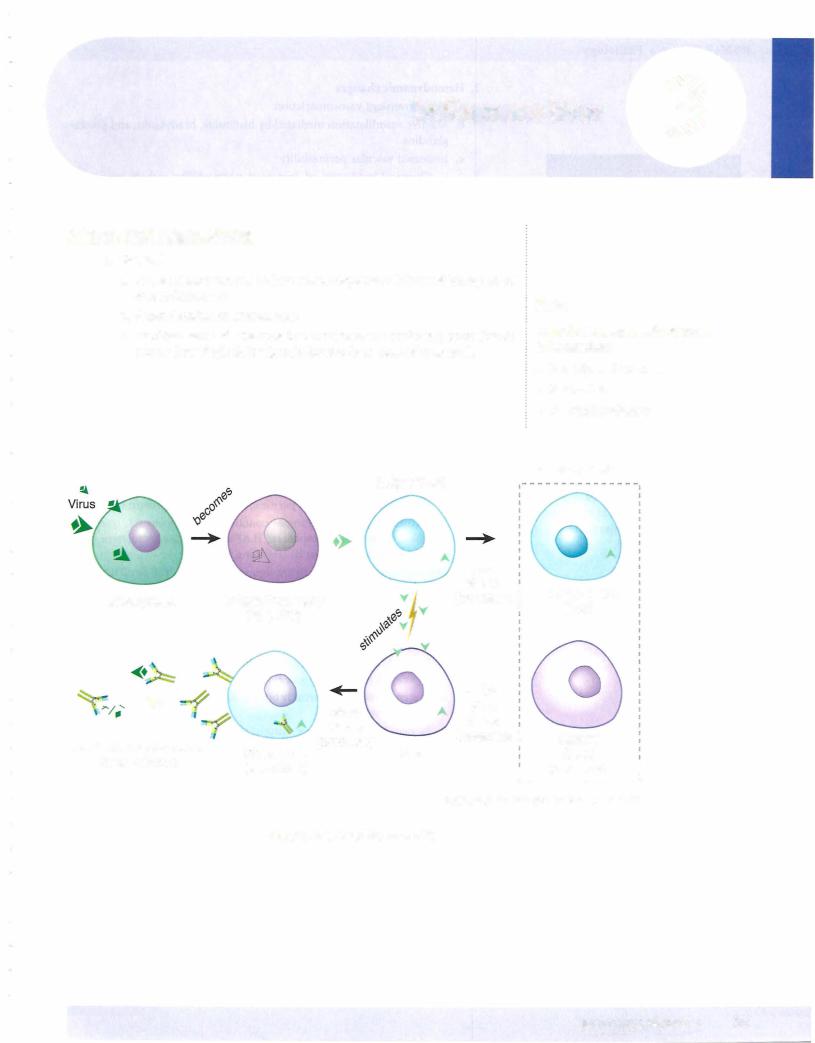
2.Apoptosis
a.Apoptosis is a specialized form of programmed cell death without an inflammatory response. It is an active process regulated by genes and involves RNA and protein synthesis that often affects only single cells or small groups of cells.
b.In morphologic appearance, the cell shrinks in size and has dense eosino philic cytoplasm. Next, nuclear chromatin condensation is seen that is followed by fragmentation of the nucleus. Cytoplasmic membrane blebs form next, leading eventually to a breakdown of the cell into fragments (apoptotic bodies). Phagocytosis of apoptotic bodies is by adjacent cells or macrophages. There is characteristically a lack of an inflammatory response.
c.Stimuli for apoptosis include cell injury and DNA damage, lack of hor mones, cytokines, or growth factors, and receptor-ligand signals such as Fas binding to the Fas ligand and Tumor necrosis factor (TNF) binding to T NF receptor 1 (TNFRl).
d.Apoptosis is regulated by genes. bcl-2 (which inhibits apoptosis) prevents release of cytochrome c from mitochondria and binds pro-apoptotic protease activating factor (Apaf-1). p53 (which stimulates apoptosis) is elevated by DNA injury and arrests the cell cycle. If DNA repair is impos sible, p53 stimulates apoptosis.
e.Execution of apoptosis is mediated by a cascade of caspases. The caspases digest nuclear and cytoskeletal proteins and also activate endonucleases.
f.Physiologic examples of apoptosis include embryogenesis (organogenesis
10 MEDICAL
USMLE Step 2. • Pathology
Clinical Correlate
Residence at high altitude, where oxygen content of air is relatively low, leads to compensatory hyperplasia of red blood cell precursors in the bone marrow and an increase in the number of circulating red blood cells (secondary polycythemia).
Clinical Correlate
Barrett's esophagus is a classic example of metaplasia. The esophageal epithelium is normally squamous, but it undergoes a change to intestinal epithelium (columnar) when it is under constant contact with gastric acid.
i.Increased mechanical demand can be physiologic (striated muscle of weight lifters) or pathologic (cardiac muscle in hypertension).
ii.Increased endocrine stimulation plays a role in puberty (growth hor mone, androgens/estrogens, etc.), gravid uterus (estrogen), and lac tating breast (prolactin and estrogen).
c.Hypertrophy is mediated by growth factors, cytokines, and other trophic stimuli and leads to increased expression of genes and increased protein synthesis.
d.Hypertrophy and hyperplasia often occur together.
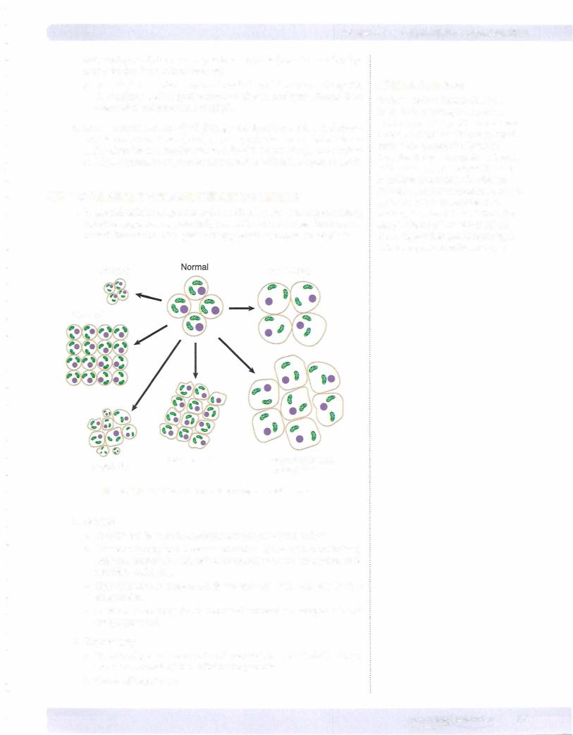
4.Hyperplasia
a.Hyperplasia is an increase in the number of cells in a tissue or organ.
b.Some cell types are unable to exhibit hyperplasia (e.g., nerve, cardiac, skeletal muscle cells).
c.Physiologic causes of hyperplasia include compensatory mechanisms (e.g., after partial hepatectomy), hormonal stimulation (e.g., breast development at puberty), and antigenic stimulation (e.g., lymphoid hyperplasia).
d.Pathologic causes of hyperplasia include endometrial hyperplasia and prostatic hyperplasia of aging.
e.Hyperplasia is mediated by growth factors, cytokines, and other trophic stimuli; increased expression of growth-promoting genes (protoonco genes); and increased DNA synthesis and cell division.
5.Metaplasia
a.Metaplasia is a reversible change of one cell type to another, usually in response to irritation. It has been suggested that the replacement cell is better able to tolerate the environmental stresses. For example, bron chial epithelium undergoes squamous metaplasia in response to the chronic irritation of tobacco smoke.
b.The proposed mechanism is that the reserve cells (or stem cells) of the irritated tissue differentiate into a more protective cell type due to the influence of growth factors, cytokines, and matrix components.
6.Dysplasia
a.Dysplasia is an abnormal proliferation of cells that is characterized by changes in cell size, shape, and loss of cellular organization.Dysplasia is not cancer but may progress to cancer (preneoplastic lesion). Examples include cervical dysplasia, actinic (solar) keratosis, and oral leukoplakia.
OTHER CELLULAR ALTERATIONS DURING INJURY
a.Lipids that can accumulate intracellularly include triglycerides (e.g., fatty change in liver cells), cholesterol (e.g., atherosclerosis, xanthomas), and complex lipids (e.g., sphingolipid accumulation).
b.Proteins can accumulate in proximal renal tubules in proteinuria and can form Russell bodies (intracytoplasmic accumulation of immuno globulins) in plasma cells.
c.Glycogen storage diseases (see Chapter 6)
d.Exogenous pigments include anthracotic pigmentation of thelung (sec ondary to the inhalation of carbon dust), tattoos, and lead that has been ingested (e.g., gingival lead line, renal tubular lead deposits).
12 MEDICAL
Chapter 2 • Cellular Injury and Adaptation
e.Endogenous pigments
i.Lipofuscin is a wear-and-tear pigment that is seen as perinuclear yellow-brown pigment. It is due to indigestible material within lyso somes and is common in the liver and heart.
ii.Melanin is a black-brown pigment derived from tyrosine found in melanocytes and substantia nigria.
iii.Hemosiderin is a golden yellow-brown granular pigment found in areas of hemorrhage or bruises. Systemic iron overload can lead to hemosiderosis (increase in total body iron stores without tissue injury) or hemochromatosis (increase in total body iron stores with tissue injury). Prussian blue stain can identify the iron in the hemo siderin.
iv.Bilirubin accumulates in newborns in the basal ganglia, causing per manent damage (kernicterus).
2.Hyaline change
a.Hyaline change is a nonspecific term used to describe any intracellular or extracellular alteration that has a pink homogenous appearance (pro teins) on H&E stains.
b.Examples of intracellular hyaline include renal proximal tubule protein reabsorption droplets, Russell bodies, and alcoholic hyaline.
c.Examples of extracellular hyaline include hyaline arteriolosclerosis, am yloid, and hyaline membrane disease of the newborn.
3.Pathologic forms of calcification
a.Dystrophic calcification is the precipitation of calcium phosphate in dy ing or necrotic tissues. Examples include fat necrosis (saponification), psammoma bodies (laminated calcifications that occur in meningiomas and papillary carcinomas of the thyroid and ovary), Monckeberg medial calcific sclerosis in arterial walls, and atherosclerotic plaques.
b.Metastatic calcification is the precipitation of calcium phosphate in normal tissue due to hypercalcemia (supersaturated solution). The many causes include hyperparathyroidism, parathyroid adeno mas, renal failure, paraneoplastic syndrome, vitamin D intoxication, Milk-alkali syndrome, sarcoidosis, paget disease, multiple myeloma, metastatic cancer to the bone. The calcifications are located in the interstitial tissues of the stomach, kidneys, lungs, and blood vessels.
MEDICAL 1 3
USMLE Step 1 • Pathology
Chapter Summary
•Cells can be damaged by a variety of mechanisms.
•Hypoxia causes a loss ofATP production secondary to oxygen deficiency and can be caused by ischemia, cardiopulmonary failure, or decreased oxygen-carrying capacity ofthe blood.
•Infections can injure cells directly, or indirectly, via toxin production or host inflammatory response.
•Hypersensitivity reactions and autoimmune diseases may kill or injure cells.
•Congenital causes of cellular injury include enzyme defects, structural protein defects, chromosomal disorders, and congenital malformations.
•Chemical agents, physical agents, and nutritional imbalances can also injure cells.
•The response of cells to an insult depends on both the state ofthe cell and the type of insult. The response can range from adaptation to reversible injury to irreversible injurywith cell death.
•Intracellular sites and systems particularly vulnerable to injury include DNA, ATP production, cell membranes, and protein synthesis.
•Reversible cell injury is primarily related to decreased ATP synthesis by oxidative phosphorylation, leading to cellular swelling and inadequate protein synthesis.
•Irreversible cell injury often additionally involves severe damage to membranes, mitochondria, lysosomes, and nucleus.
•Death oftissues (necrosis) can produce a variety of histologic patterns, including coagulative necrosis, liquefaction necrosis, caseous necrosis, fibrinoid necrosis, and gangrenous necrosis, often with an inflammatory response.
•Apoptosis is a specialized form of programmed cell death that can be regulated genetically or by cellular or tissue triggers without an inflammatory response.
14 MEDICAL
USMLE Step 1 • Pathology
Source: commons.wikimedia.org (Mgiganteus)
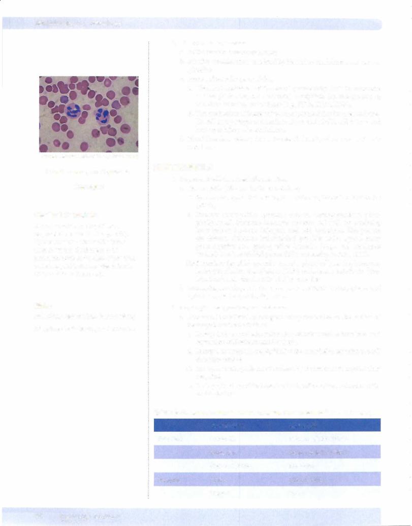
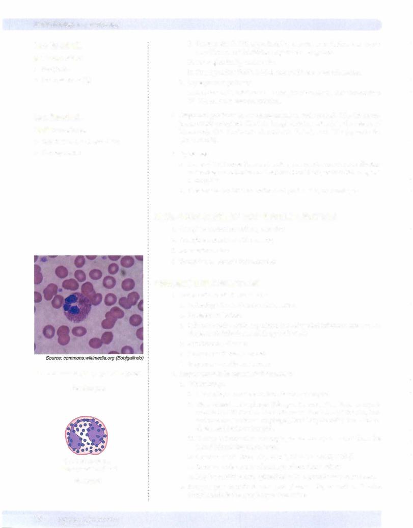
Lobed nucleus, small granules
Neutrophil
Clinical Correlate
A normal mature neutrophil has a segmented nucleus (3-4 segments). Hypersegmented neutrophils (more than 5) are usually thought to be pathognomonic of the class of anemias called megaloblastic anemias (vitamin 812 or folate deficiencies).
Note
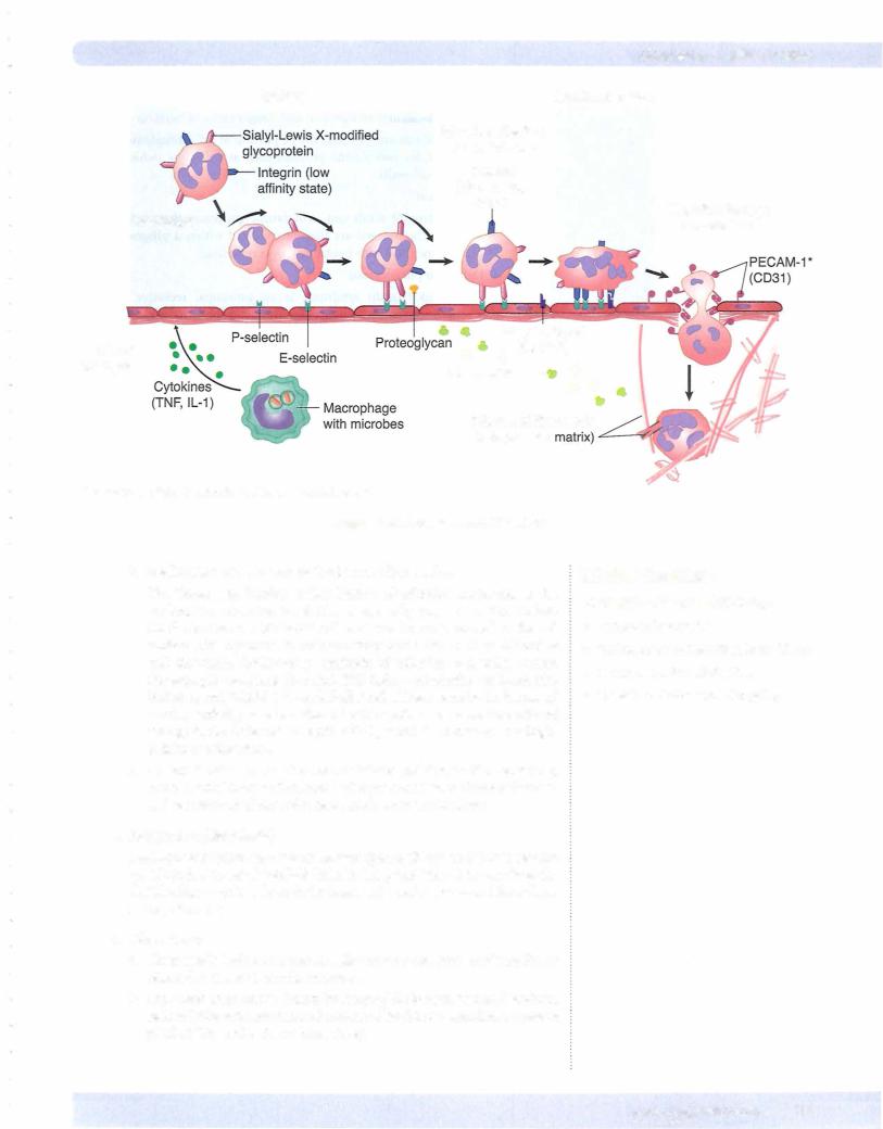
Selectins: weak binding; initiate rolling
lntegrins: stable binding and adhesion
2.Hemodynamic changes
a.Initial transient vasoconstriction
b.Massive vasodilatation mediated by histamine, bradykinin, and prosta glandins
c.Increased vascular permeability
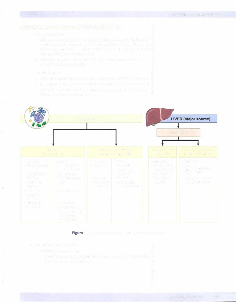
I.Chemical mediators of increased permeability include vasoactive amines (histamine, and serotonin), bradykinin (an end-product of the kinin cascade), leukotrienes (e.g., LTC4, LTD4, LTE4).
ii. The mechanism ofincreased vascular permeability involves endothe lial cell and pericyte contraction; direct endothelial cell injury; and leukocyte injury of endothelium.
d.Blood flow slows (stasis) due to increased viscosity, allows neutrophils to marginate
NEUTROPHILS
1.Important cells in acute inflammation
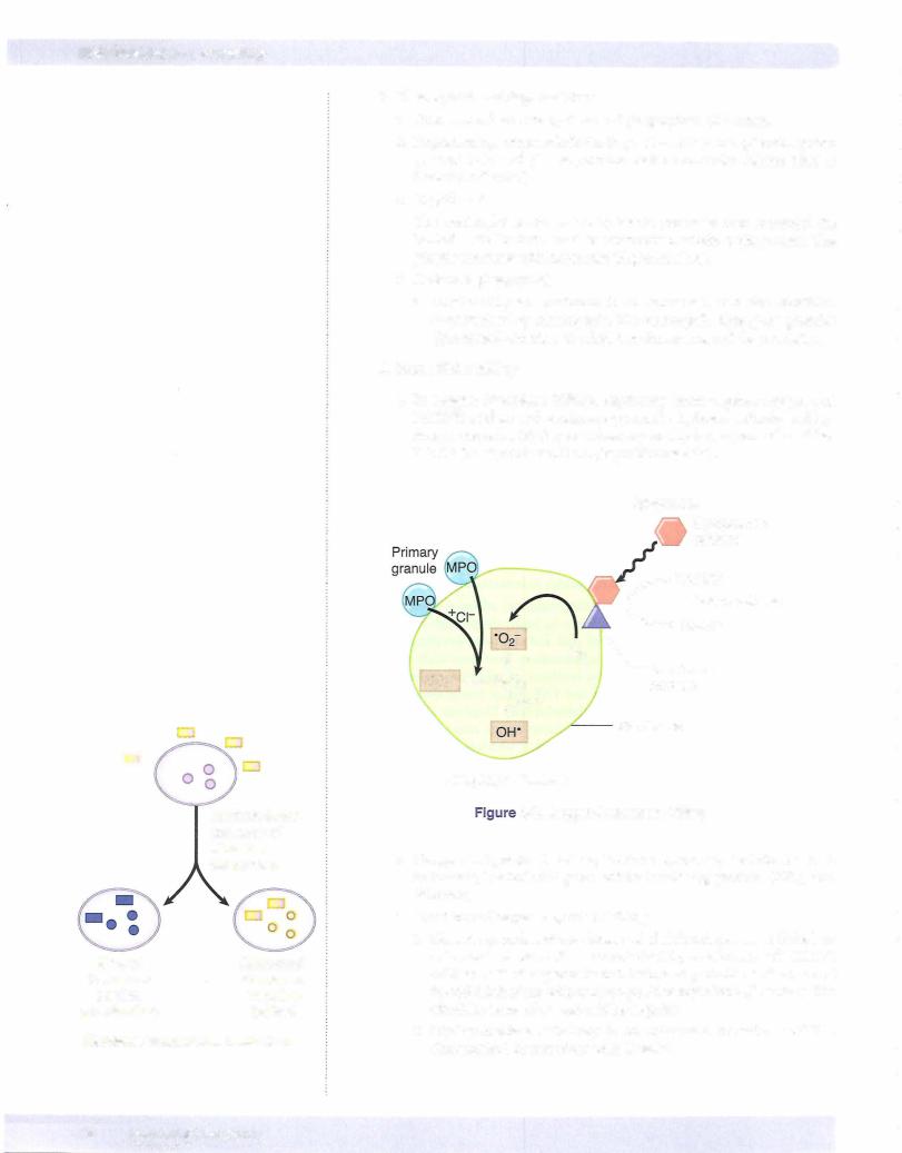
a.Neutrophils (life span in tissue 1-2 days)
i.Synonyms: segmented neutrophils, polymorphonuclear leukocytes (PMN)
ii.Primary (azurophilic) granules contain myeloperoxidase, phos pholipase A2, lysozyme (damages bacterial cell walls by catalyzing hydrolysis of 1,4-beta- linkages), and acid hydrolases. Also present are elastase, defensins (microbicidal peptides active against many gram-negative and gram-positive bacteria, fungi, and enveloped viruses), and bactericidal permeability increasing protein (BPI).
iii.Secondary (specific) granules contain phospholipase A2, lysozyme, leukocyte alkaline phosphatase (LAP), collagenase, lactoferrin (che lates iron), and vitamin Bl2-binding proteins.
b.Macrophages (life span in tissue compartment is 60-120 days) have acid hydrolases, elastase, and collagenase.
2.Neutrophil margination and adhesion
a.Adhesion is mediated by complementary molecules on the surface of neutrophils and endothelium.
i.In step l, the endothelial cells at sites ofinflammation have increased expression ofE-selectin and P-selectin.
ii.In step 2, neutrophils weakly bind to the endothelial selectins and roll along the surface.
iii.In step 3, neutrophils are stimulated by chemokines to express their integrins.
iv.In step 4, binding ofthe integrins firmly adheres the neutrophil to the endothelial cell.
Table 3-1. Selectin and lntegrin Distribution in the Endothelium and Leukocyte
|
Endothelium |
Leukocyte |
Selectins |
P-Selectin |
Sialyl-Lewis X & PSGL-1 |
|
E-Selectin |
Sialyl-Lewis X & PSGL-1 |
|
GlyCam-1/CD34 |
L-Selectin |
lntegrins |
ICAM-1 |
LFA-1 & MAC-1 |
|
VCAM-1 |
VLA-4 |
16 MEDICAL
USMLE Step i. • Pathology
In a Nutshell
Mediators of Pain
•Bradykinin
•Prostaglandins (E2)
In a Nutshell
Mediators of Fever
•Cytokines IL-1, IL-6, and TNF-a
•Prostaglandins
Bilobed nucleus, large granules (pink)
Eosinophil
Bilobed nucleus, large granules (blue)
Basophil
ii.Prostacyclin (PGI2) is produced by vascular endothelium and causes vasodilation and inhibition of platelet aggregation.
iii.Prostaglandin E2 causes pain.
iv.Prostaglandins PGE2, PGD2, and PGF2 cause vasodilatation.
b.Lipoxygenase pathway
Leukotriene B4 (LTB4) causes neutrophil chemotaxis, while leukotriene C4, D4, E4 cause vasoconstriction.
4.Important products in the complement cascade include C5b-C9 (mem brane attack complex), C3a,C5a (anaphylotoxins stimulate the release of histamine), CSa (leukocyte chemotactic factor), and C3b (opsonin for phagocytosis).
5.Cytokines
a.IL-1 and TNF cause fever and acute phase reactants; enhance adhesion molecules; and stimulate and activate fibroblasts, endothelial cells, and neutrophils.
b.IL-8 is a neutrophil chemoattractant produced by macrophages.
FOUR OUTCOMES OF ACUTE INFLAMMATION
1. Complete resolution with regeneration
2.Complete resolution with scarring
3.Abscess formation
4.Transition to chronic inflammation
CHRONIC INFLAMMATION
1. Causes ofchronic inflammation
a.Following a bout of acute inflammation
b.Persistent infections
c.Infections with certain organisms, including viral infections, mycobacte- ria, parasitic infections, and fungal infections
d.Autoimmune diseases
e.Response to foreign material
f.Response to malignant tumors
2.Important cells in chronic inflammation
a.Macrophages
i.Macrophages are derived from blood monocytes.
ii.Tissue-based macrophages (life span in connective tissue compart ment is 60-120 days) are found in connective tissue (histiocyte), lung (pulmonary alveolar macrophages), liver (Kupffer cells), bone (osteo clasts), and brain (microglia).
iii.During inflammation macrophages are mainly recruited from the blood (circulating monocytes).
iv.Chemotactic factors: CSa, MCP-1, MIP-la, PDGF, TGF-
v.Secrete a wide variety of active products (monokines)
vi.May be modified into epithelioid cells in granulomatous processes
b.Lymphocytes include B cells and plasma cells, as well as T cells. Lymphotaxin is the lymphocyte chemokine.
20 MEDICAL
c.Eosinophilsplay animportant role inparasitic infections and IgE-mediated· allergic reactions. The eosinophilic chemokine is eotaxin. Eosinophil granules contain major basic protein, which is toxic to parasites.
d.Basophils contain similar chemical mediators as mast cells in their granules. Mast cells are present in high numbers in the lung and skin. Both basophils and mast cells play an important role in IgE-mediated reactions (allergies and anaphylaxis) and can release histamine.
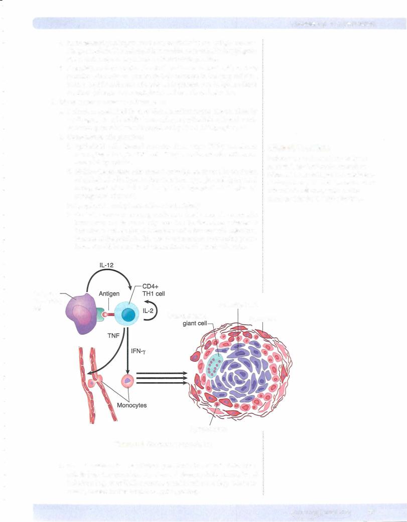
3.Chronic granulomatous inflammation
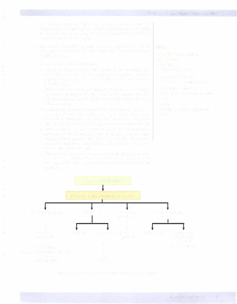
a.Definition: specialized form of chronic inflammation characterized by small aggregates of modified macrophages (epithelioid cells and multi nucleated giant cells) usually populated by CD4+ Thl lymphocytes
b.Composition of a granuloma
i.Epithelioid cells, located centrally, form when IFN-y transforms macrophages to epithelioid cells. They are enlarged cells with abun dant pink cytoplasm.
11.Multinucleated giant cells, located centrally, are formed by the fusion of epithelioid cells. Types include Langhans-type giant cell (peripheral arrangement of nuclei) and foreign body type giant cell (haphazard arrangement of nuclei).
iii.Lymphocytes and plasma cells at the periphery
iv.Central necrosis occurs in granulomata due to excessive enzymatic breakdown and is commonly seen in Mycobacterium tuberculosis infection as well as fungal infections and a few bacterial infections. Because of the public health risk of tuberculosis, necrotizing granu lomas should be considered tuberculosis until proven otherwise.
Chapter 3 • Inflammation
Clinical Correlate
Patients who are to be placed on tumor necrosis factor (TNF) inhibitors such as infliximab must undergo a PPD test before starting therapy. The PPD checks for latent tuberculosis infection, which can be reactivated by the TNF alpha inhibitor.
Antigen |
|
|
presenting |
|
Epithelioid cell |
cell |
|
|
Multinucleated |
|
|
|
Fibroblast |
|
|
|
Lymphocytes
Figure 3-5. Granuloma Formulation
c.Granulomatous diseases of which you should be aware include tuber culosis (caseating granulomas), cat-scratch fever, syphilis, leprosy, fungal infections (e.g., coccidioidomycosis), parasitic infections (e.g., schistoso miasis), foreign bodies, beryllium, and sarcoidosis.
MEDICAL 21
USMLE Step 1 • Pathology
Epithelioid cells
Multinucleated giant cell
© Henry Sanchez, M.D. Used with permission.
Figure 3-6. A Granuloma Is Seen in the Large, Poorly
Circumscribed Nodule in the Center of the Field
TISSUE RESPONSES TO INFECTIOUS AGENTS
1.General
Infectious diseases are very prevalent worldwide and are a major cause of morbidity and mortality. Infectious agents tend to have tropism for specific tissues and organs.
2.Six major histologic patterns
a.Exudative inflammation is acute inflammatory response with neutro phils. Examples include bacterial meningitis, bronchopneumonia, and abscess.
b.Necrotizing inflammation occurs when a virulent organism produces severe tissue damage and extensive cell death. Examples include necro tizing fasciitis and necrotizing pharyngitis.
c.Granulomatous inflammation
Granulomatous response predominates with slow-growing organisms such as mycobacteria, fungi, and parasites.
d.Interstitial inflammation is a diffuse mononuclear interstitial infiltrate that is a common response to viral infectious agents. Examples include myocarditis (Coxsackie virus) and viral hepatitis.
e.Cytopathic/cytoproliferative inflammation refers to inflammation in which the infected/injured cell is altered. The changes may include intranuclear/cytoplasmic inclusions (cytomegalic inclusion disease, rabies [Negri body] ); syncytia formation (respiratory syncytial virus and herpes virus); and apoptosis (Councilman body in viral hepatitis).
f.No inflammation
i.No evidence ofan inflammatory response to presence ofmicrobes can occur in severely immunosuppressed individuals due to primaryim munodeficiencies or acquired immunodeficient states (e.g., AIDS).
22 MEDICAL
Chapter 3 • Inflammation
Chapter Summary
•Acute inflammation is an immediate response to injury that can cause redness, heat, swelling, pain, and loss of function.
•Hemodynamic changes in acute inflammation are mediated by vasoactive chemicals and, after a transient initial vasoconstriction, produce massive dilation with increased vascular permeability.
•Neutrophils are important white blood cells in acute inflammation that contain granules with many degradative enzymes.
•Neutrophils leave the bloodstream in a highly regulated process involving margination (moving toward the vessel wall), adhesion (bindingto the endothelium), and emigration (moving between endothelial cells to leave the postcapillary venule). Defects in adhesion can contribute to the immunosuppression seen in diabetes mellitus and corticosteroid use.
•Chemotaxis is the attraction ofcells toward a chemical mediator, which is released in the area of inflammation.
•The phagocytosis of bacteria by neutrophils is improved ifopsonins, such as the Fe portion of immunoglobulin (lg) G or the complement product C3B, are bound to the surface ofthe bacteria. Chediak-Higashi syndrome is an example of a genetic disease with defective neutrophil phagocytosis.
•Once a bacterium has been phagocytized, both oxygen-requiring and oxygen independent enzymes can contribute to the killing ofthe bacteria. Chronic granulomatous disease of childhood and myeloperoxidase deficiency are genetic immunodeficiencies related to a deficiency of oxygen-dependent killing.
•Chemical mediators of inflammation include vasoactive amines, the kinin system, arachidonic acid products, the complement cascade, coagulation/ fibrinolytic cascade, and cytokines.
•Acute inflammation may lead to tissue regeneration, scarring, abscess formation, or chronic inflammation.
•Cells important in chronic inflammation include macrophages, lymphocytes, eosinophils, and basophils.
•Chronic granulomatous inflammation is a specialized form of chronic inflammation with modified macrophages (epithelioid cells and multinucleated giant cells) usually surrounded by a rim of lymphocytes. A wide variety of diseases can cause chronic granulomatous inflammation, most notably tuberculosis, syphilis, leprosy, and fungal infections.
•Patterns oftissue response to infectious agents can include exudative inflammation, necrotizing inflammation, granulomatous inflammation, interstitial inflammation, cytopathic/cytoproliferative inflammation, and no inflammatory response.
MEDICAL 23
