

2 |
1 Introduction, Development of the Algorithm, RoboChest Introduction, Additional Tools |
Fundamentals > Chest Primer Presentation
This work presents a structured lexicon for use by students of radiology to reproducibly describe abnormalities detected on plain CXRs. The lexicon is designed to provide the students with clinically significant differentiation of abnormalities detected. The content is chunked (displayed) in a directory structure format that relates specific combinations of distinct radiographic findings to classes/groupings of pathological etiologies of those findings. Recognizing the individual findings and identifying their combination or lack of combination with other findings allows one to create effective differential diagnoses that can then be further evaluated using other imaging procedures and/or non-radiographic clinical information. Radiology can be viewed as a descriptive art; where radiologists assign text to the imaging studies. The student of radiology attempts the same process in school, conference, rounds and direct patient care.
Included in this work are hundreds of images including X-rays, Computerized Tomography (CT) images, graphics, analogous models, and animations, to help teach otherwise complex processes and radiographic principles. This material is by no means comprehensive, rather is designed as a teaching tool and entry level information outlined to select detail and references. This material was not created for medical diagnosis and should not be used in isolation. This book and website can be a launching platform for information on chest imaging.
This directory structure method has been taught in the National Capital Area medical schools and radiology residency programs in the form of categories for years, from the introduction of chest imaging to students, to preparing for boards for senior residents. This method may also be helpful for General Medical Officers (GMOs) in deployed or remote locations without other available references. I have found the intensive care and infectious disease fellows enjoy this organized high-level format and method of solidifying concepts.
Chest X-Ray Interpretation Self-Study Instructions
This Guide organizes the abnormal Chest X-Ray (CXR) into anatomic regions and processes within these regions. This book begins with the search pattern, followed by normal anatomy found on the CXR. A mnemonic-driven approach to the search pattern and diagnostic process allows students, residents, and fellows alike to methodically approach, interpret, and profess the CXR in a quick, efficient, and consistent manner.
Since the CXR has the most anatomic noise (overlapping thoracic skeletal structures, soft tissues, mediastinum, etc.) of any radiographic procedure, the best way I have found to organize this material is to separate out the lungs from everything outside the lungs. Content relating to the lungs can be found in the Abnormal Lung Parenchyma section of the Guide, and content relating to the area outside the lungs is broken up into
Using the RoboChest Website |
3 |
Fig. 1.1 The contents directory is on the left and main screen on the right. At any time, the reader has a breadcrumb trail to show where they are in the algorithm. Image created by USUHS ETI support office
anatomic regions: Abnormal Pleura, Abnormal Mediastinum, and Abnormal Bones, Soft Tissue, and Other Findings. There is also a section about Trauma.
This book and browser tool (available at www.robochest.com) contains search pattern information to determine abnormality and become familiar with abnormal CXRs. Increased familiarity with abnormal CXRs should help guide identification of the general location of the abnormality, which is key to narrowing the differential diagnosis. The tool contains many cases that highlight regional abnormalities. You can match these cases with unknown images in self-study or while on clinical or subspecialty radiology rotations.
Using the RoboChest Website
See Fig. 1.1 for an example screen shot of the RoboChest website. Depending on your browser/security settings, you may need to click a yellow bar at the top of the screen if it appears when you open RoboChest. Clicking this bar and selecting “Allow Blocked Content” will enable your computer to run the ActiveX controls and scripts that are necessary to use RoboChest. The following image shows you an example RoboChest screen described in these instructions.
Once RoboChest is opened, you will note three tabs in the upper left called “Contents,” “Index,” and “Search.” The default tab is “Contents” and will show the directory structure. The Index tab is not yet operational; however, key word searches can be accomplished by using the “Search” tab or the blank text entry on the right top screen.
4 |
1 Introduction, Development of the Algorithm, RoboChest Introduction, Additional Tools |
You will note that the Contents directory works like an interactive table of contents. The book graphics indicate major sections and are “closed” until clicked on, then are “open,” with a list of available topic areas or other major headings underneath.
The main screen to the right of the Contents directory structure will display text and graphics aligned with the word or words selected in the Contents directory structure. NOTE: Links open in the main window.
If you click a link (blue, underlined text) on the main screen, the linked content will appear in the main screen, regardless of whether the link takes you to another page in RoboChest or to an outside site. Clicking the back button of your browser will allow you to return to the page that contained the link you clicked.
The main screen will function independently as its own browser window as you dig deeper into the content of your search. No matter what is visible in the main screen, the Contents directory structure will remain on the left and can serve as an outline to keep you focused on the CXR. If you accidentally close it, you can reopen it by clicking the “Contents” tab.
You will note a “bread crumb” path with hyperlinked text within the USU blue banner across the main screen. This path will show your current location in the directory structure while allowing you to click on any higher level for a quick way to see a more broad description of whatever topic you have delved into.
Parenchymal Diagnostic Algorithm
(Chest Imaging Diagnostic Algorithm)
The five major lung parenchymal patterns are covered in detail in Chap. 4. Once a student or resident realizes there is a parenchymal process (as opposed to nonparenchymal), then the following patterns help narrow the differential diagnosis. This work was first introduced to the literature in 1993 by Feigin [2].
•Mass: Any localized opacity not completely bordered by fissures/pleura
•Consolidative: Fluffy, cloud-like, coalescent opacities
•Interstitial: Thickening of peribronchial, perivascular, alveolar wall, and/or subpleural areas; thick-walled cystic spaces (honeycomb)
•Vascular: Change in diameter of vessels, whether intrinsically (vascular volume) or extrinsically (compression such as emphysema)
•Airway: Thick-walled airways (circular on end or tram-track), segmental or lobar atelectasis, and, lastly, bronchiectasis
Decision Tree Algorithms to Help Solidify Concepts
Prior work has resulted in two extensive charts in Microsoft® Excel® to help convey the complexity of chest imaging. When one breaks the CXR into components and takes one finding at a time the diagnostic process is simplified.
Decision Tree Algorithms to Help Solidify Concepts |
5 |
Chest Imaging Diagnositc Portal
Instructions:
Follow the path based on image findings. More than one path may be justified.
Cells with red corners have hidden comment boxes. Place the curser over those to see the comment.
Chest abnormality  1 0
1 0
Handout
SHIFT OF THE MEDIASTINUM
Decreased Lung Volume
Increased Lung Volume
Swyer-James syn
Pleural Space Abnormalities
Partial absence of the pericardium
Airway obstruction (especially peds)
Lung parenchyma |
|
See CIDA algorithm |
|
|
|
1 |
|
0 |
0 |
0 |
0 |
|
|
|
PLEURAL MASSES |
|
|
|
Solitary Pleural Density |
|
|
|
Loculated pleural effusion |
|
|
Mass |
Mesothelioma |
|
|
|
Multiple Pleural Densities |
|
|
|
Loculated pleural effusion |
|
|
0 |
Metastases0 |
|
|
|
Mesothelioma |
|
|
|
Pancoast tumor |
Pleura |
|
Thickening |
Plueral plaques |
|
(Asbestos pleura disease, calcified) |
||
|
|
1 |
Splenosis |
0 |
0 |
0 |
Subpleural Parenchymal Lung Densities |
0 |
|||
Infarct
Rheumatoid nodule
Round atelectasis
PLEURAL THICKENING
Trauma
Infection
Neoplasm
Inhalation Dz
Connective Tissue Dz
PNEUMOTHORAX
Eosinophilic Granuloma
Alveolar Proteinosis
Fibrothorax: scarring of the pleura occasionally seen with calcifications from prior empyema or hemothorax.
|
|
Fluid, air |
|
|
|
|
|
|
|
|
|
0 |
0 |
|
|
|
Thymic lesions |
||
|
|
|
|
|
Thyroid lesions |
||||
|
|
|
|
|
|
|
|
||
|
|
|
|
|
|
|
|
Terrible lyphomas |
|
|
|
|
|
|
Anterior |
Borders |
The 6 "T's" |
Teratomas, Dermoid cyst |
|
|
Consider lung mass as well |
|
|
|
|
|
Tortuous vessels |
||
|
(see CIDA algorithm) |
|
|
0 |
0 |
|
Trauma |
||
|
|
|
|
|
|
|
|
|
|
|
|
Mass |
|
Middle |
Borders |
|
Nerve root tumors |
|
|
|
|
|
|
Ganglion Tumors |
|
||||
|
|
|
|
|
1 |
|
|
Paragangliomas |
|
|
|
0 |
0 |
0 |
0 |
|
Lateral Meningocele |
|
|
|
|
|
|
|
|
|
|
|
|
|
|
|
|
|
|
|
|
NeuralLesions |
|
|
|
|
|
|
Posterior |
Borders |
|
EsophagealLesions |
|
|
|
|
|
|
|
|
>>>>>>> |
Cysts |
|
Mediastinum |
|
|
|
0 |
>>>>>>> |
|
Other |
||
|
|
1 |
|
|
|
|
|
|
|
0 |
0 |
|
|
|
WIDENING OF THE MEDIASTINUM |
||||
|
|
Enlargement, cardiac |
See also 6 "T's" |
Technique error |
|
|
|||
|
|
Vascular Structures |
|
|
|||||
|
|
|
|
|
|
Dissection |
|
|
|
|
|
0 |
0 |
|
Trauma |
|
|
|
|
|
|
|
|
|
|
Neoplasm |
|
|
|
|
|
|
|
|
|
Pneumomediastinum |
|
|
|
|
|
Fluid, blood, shift, silhouette |
Pneumopericardium |
|
|
||||
|
|
Mediastinitis |
|
|
|||||
|
|
|
|
|
|
Acute secondary to bacterial |
|||
|
|
0 |
0 |
|
Chronic Sclerosing Mediastinitis |
||||
|
|
|
Lipomatosis |
|
|
||||
Multiple symmetric Lipomatosis
Achalasia or Hiatal hernia
Also think of:
Cystic Hygroma
Mesenchymal Tumors:
>>>>>Germcell tumors Aortic abnormalities
Esophageal Carcinoma
Benign Esophageal Neoplasm
Hiatal Hernia
Pulsion Diverticuli
Achalasia
Esophageal Varices
Duplication cysts
Enteric cysts
Neurenteric cyst
Bronchogenic cysts
Extralobar sequestration - 2/3 in lower lobes Inflammation
Vascular lesions Trauma
Abdominal Origin Posterior Masses:
Bochdalek hernia
Bone, soft-tissue, other |
See appropriate algorithm (draft pending) |
||
|
|
1 |
|
0 |
0 |
0 |
0 |
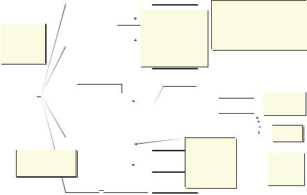
Fig. 1.2 This screen-shot of the higher level algorithm shows all the major anatomic regions where abnormalities can occur in chest imaging. At the top, the “Lung Parenchyma” takes one to the CIDA algorithm (figure 1.3). Both of these algorithms are available on Robochest under “CIDA”
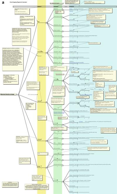
The Chest Imaging Diagnostic Assistant (CIDA) Portal (Fig. 1.2) displays the possible diagnoses for CXR abnormalities. I call this the high level since it leads the other, even more complex algorithm [3]. Abnormalities outside the lungs themselves include the pleural space, mediastinum and components, visualized soft tissues of the chest wall and the included bones. See Fig. 1.3a and b for flowchart for patterns seen in lungs.
You will need to have Microsoft® Excel® installed to view this document in its entirety. You can view this document on www.robochest.com by searching for CIDA. You can then open the document in a new window by clicking the Excel CIDA link and clicking the “Open” button on the download screen. You can also choose to save the document to your computer and view it at a later time.
If you do not have Excel installed but you have a Portable Document Format (PDF) reader such as Adobe® Acrobat®, you can view a PDF version of this document by clicking this PDF CIDA link. Please note, however, that some of the interactive content is not visible in the PDF version.
In summary, this book, along with the RoboChest website, and tools within are intended to enhance the ability of medical students and residents to learn, recall, and
6 |
1 Introduction, Development of the Algorithm, RoboChest Introduction, Additional Tools |
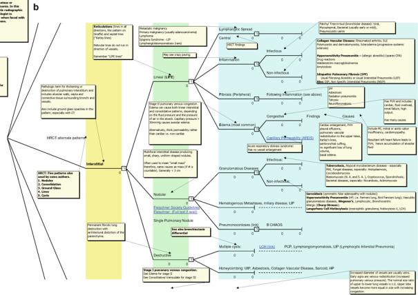
Fig. 1.3 Screen shot (a) shows the online Excel® document breaking down the five major lung patterns seen on CXR (Mass, Consolidation, Interstitial, Vascular and Airway patterns). CT and High Resolution CT. Note: this figure is for reference and not meant to be legible. Please refer to the actual document in RoboChest.com under “CIDA”
Concepts Solidify Help to Algorithms Tree Decision
7
Fig. 1.3 (b) Enlargement of a section of the parenchymal spreadsheet, in this case showing the interstitial pathway magnified section view of the larger chart