

112 |
5 Abnormalities Involving the Pleura |
Fig. 5.3 Continued
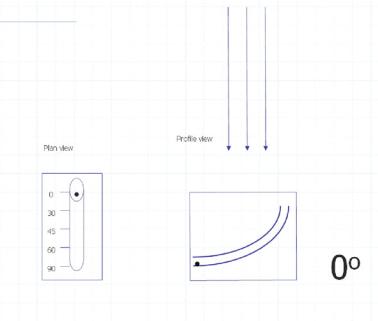
Loculated Fluid/Pseudotumor
Case 5.3
See Fig. 5.4 in a patient with both a peripheral loculated effusion and pseudotumor, or loculated fluid collection within a fissure.
Case 5.4
The following is a case where a young male was shot in the upper left chest by a sniper. More information can be obtained for a case report [4]. Case and images reprinted with permission from Military Medicine: International Journal of AMSUS.
Findings: Loss of left paratracheal stripe and pleural thickening consistent with pleural cap in upper lung field. In addition, left upper lung contusion and metallic fragmentation from bullet is noted.
Location: Loculated pleural fluid: blood from vascular injury.
Differential Diagnosis: Loculated blood in left upper pleural space.