- •Chest Imaging
- •Foreword
- •Preface
- •Educational Support and Funding
- •Acknowledgments
- •Contents
- •Fundamentals > Chest Primer Presentation
- •Chest X-Ray Interpretation Self-Study Instructions
- •Using the RoboChest Website
- •Decision Tree Algorithms to Help Solidify Concepts
- •References
- •Comprehensive Review of Search Patterns
- •Search Pattern Mnemonic
- •Interpretive Approach to CXR
- •Applying the Mnemonic to the Search Pattern
- •Chest Primer Presentation
- •References
- •Introduction and Terminology
- •Chest Imaging Terminology
- •Mach Effect on CXR
- •Trachea and Lungs on CXR
- •Mediastinal Anatomy on CXR
- •The Hilum (Plural: Hila)
- •Pulmonary Arteries and Veins
- •Normal Lung Markings
- •Vessel Size
- •Quiz Yourself: Mediastinum Lines, Edges
- •Shoulder Anatomy
- •Reference
- •Abnormal Lung Parenchyma
- •Mass
- •Mass Considerations
- •Size
- •Mass Characteristics
- •Malignancy
- •Case 4.1
- •Metastatic
- •Case 4.2
- •Bronchial Carcinoid
- •Radiological Signs
- •Case 4.3
- •Granulomatous Disease
- •Infectious Granulomatous Disease
- •Case 4.4
- •Non-infectious Granulomatous Disease
- •Benign Neoplasm
- •Hamartoma
- •Case 4.6
- •Congenital Abnormality
- •Pulmonary Arteriovenous Malformations
- •Case 4.7
- •Consolidation
- •Consolidative Radiological Findings/Distribution
- •Consolidative Model
- •Blood (Hemorrhage)
- •Case 4.8
- •Pus (Exudate)
- •Case 4.9
- •Case 4.10
- •Water (Transudate)
- •Pulmonary Edema
- •Case 4.11
- •Case 4.12
- •Protein (Secretions)
- •Case 4.13 (see Figs. 4.38 and 4.39)
- •Cells (Malignancy)
- •Interstitial
- •Radiological Signs
- •Linear Form: Lines
- •Case 4.14
- •Nodular Form: Dots
- •Case 4.15
- •Reticulo-Nodular Form
- •Pneumoconiosis
- •Case 4.16
- •Case 4.17
- •Destructive Fibrotic Lung
- •Case 4.18
- •Langerhans Cell Histiocytosis
- •Case 4.19
- •Vascular Pattern
- •Normal Pulmonary Vascular Anatomic Review
- •Radiological Signs in the Vascular Pattern
- •Mechanism
- •Vascular Examples
- •Pulmonary Arterial Hypertension (PAH)
- •Case 4.20
- •Pulmonary Venous Congestion
- •Pulmonary Venous Congestion: Edema
- •Emphysema
- •Airway (Bronchial) Patterns
- •Complete Obstruction
- •Lobar Atelectasis (Collapse)
- •Signs
- •Lobar Atelectasis Patterns
- •Complete Obstruction: Case Study
- •Partial Obstruction
- •Radiological Signs
- •Bronchial Wall Thickening
- •Bronchial Wall Thickening Causes
- •Bronchial Wall Thickening Model
- •Bronchiolar
- •Case 4.21
- •References
- •Pleural Effusion
- •Case 5.1
- •Technique and Positioning Revisited
- •Case 5.2
- •Comparison of Effusions over Time
- •Loculated Fluid/Pseudotumor
- •Case 5.3
- •Case 5.4
- •Thickening
- •Pneumothorax
- •Fluid and Air
- •Analogous Model
- •References
- •Anterior Mediastinal Mass
- •Case 6.1
- •Middle Mediastinal Mass
- •Posterior Mediastinal Mass
- •Case 6.2
- •Mediastinal Enlargement
- •Case 6.3
- •Reference
- •Case 7.1
- •Lines and Tubes
- •References
- •Appendix
- •Appendix 1: Glossary and Abbreviations
- •Appendix 2: Sources and Additional References
- •Text Sources
- •Image Sources
- •Additional References
- •Chest Imaging References
- •Chest Imaging Online References
- •Index
78 |
4 Abnormal Lung Patterns |
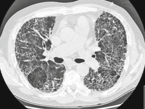
Fig. 4.57 CT of destructive cyst formation, honeycomb pattern in periphery
Langerhans Cell Histiocytosis
When you see lines, dots, and holes together as in the images below, think about Langerhans cell histiocytosis (LCH). One can consider the differential for each set of lines, dots, and holes; however, this would be an extensive differential diagnosis. Considerations can be narrowed based on the severity of findings, the mixture of each entity, and the clinical history.
Case 4.19
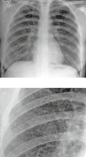
Figures 4.58 and 4.59 are from a case of LCH.
Findings: Lines and dots diffusely distributed, with holes between lines. Pattern: Interstitial, specifically lines, dots, and holes.
Differential Diagnosis: Langerhans cell histiocytosis (LCH) and LAM (lymphangioleiomyomatosis). Again, one could consider an exhaustive differential (all of interstitial); however, the combination of lines, dots, and holes is typically seen in LCH.
Open lung biopsy supported LCH.
Vascular Pattern
Normal Pulmonary Vascular Anatomic Review
As mentioned previously, normal pulmonary markings (vessels, arteries for the most part) can typically be followed from the hilum toward the lung periphery in all directions. They branch at acute angles, taper, and diverge toward the periphery.
Vascular Pattern |
79 |
Fig. 4.58 CXR showing lines, dots, and holes together
Fig. 4.59 Close-up of CXR showing lines, dots, and holes
Pulmonary markings on-end appear as small masses or nodules. They are recognized as normal vessels by the fact that they are often superimposed upon vessels of the same diameter branching in other directions. They are also larger toward the hilum and similarly sized to vessels nearby.
Radiological Signs in the Vascular Pattern
Abnormal diameter or distribution/ distortion of pulmonary vasculature.