- •Chest Imaging
- •Foreword
- •Preface
- •Educational Support and Funding
- •Acknowledgments
- •Contents
- •Fundamentals > Chest Primer Presentation
- •Chest X-Ray Interpretation Self-Study Instructions
- •Using the RoboChest Website
- •Decision Tree Algorithms to Help Solidify Concepts
- •References
- •Comprehensive Review of Search Patterns
- •Search Pattern Mnemonic
- •Interpretive Approach to CXR
- •Applying the Mnemonic to the Search Pattern
- •Chest Primer Presentation
- •References
- •Introduction and Terminology
- •Chest Imaging Terminology
- •Mach Effect on CXR
- •Trachea and Lungs on CXR
- •Mediastinal Anatomy on CXR
- •The Hilum (Plural: Hila)
- •Pulmonary Arteries and Veins
- •Normal Lung Markings
- •Vessel Size
- •Quiz Yourself: Mediastinum Lines, Edges
- •Shoulder Anatomy
- •Reference
- •Abnormal Lung Parenchyma
- •Mass
- •Mass Considerations
- •Size
- •Mass Characteristics
- •Malignancy
- •Case 4.1
- •Metastatic
- •Case 4.2
- •Bronchial Carcinoid
- •Radiological Signs
- •Case 4.3
- •Granulomatous Disease
- •Infectious Granulomatous Disease
- •Case 4.4
- •Non-infectious Granulomatous Disease
- •Benign Neoplasm
- •Hamartoma
- •Case 4.6
- •Congenital Abnormality
- •Pulmonary Arteriovenous Malformations
- •Case 4.7
- •Consolidation
- •Consolidative Radiological Findings/Distribution
- •Consolidative Model
- •Blood (Hemorrhage)
- •Case 4.8
- •Pus (Exudate)
- •Case 4.9
- •Case 4.10
- •Water (Transudate)
- •Pulmonary Edema
- •Case 4.11
- •Case 4.12
- •Protein (Secretions)
- •Case 4.13 (see Figs. 4.38 and 4.39)
- •Cells (Malignancy)
- •Interstitial
- •Radiological Signs
- •Linear Form: Lines
- •Case 4.14
- •Nodular Form: Dots
- •Case 4.15
- •Reticulo-Nodular Form
- •Pneumoconiosis
- •Case 4.16
- •Case 4.17
- •Destructive Fibrotic Lung
- •Case 4.18
- •Langerhans Cell Histiocytosis
- •Case 4.19
- •Vascular Pattern
- •Normal Pulmonary Vascular Anatomic Review
- •Radiological Signs in the Vascular Pattern
- •Mechanism
- •Vascular Examples
- •Pulmonary Arterial Hypertension (PAH)
- •Case 4.20
- •Pulmonary Venous Congestion
- •Pulmonary Venous Congestion: Edema
- •Emphysema
- •Airway (Bronchial) Patterns
- •Complete Obstruction
- •Lobar Atelectasis (Collapse)
- •Signs
- •Lobar Atelectasis Patterns
- •Complete Obstruction: Case Study
- •Partial Obstruction
- •Radiological Signs
- •Bronchial Wall Thickening
- •Bronchial Wall Thickening Causes
- •Bronchial Wall Thickening Model
- •Bronchiolar
- •Case 4.21
- •References
- •Pleural Effusion
- •Case 5.1
- •Technique and Positioning Revisited
- •Case 5.2
- •Comparison of Effusions over Time
- •Loculated Fluid/Pseudotumor
- •Case 5.3
- •Case 5.4
- •Thickening
- •Pneumothorax
- •Fluid and Air
- •Analogous Model
- •References
- •Anterior Mediastinal Mass
- •Case 6.1
- •Middle Mediastinal Mass
- •Posterior Mediastinal Mass
- •Case 6.2
- •Mediastinal Enlargement
- •Case 6.3
- •Reference
- •Case 7.1
- •Lines and Tubes
- •References
- •Appendix
- •Appendix 1: Glossary and Abbreviations
- •Appendix 2: Sources and Additional References
- •Text Sources
- •Image Sources
- •Additional References
- •Chest Imaging References
- •Chest Imaging Online References
- •Index
Chapter 5
Abnormalities Involving the Pleura
Abnormalities involving the pleura include pathologic processes (masses, calcifications, infections, thickening) of the pleura, in addition to fluid and air collections within the pleural space. This chapter will start off with more common conditions such as pleural effusions and pneumothorax, followed by less common/ more complex processes.
Pleural Effusion
Definition: fluid in the potential space between the parietal and visceral pleura. This usually results in obliteration part or all of the hemidiaphragms (silhouette sign), blunting of the costophrenic angles both peripherally on the frontal projection and posteriorly on the lateral.
Case 5.1
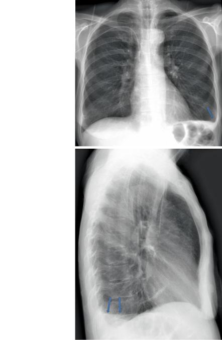
Figure 5.1 demonstrates a left pleural effusion on interdepartmental PA and lateral, a portable projection on same patient next day, then on CT.
Technique and Positioning Revisited
In the Chap. 2 of this book, we reviewed how technical factors such as rotation, X-ray energy, and markers are important. The preceding and following examples of the appearance of effusion also demonstrate how technique, especially portable positioning, is important in evaluating effusions, and differentiating them from other conditions such as consolidations.
L.R. Folio, Chest Imaging, DOI 10.1007/978-1-4614-1317-2_5, |
107 |
© Henry M. Jackson Foundation for the Advancement of Military Medicine, Inc. 2012 |
|
108
Fig. 5.1a,b PA (a) and lateral (b) erect projections of the chest demonstrate blunting of left costophrenic angle with a meniscus peripherally on the PA and posteriorly on the lateral (arrows). Also note the left PICC line with tip in SVC
5 Abnormalities Involving the Pleura
a
b