
- •Chest Imaging
- •Foreword
- •Preface
- •Educational Support and Funding
- •Acknowledgments
- •Contents
- •Fundamentals > Chest Primer Presentation
- •Chest X-Ray Interpretation Self-Study Instructions
- •Using the RoboChest Website
- •Decision Tree Algorithms to Help Solidify Concepts
- •References
- •Comprehensive Review of Search Patterns
- •Search Pattern Mnemonic
- •Interpretive Approach to CXR
- •Applying the Mnemonic to the Search Pattern
- •Chest Primer Presentation
- •References
- •Introduction and Terminology
- •Chest Imaging Terminology
- •Mach Effect on CXR
- •Trachea and Lungs on CXR
- •Mediastinal Anatomy on CXR
- •The Hilum (Plural: Hila)
- •Pulmonary Arteries and Veins
- •Normal Lung Markings
- •Vessel Size
- •Quiz Yourself: Mediastinum Lines, Edges
- •Shoulder Anatomy
- •Reference
- •Abnormal Lung Parenchyma
- •Mass
- •Mass Considerations
- •Size
- •Mass Characteristics
- •Malignancy
- •Case 4.1
- •Metastatic
- •Case 4.2
- •Bronchial Carcinoid
- •Radiological Signs
- •Case 4.3
- •Granulomatous Disease
- •Infectious Granulomatous Disease
- •Case 4.4
- •Non-infectious Granulomatous Disease
- •Benign Neoplasm
- •Hamartoma
- •Case 4.6
- •Congenital Abnormality
- •Pulmonary Arteriovenous Malformations
- •Case 4.7
- •Consolidation
- •Consolidative Radiological Findings/Distribution
- •Consolidative Model
- •Blood (Hemorrhage)
- •Case 4.8
- •Pus (Exudate)
- •Case 4.9
- •Case 4.10
- •Water (Transudate)
- •Pulmonary Edema
- •Case 4.11
- •Case 4.12
- •Protein (Secretions)
- •Case 4.13 (see Figs. 4.38 and 4.39)
- •Cells (Malignancy)
- •Interstitial
- •Radiological Signs
- •Linear Form: Lines
- •Case 4.14
- •Nodular Form: Dots
- •Case 4.15
- •Reticulo-Nodular Form
- •Pneumoconiosis
- •Case 4.16
- •Case 4.17
- •Destructive Fibrotic Lung
- •Case 4.18
- •Langerhans Cell Histiocytosis
- •Case 4.19
- •Vascular Pattern
- •Normal Pulmonary Vascular Anatomic Review
- •Radiological Signs in the Vascular Pattern
- •Mechanism
- •Vascular Examples
- •Pulmonary Arterial Hypertension (PAH)
- •Case 4.20
- •Pulmonary Venous Congestion
- •Pulmonary Venous Congestion: Edema
- •Emphysema
- •Airway (Bronchial) Patterns
- •Complete Obstruction
- •Lobar Atelectasis (Collapse)
- •Signs
- •Lobar Atelectasis Patterns
- •Complete Obstruction: Case Study
- •Partial Obstruction
- •Radiological Signs
- •Bronchial Wall Thickening
- •Bronchial Wall Thickening Causes
- •Bronchial Wall Thickening Model
- •Bronchiolar
- •Case 4.21
- •References
- •Pleural Effusion
- •Case 5.1
- •Technique and Positioning Revisited
- •Case 5.2
- •Comparison of Effusions over Time
- •Loculated Fluid/Pseudotumor
- •Case 5.3
- •Case 5.4
- •Thickening
- •Pneumothorax
- •Fluid and Air
- •Analogous Model
- •References
- •Anterior Mediastinal Mass
- •Case 6.1
- •Middle Mediastinal Mass
- •Posterior Mediastinal Mass
- •Case 6.2
- •Mediastinal Enlargement
- •Case 6.3
- •Reference
- •Case 7.1
- •Lines and Tubes
- •References
- •Appendix
- •Appendix 1: Glossary and Abbreviations
- •Appendix 2: Sources and Additional References
- •Text Sources
- •Image Sources
- •Additional References
- •Chest Imaging References
- •Chest Imaging Online References
- •Index

Airway (Bronchial) Patterns |
87 |
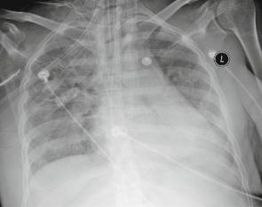
Fig. 4.64 Interstitial edema, note the fine interstitial markings in increasing opacifying lungs. Note adequately placed ET and NG tubes. Right IJ line in SVC (rotated projection)
Pulmonary Venous Congestion: Edema
Figure 4.64 depicts a diffuse, fine reticular pattern with a differential diagnosis of exudates, transudate, hemorrhage, secretions, or malignancy. Although not a vascular pattern, the interstitial pattern is compatible with ARDS.
Emphysema
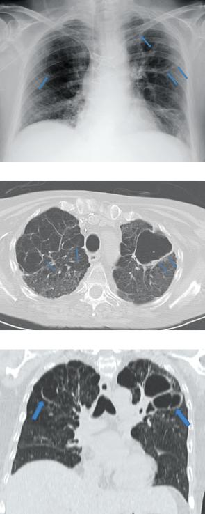
Although not primarily a vascular condition, emphysema can cause extrinsic pressure to vasculature, causing a compressed and distorted appearance to the pulmonary vasculature in the apicies leading to this diagnosis.
Figure 4.65a provides an example of compressed vessels; decreased diameter of vessels is caused by excessive air pressure around them.
Findings: Upper lobe diminished and distorted vessels. Pattern: Vascular.
Differential Diagnosis: Bullous emphysema, COPD.
Airway (Bronchial) Patterns
For a brief description and graphic of normal airways, see the normal lung markings page of this book.
Mechanisms
•Complete or partial obstruction of airways
•Thickening of airway walls
•Atelectasis literally means incomplete expansion or loss of volume

88 |
4 Abnormal Lung Patterns |
Fig. 4.65a CXR showing diminished and distorted vasculature in upper lobes (arrows)
Fig. 4.65b Axial CT showing better detail of compressed and distorted vessels due to biapical bullous emphysema
Fig. 4.65c Coronal reformat again demonstrating compressed, distorted pulmonary vasculature (arrows)