
- •Chest Imaging
- •Foreword
- •Preface
- •Educational Support and Funding
- •Acknowledgments
- •Contents
- •Fundamentals > Chest Primer Presentation
- •Chest X-Ray Interpretation Self-Study Instructions
- •Using the RoboChest Website
- •Decision Tree Algorithms to Help Solidify Concepts
- •References
- •Comprehensive Review of Search Patterns
- •Search Pattern Mnemonic
- •Interpretive Approach to CXR
- •Applying the Mnemonic to the Search Pattern
- •Chest Primer Presentation
- •References
- •Introduction and Terminology
- •Chest Imaging Terminology
- •Mach Effect on CXR
- •Trachea and Lungs on CXR
- •Mediastinal Anatomy on CXR
- •The Hilum (Plural: Hila)
- •Pulmonary Arteries and Veins
- •Normal Lung Markings
- •Vessel Size
- •Quiz Yourself: Mediastinum Lines, Edges
- •Shoulder Anatomy
- •Reference
- •Abnormal Lung Parenchyma
- •Mass
- •Mass Considerations
- •Size
- •Mass Characteristics
- •Malignancy
- •Case 4.1
- •Metastatic
- •Case 4.2
- •Bronchial Carcinoid
- •Radiological Signs
- •Case 4.3
- •Granulomatous Disease
- •Infectious Granulomatous Disease
- •Case 4.4
- •Non-infectious Granulomatous Disease
- •Benign Neoplasm
- •Hamartoma
- •Case 4.6
- •Congenital Abnormality
- •Pulmonary Arteriovenous Malformations
- •Case 4.7
- •Consolidation
- •Consolidative Radiological Findings/Distribution
- •Consolidative Model
- •Blood (Hemorrhage)
- •Case 4.8
- •Pus (Exudate)
- •Case 4.9
- •Case 4.10
- •Water (Transudate)
- •Pulmonary Edema
- •Case 4.11
- •Case 4.12
- •Protein (Secretions)
- •Case 4.13 (see Figs. 4.38 and 4.39)
- •Cells (Malignancy)
- •Interstitial
- •Radiological Signs
- •Linear Form: Lines
- •Case 4.14
- •Nodular Form: Dots
- •Case 4.15
- •Reticulo-Nodular Form
- •Pneumoconiosis
- •Case 4.16
- •Case 4.17
- •Destructive Fibrotic Lung
- •Case 4.18
- •Langerhans Cell Histiocytosis
- •Case 4.19
- •Vascular Pattern
- •Normal Pulmonary Vascular Anatomic Review
- •Radiological Signs in the Vascular Pattern
- •Mechanism
- •Vascular Examples
- •Pulmonary Arterial Hypertension (PAH)
- •Case 4.20
- •Pulmonary Venous Congestion
- •Pulmonary Venous Congestion: Edema
- •Emphysema
- •Airway (Bronchial) Patterns
- •Complete Obstruction
- •Lobar Atelectasis (Collapse)
- •Signs
- •Lobar Atelectasis Patterns
- •Complete Obstruction: Case Study
- •Partial Obstruction
- •Radiological Signs
- •Bronchial Wall Thickening
- •Bronchial Wall Thickening Causes
- •Bronchial Wall Thickening Model
- •Bronchiolar
- •Case 4.21
- •References
- •Pleural Effusion
- •Case 5.1
- •Technique and Positioning Revisited
- •Case 5.2
- •Comparison of Effusions over Time
- •Loculated Fluid/Pseudotumor
- •Case 5.3
- •Case 5.4
- •Thickening
- •Pneumothorax
- •Fluid and Air
- •Analogous Model
- •References
- •Anterior Mediastinal Mass
- •Case 6.1
- •Middle Mediastinal Mass
- •Posterior Mediastinal Mass
- •Case 6.2
- •Mediastinal Enlargement
- •Case 6.3
- •Reference
- •Case 7.1
- •Lines and Tubes
- •References
- •Appendix
- •Appendix 1: Glossary and Abbreviations
- •Appendix 2: Sources and Additional References
- •Text Sources
- •Image Sources
- •Additional References
- •Chest Imaging References
- •Chest Imaging Online References
- •Index

Consolidation |
57 |
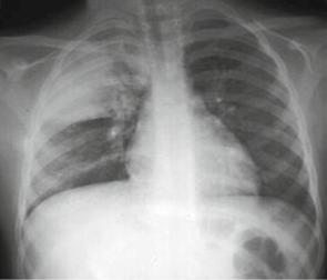
Fig. 4.31 Opacification of the right upper lung field representing a consolidation pattern in bacterial pneumonia in the RUL. Location is confirmed by horizontal fissure inferiorly on the PA and lateral, and the major fissure on the lateral
Pus (Exudate)
Pus from a variety of infections can cause pneumonia. Pneumonia generally respects lobes; hence distribution is lobar or multilobar. However, atypical pneumonia may be diffuse and bilateral.
Some pneumonias originate in the lung periphery where the Streptococcus pneumoniae reaches the lung via the airway. In lower right lobe pneumonia especially, aspiration should be considered since the right mainstem bronchus and bronchus intermedius are more vertical than on the left.
Bacterial Pneumonia
•Is most commonly caused by Streptococcus pneumoniae
•May present with mild to severe symptoms, including shaking chills, chattering teeth, severe chest pain, and a cough productive of rust-colored or greenish sputum
•May be febrile, diaphoretic, tachypneic, dyspneic, and/or cyanotic
Case 4.9
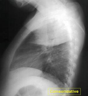
Figures 4.31 and 4.32 depict lobar consolidation in the RUL, with fissures limiting extension.
Findings
RUL: Large area of airspace opacification on the frontal view has both major and minor fissures as its inferior border. The lateral view demonstrates nicely the fissures of the right lung. Both RML and RLL remain well aerated.
Pattern: Consolidation.
Differential Diagnosis: Bacterial Lobar Pneumonia.

58 |
4 Abnormal Lung Patterns |
Fig. 4.32 Lobar consolidation pattern
on lateral. Note the airspace opacity limited inferiorly by the minor fissue anteriorly and the major fissure posteriorly
Case 4.10
In some cases, a consolidation can resemble a mass, as shown on the CXR in this case. Additionally, this case demonstrates radiopaque markers that attempt to show degree of inclination (however, unsuccessfully). Lastly, this AP portable is an example of a rotated right projection. (Note the clavicles are to the right of the spine.)
Findings: RUL opacity bordering minor fissure inferiorly, however, maintains fluffy superior margin.
Pattern: Consolidation.
Note: Mass could also be considered, however, since abutment to horizontal fissure inferiorly gives the impression of mass (such as malignancy). A follow-up CXR documenting the resolution is paramount to rule out mass in cases like this.
Differential Diagnosis: Pneumonia, loculated fluid in horizontal fissure (pseudomass); mass differential can be considered until ruled out by CT or follow-up CXRs.
Diagnosis: RUL bacterial lobar pneumonia, confirmed by resolution with antibiotics and follow-up CXR.
Note the small ball bearings (BBs) in the left marker indicating an upright projection. Experience with these, however, demonstrates that the BBs fall to this location starting at 30° of inclination. Work is being done to achieve higher accuracy (discussed in the pleural section of this book).