

Autonomic Nervous System: General Topography
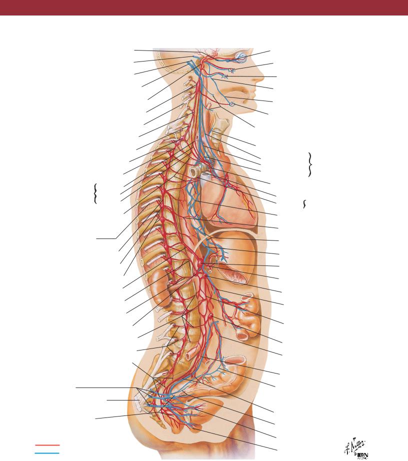
Oculomotor nerve (III)
Facial nerve (VII)
Glossopharyngeal nerve (IX)
Vagus nerve (X)
Internal carotid nerve and plexus
Superior cervical sympathetic ganglion
C4 spinal nerve
Middle cervical sympathetic ganglion
Vertebral ganglion
Cervicothoracic (stellate) ganglion
|
Sympathetic trunk |
||
Cervical (sympathetic) |
Superior |
||
Middle |
|||
cardiac nerves |
|
||
|
Inferior |
||
|
|
||
Thoracic (sympathetic) cardiac nerves
6th intercostal nerve (ventral ramus of T6 spinal nerve)
Sympathetic trunk
6th thoracic sympathetic ganglion
Gray and white rami communicantes
Greater splanchnic nerve
Lesser splanchnic nerve
Least splanchnic nerve
Aorticorenal ganglion
Lumbar splanchnic nerves (sympathetic)
Gray rami communicantes
Sacral splanchnic nerves (sympathetic)
Pelvic splanchnic nerves (sacral parasympathetic outflow)
Sciatic nerve
Inferior hypogastric (pelvic) plexus
Sympathetic fibers
Parasympathetic fibers
NEUROANATOMY
Ciliary ganglion
Pterygopalatine ganglion
Otic ganglion
Chorda tympani nerve
Lingual nerve
Submandibular ganglion
Pharyngeal and superior laryngeal branches of vagus nerve
Recurrent laryngeal branch of vagus nerve
Superior cervical |
Cardiac branches |
|
Inferior cervical |
||
of vagus nerve |
||
Thoracic |
||
|
||
Cardiac plexus |
|
Anterior  Pulmonary plexuses
Posterior
Esophageal plexus
Thoracic aortic plexus
Anterior vagal trunk
Posterior vagal trunk
Celiac ganglion
Celiac trunk and plexus
Superior mesenteric ganglion
Superior mesenteric artery and plexus
Intermesenteric (abdominal aortic) plexus
Inferior mesenteric ganglion
Inferior mesenteric artery and plexus
Superior hypogastric plexus
Parasympathetic branch from inferior hypogastric plexus to descending colon
Hypogastric nerves
Rectal plexus
Vesical plexus
Prostatic plexus
23
NEUROANATOMY |
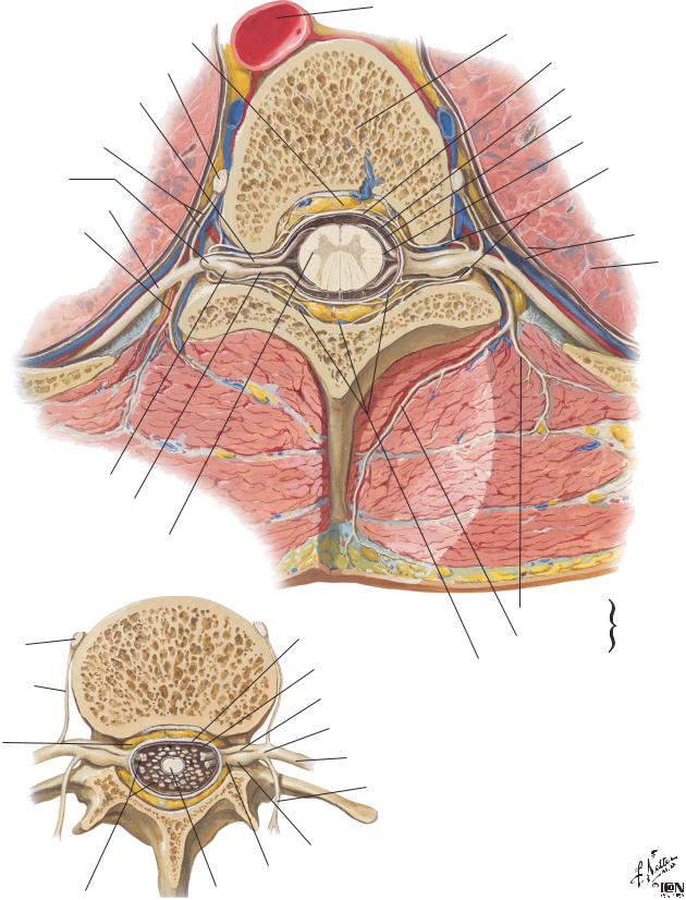
Spinal Nerve Origin: Cross Sections |
Section through thoracic vertebra
Fat in epidural space
Sympathetic ganglion
Ventral root
White and gray rami communicantes
Spinal nerve
Ventral ramus (intercostal nerve)
Dorsal ramus
Spinal sensory (dorsal root) ganglion
Dorsal root
Lateral horn of
gray matter of spinal cord
Section through lumbar vertebra
Sympathetic ganglion
Gray ramus communicans
Fat in epidural space
Dorsal and ventral roots of lumbar and sacral spinal nerves forming cauda equina
Aorta
Body of vertebra
Dura mater
Arachnoid mater*
Subarachnoid space
Pia mater*
Recurrent meningeal branches of spinal nerve
Pleura
Lung
|
Lateral branch |
of dorsal |
|
|
ramus |
||
Dura mater |
Medial branch |
of spinal |
|
nerve |
|||
|
|
||
Arachnoid mater |
Internal vertebral |
|
|
|
(epidural) venous plexus |
|
|
Ventral root |
|
|
Spinal nerve
Ventral ramus (contributes to lumbar plexus)
Dorsal ramus
Spinal sensory (dorsal root) ganglion
Dorsal root
Conus medullaris
*Leptomeninges
24
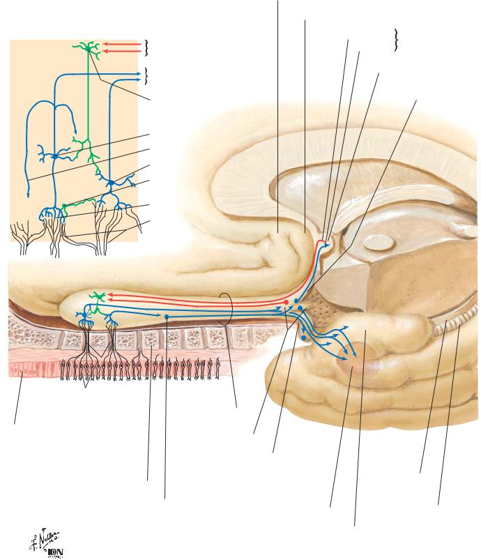
Olfactory Nerve (I): Schema |
NEUROANATOMY |
|
Subcallosal (parolfactory) area |
|
Olfactory bulb cells: schema |
Septal area and nuclei |
|
|
|
|
Efferent fibers to |
Fibers from |
Contralateral |
Fibers to |
olfactory bulb |
|
olfactory bulb |
|
|
Afferent fibers from bulb |
Anterior commissure |
|
to central connections |
|
|
and contralateral bulb |
|
Medial |
Granule cell (excited by |
|
olfactory stria |
|
|
|
and inhibiting to mitral |
|
|
and tufted cells) |
|
|
Mitral cell
Recurrent process
Tufted cell
Periglomerular cell
Glomerulus
Olfactory nerve fibers
|
|
|
|
Olfactory tract |
|
|
|
|
|
|
|
|
|
|
|
|
|
|
|
|
|
|
|
|
|
|
|
|
|
|
|
|
|
|
|
|
|
|
|
|
|
|
|
|
|
|
|
|
|
|
|
|
|
|
Olfactory cells |
|
|
|
|
|
|
|
|
||
Olfactory mucosa |
|
|
Olfactory trigone |
|
|
|
|
|
|
|
|
|
|
|
and olfactory tubercle |
|
|
|
|
|
|
Olfactory nerves (I) |
|
Lateral olfactory stria |
|
|
|
|
|
|
||
Olfactory bulb |
|
|
|
Uncus |
|
|
|
|||
|
Lateral olfactory tract nucleus |
|
|
Hippocampal |
|
|
||||
Cribriform plate of ethmoid bone |
|
|
|
|
||||||
|
|
|
|
|
||||||
|
|
|
fimbria |
|
|
|||||
Anterior olfactory nucleus |
Anterior perforated substance |
|
|
|||||||
|
|
|
|
|||||||
Amygdaloid body (phantom) |
Dentate gyrus |
|
|
|||||||
|
|
|
|
|
|
|||||
|
|
|
|
|
Piriform lobe |
Parahippocampal gyrus |
|
|
||
|
|
|
|
|
||||||
25
NEUROANATOMY |
Optic Nerve (II) (Visual Pathway): Schema |
G
G
A
A
BB
H H
R R C C
P P
Choroid Choroid
Periphery Macula
Structure of retina: schema
A Amacrine cells
B Bipolar cells
CCones
G Ganglion cells
H Horizontal cells
P Pigment cells
RRods
Calcarine sulcus
Projection on left occipital lobe
|
Central darker |
|
|
circle represents |
|
|
macular zone |
|
Overlapping |
Lighter shades |
|
visual fields |
||
represent |
||
|
||
|
monocular fields |
|
|
Each quadrant |
|
|
a different color |
Projection on |
Projection on |
|
right retina |
||
left retina |
||
|
||
|
Optic nerves (II) |
|
|
Optic chiasm |
|
Projection on left |
Projection on right |
|
dorsal lateral |
dorsal lateral |
|
geniculate nucleus |
geniculate nucleus |
|
|
Optic tracts |
Lateral geniculate bodies
Optic radiation |
Optic radiation |
Calcarine sulcus
Projection on right occipital lobe
26