

NEUROPHYSIOLOGY |
Hypothalamus |
|
Septum |
|
|
|
Corpus callosum |
|||
|
|
|
|
Fornix |
|
|
|
|
|
pellucidum |
|
|
|
|
|
|
|
|
|
|
|
|
|
|
|
|
Lateral |
|
|
|
|
|
|
|
|
ventricle |
|
|
|
|
|
|
|
|
|
Thalamus |
|
|
From |
||||
|
|
|
hippocampal |
|||||
|
|
|
|
Interthalamic |
formation |
|||
|
Lateral |
|
|
|
|
|
||
|
|
|
adhesion |
|
|
|
||
|
hypothalamic area |
|
|
|
|
|
||
Medial |
Paraventricular nucleus |
|
|
|
||||
Anterior |
Anterior hypothalamic area |
|
|
|
||||
forebrain |
commissure |
Dorsal hypothalamic area |
|
|
|
|||
bundle |
|
|
|
|||||
|
|
Dorsomedial nucleus |
|
|
|
|||
|
|
|
|
|
|
|||
|
|
|
|
|
Mamillothalamic tract |
|||
|
|
|
|
|
Posterior area |
|||
Lateral |
|
|
|
|
Periventricular |
|||
preoptic |
Medial |
|
|
|
nucleus |
|||
nucleus |
preoptic |
|
|
|
Nucleus |
|||
|
nucleus |
|
|
|
intercalatus |
|||
Olfactory |
|
|
|
|
|
|
|
|
tract |
|
|
|
|
|
|
|
|
|
|
|
Fornix |
Red nucleus |
Cerebral |
|||
|
|
|
|
|
peduncle |
|||
|
|
Ventromedial |
|
|
|
|||
|
|
nucleus |
|
Mamillary |
|
|
|
|
Optic (II) |
|
|
|
|
complex |
Dorsal |
||
nerve |
|
Tuberohypophyseal tract |
||||||
Optic chiasm |
|
|
|
|
longitudinal |
|||
|
Oculomotor (III) nerve |
fasciculus |
||||||
|
|
|
||||||
|
|
Supraoptic nucleus |
Descending |
|||||
|
|
Supraopticohypophyseal |
hypothalamic |
|||||
|
|
connections |
||||||
|
|
tract |
|
|
Pons |
|||
|
|
Posterior lobe of pituitary |
||||||
Anterior |
|
Reticular |
|
|
||||
|
|
|
|
|
|
|
||
lobe of |
|
|
|
|
|
formation |
|
|
pituitary |
|
|
|
|
|
|
© |
|
CHART 2.3 MAJOR FUNCTIONS OF THE HYPOTHALAMUS |
|
|
|
|
|
|||
|
|
|
|
|
||||
|
|
|
|
|
|
|
|
|
Hypothalamic Area |
|
|
|
Major Functions* |
|
|
|
|
Preoptic and anterior |
|
|
|
Heat loss center: cutaneous vasodilation and sweating |
||||
Posterior |
|
|
|
Heat conservation center: cutaneous vasoconstriction and shivering |
||||
Lateral |
|
|
|
Feeding center: eating behavior |
||||
Ventromedial |
|
|
|
Satiety center: inhibits eating behavior |
||||
Supraoptic (subfornical organ and organum vasculosum) |
|
ADH and oxytocin secretion (sensation of thirst) |
||||||
Paraventricular |
|
|
|
ADH and oxytocin secretion |
||||
Periventricular |
|
|
|
Releasing hormones for the anterior pituitary |
||||
|
|
|
|
|
|
|
|
|
*Stimulation of the center causes the responses listed. |
|
|
|
|
|
|
|
|
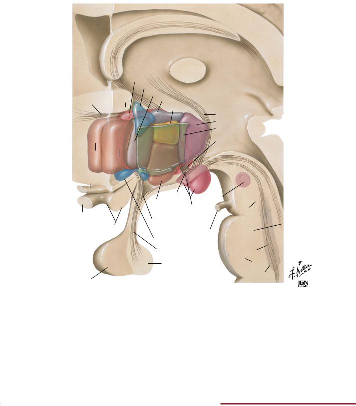
FIGURE 2.17 SCHEMATIC RECONSTRUCTION OF THE HYPOTHALAMUS•
The hypothalamus, part of the diencephalon, controls a number of important homeostatic systems within the body, including temperature regulation, food intake, water intake, many of the endocrine systems (see Chapter 8), motivation, and emotional behavior. It receives inputs from the reticular formation (sleep/wake cycle
information), the thalamus (pain), the limbic system (emotion, fear, anger, smell), the medulla oblongata (blood pressure and heart rate), and the optic system, and it integrates these inputs for regulation of the functions listed.
68
Limbic System |
NEUROPHYSIOLOGY |
Columns of fornix
Genu of corpus callosum
Head of caudate nucleus
Columns of fornix
Body of fornix
Thalamus
Uncus
Crura of fornix
Fimbria of hippocampus
Hippocampus
Commissure of fornix
Splenium of corpus callosum
Lateral ventricle
Body of fornix
Commissure of fornix
Crura of fornix
Mamillary bodies
|
Hippocampus |
|
Amygdaloid bodies |
with fimbria |
© |
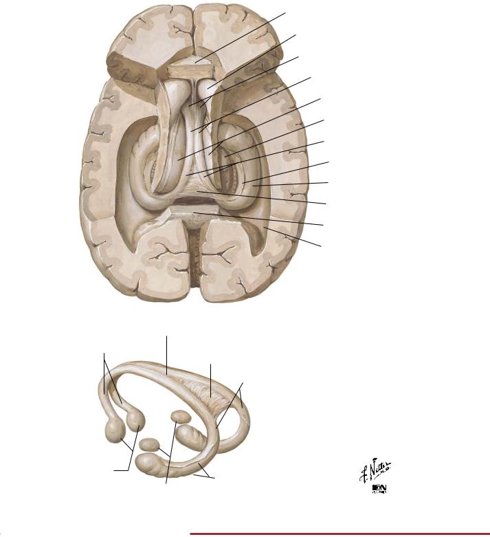
FIGURE 2.18 HIPPOCAMPUS AND FORNIX•
The limbic system includes the hypothalamus and a collection of interconnected structures in the telencephalon (cingulate, parahippocampal, and subcallosal gyri), as well as the amygdala and hip-
pocampal formation. The limbic system functions in linking emotion and motivation (amygdala), learning and memory (hippocampal formation), and sexual behavior (hypothalamus).
69
NEUROPHYSIOLOGY |
The Cerebral Cortex |
Ms I
Motor Premotor; orientation; Ms II
eye and head movements
Prefrontal; inhibitory control of behavior; higher intelligence
Motor control of speech
Motor
Premotor
Prefrontal; inhibitory control of behavior; higher intelligence
Cingulate gyrus (emotional behavior) and cingulum
Olfactory
Ms I
Ms II
Sm I
Sm II Sensory
Sensory analysis
Visual III
Visual II
Visual I
Language; reading; speech
Auditory I
Auditory II
Sm I
Sm III Sensory
?
Visual III
Visual II
Visual I
©
Corpus callosum
Hippocampal commissure
Anterior commissure
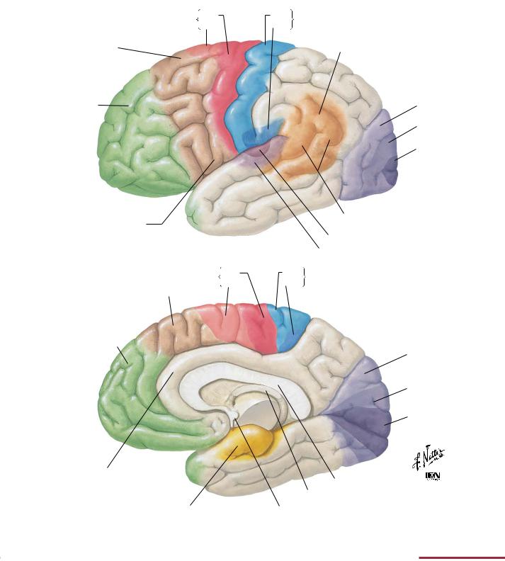
FIGURE 2.19 CEREBRAL CORTEX: LOCALIZATION OF FUNCTION AND ASSOCIATION PATHWAYS•
The cerebral cortex is organized into functional regions. In addition to specific areas devoted to sensory and motor functions, there are areas that integrate information from multiple sources. The cerebral cortex participates in advanced intellectual functions,
including aspects of memory storage and recall, language, higher cognitive functions, conscious perception, sensory integration, and planning/execution of complex motor activity. General cortical areas associated with these functions are illustrated.
70
Descending Motor Pathways |
NEUROPHYSIOLOGY |
Motor cortex
Internal capsule
Midbrain
Pons
Medulla
Medulla
Above midthoracic level
Spinal
cord Below midthoracic
level
Hip
Knee
Ankle
Toes
Trunk
Shoulder
Elbow
W rist Fingers Thumb Neck
Brow
Eyelid
Nares
Lips
Tongue
Larynx
Lateral aspect of cerebral cortex to show topographic projection of motor centers on precentral gyrus
Basis |
Motor system |
|
Fibers originate in motor cortex and |
||
pedunculi |
||
|
descend via posterior limb of internal |
|
|
capsule to basis pedunculi of midbrain |
|
|
Longitudinal bundles branch upon |
|
|
entering basis pontis and rejoin to |
|
Basis |
enter pyramids of medulla |
|
pontis |
At lower medulla, bulk of fibers cross |
|
|
||
|
median plane to form lateral |
|
|
corticospinal tract; some fibers |
|
|
continue downward in ipsilateral |
|
|
lateral corticospinal tract; others |
|
Pyramids |
descending ipsilateral anterior |
|
corticospinal tract |
||
|
Synapse occurs at spinal level: Lateral |
|
|
corticospinal fibers synapse on |
|
|
ipsilateral anterior horn cells; anterior |
|
|
corticospinal fibers synapse on |
|
Decussation |
contralateral anterior horn cells |
|
|
||
of pyramids |
|
Motor endplate
Anterior corticospinal tract
Lateral corticospinal tract
Motor
endplate
©
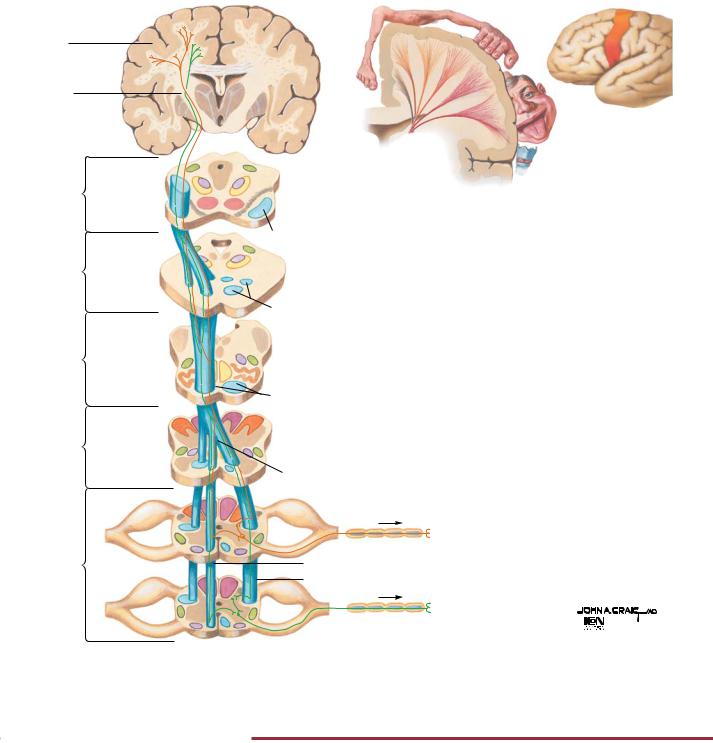
FIGURE 2.20 CORTICOSPINAL TRACTS•
The corticospinal, or pyramidal, tract is the major motor tract that controls voluntary movement of the skeletal muscles, especially skilled movements of distal muscles of the limbs. All structures from the cerebral cortex to the anterior horn cells in the spinal
cord constitute the upper portion of the system (upper motor neuron). The anterior horn cells and their associated axons constitute the lower portion of the system (lower motor neuron).
71