


NEUROPHYSIOLOGY |
Organization of the Brain: Cerebrum |
Central sulcus (Rolando)
Precentral gyrus
Precentral sulcus
Frontal pole
Lateral sulcus (Sylvius)
Temporal pole |
Superior temporal gyrus |
|
|
|
Middle temporal gyrus |
Frontal lobe
Parietal lobe
Temporal lobe
Occipital lobe
Insula (island of Reil)
Postcentral gyrus
Postcentral sulcus
Superior parietal lobule
Inferior parietal lobule
Supramarginal gyrus
Angular gyrus
Parietooccipital sulcus
Occipital pole
Calcarine sulcus
Inferior temporal gyrus
©
FIGURE 2.1 ORGANIZATION OF THE BRAIN: CEREBRUM•
The cerebral cortex represents the highest center for sensory and motor processing. In general, the frontal lobe processes motor, visual, speech, and personality modalities. The parietal lobe processes sensory information; the temporal lobe, auditory and memory modalities; and the occipital lobe, vision. The cerebellum
coordinates smooth motor activities and processes muscle position. The brainstem (medulla, pons, midbrain) conveys motor and sensory information and mediates important autonomic functions. The spinal cord receives sensory input from the body and conveys somatic and autonomic motor information to peripheral targets (muscles, viscera).
52
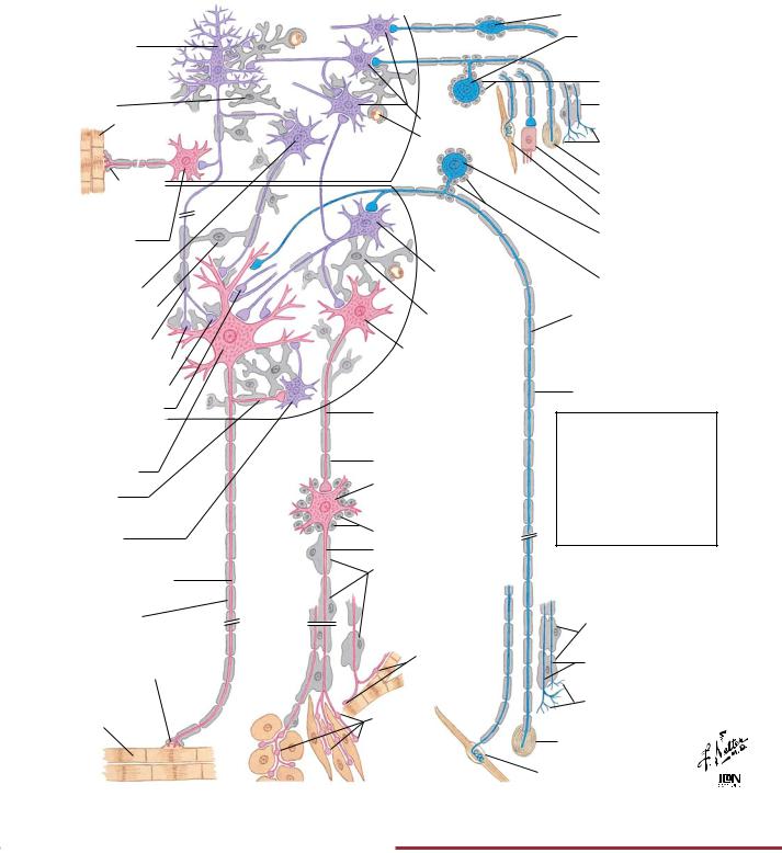
Organization of the Brain: Cell Types |
NEUROPHYSIOLOGY |
Multipolar (pyramidal) cell of cerebral
motor cortex
Astrocyte
Striated (somatic) muscle
Motor endplate
Multipolar somatic motor cell of nuclei of cranial nn.
Multipolar cell of lower brain motor centers
Oligodendrocyte
Corticospinal (pyramidal) fiber
Axodendritic ending
Axosomatic ending
Axoaxonic ending
Multipolar somatic motor cell of anterior horn
of spinal cord
Collateral
Renshaw interneuron (feedback)
Myelinated somatic motor fiber of spinal nerve
Myelin sheath
Motor endplate with Schwann cell cap
Striated (voluntary) muscle
Interneurons
Blood vessel
Interneuron
Astrocyte
Multipolar visceral motor (autonomic) cell of spinal cord
Autonomic preganglionic (sympathetic or parasympathetic) nerve fiber
Myelin sheath
Autonomic postganglionic neuron of sympathetic or parasympathetic ganglion
Satellite cells
Unmyelinated nerve fiber
Schwann cells
Endings on cardiac muscle or nodal cells
Beaded varicosities and endings on smooth muscle and gland cells
Bipolar cell of cranial n.
Unipolar cell of sensory ganglia of cranial nn.
Satellite cells Schwann cell
Free nerve endings (unmyelinated fibers)
Encapsulated ending Specialized ending Muscle spindle
Unipolar sensory cell of dorsal spinal
root ganglion Satellite cells
Myelinated afferent fiber of spinal nerve
Myelin sheath
Red: Motor neuron Blue: Sensory neuron
Purple: Interneuron
Gray: Glial and neurilemmal cells and myelin
Note: Cerebellar cells not shown here
Schwann cells
Unmyelinated fibers
Free nerve endings
Encapsulated ending
Muscle spindle |
© |
FIGURE 2.2 ORGANIZATION OF THE BRAIN: CELL TYPES•
Neurons form the functional cellular units responsible for communication, and throughout the nervous system, they are characterized by their distinctive size and shapes (e.g., bipolar, unipolar, multipolar). Supporting cells include the neuroglia
(e.g., astrocytes, oligodendrocytes), satellite cells, and other specialized cells that optimize neuronal function, provide maintenance functions, or protect the nervous system.
53
NEUROPHYSIOLOGY |
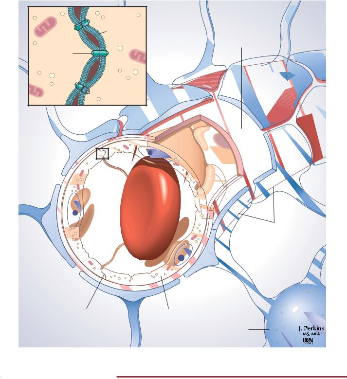
Blood-Brain Barrier |
|
Cell |
|
membrane |
|
Basement |
Tight |
membrane |
|
|
junction |
|
proteins |
|
|
Cytoplasm |
Red blood cell
Capillary |
Astrocyte |
foot processes |
|
lumen |
|
Tight |
Capillary |
|
junction |
endothelial |
|
|
cell |
Astrocyte |
|
|
FIGURE 2.3 BLOOD-BRAIN BARRIER•
The blood-brain barrier (BBB) is the cellular interface between the blood and the central nervous system (CNS; brain and spinal cord). It serves to maintain the interstitial fluid environment to ensure optimal functionality of the neurons. This barrier consists of the capillary endothelial cells with an elaborate network of tight junctions and astrocytic foot processes that abut the endothelium and its basement membrane. The movement of large molecules and
other substances (including many drugs) from the blood to the interstitial space of the CNS is restricted by the BBB. CNS endothelial cells also exhibit a low level of pinocytotic activity across the cell, so specific carrier systems for the transport of essential substrates of energy and amino acid metabolism are characteristic of these cells. The astrocytes help transfer important metabolites from the blood to the neurons and also remove excess K and neurotransmitters from the interstitial fluid.
54
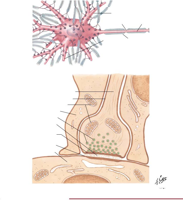
Synaptic Transmission: Morphology of Synapses |
NEUROPHYSIOLOGY |
Dendrite
Node |
Axon |
Dendrites |
Myelin sheath |
|
Numerous boutons (synaptic knobs) of presynaptic neurons terminating on a motor neuron and its dendrites
Enlarged section of bouton
Axon (axoplasm)
Axolemma
Mitochondria
Glial process
Synaptic vesicles
Synaptic cleft
Presynaptic membrane (densely staining)
Postsynaptic membrane (densely staining)
Postsynaptic cell
©

FIGURE 2.4 MORPHOLOGY OF SYNAPSES•
Neurons communicate with each other and with effector targets at specialized regions called synapses. The top figure shows a typical motor neuron that receives numerous synaptic contacts on its cell body and associated dendrites. Incoming axons lose their myelin sheaths, exhibit extensive branching, and terminate as synaptic boutons (synaptic terminals or knobs) on the motor neuron. The
lower figure shows an enlargement of one such synaptic bouton. Chemical neurotransmitters are contained in synaptic vesicles, which can fuse with the presynaptic membrane, release the transmitters into the synaptic cleft, and then bind to receptors situated in the postsynaptic membrane. This synaptic transmission results in excitatory, inhibitory, or modulatory effects on the target cell.
55