

NEUROPHYSIOLOGY |
Synaptic Transmission: Neuromuscular Junction |
Structure of Neuromuscular Junction
Active zone
Schwann cell process
Acetylcholine receptor sites
Myofibrils
Synaptic cleft
Postsynaptic membrane
Junctional fold
Sarcoplasm
Acetylcholine receptor sites
Myelin sheath
Neurilemma
Axoplasm
Schwann cell
Mitochondria
Basement membrane
Nucleus of Schwann cell
Presynaptic membrane
Active zone
Synaptic vesicles
Synaptic trough
Basement membrane
Sarcolemma
Nucleus of muscle cell
©
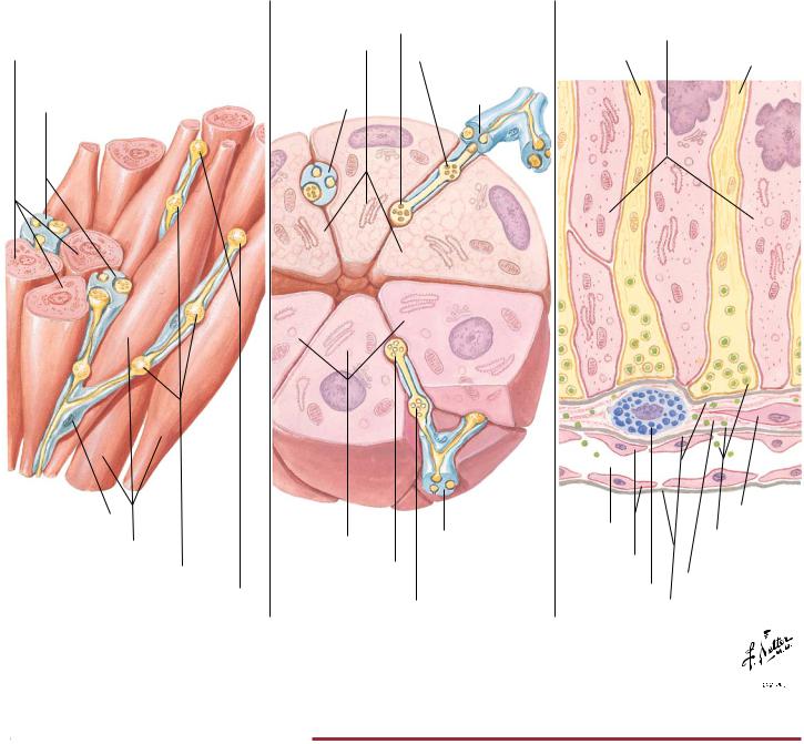
FIGURE 2.5 STRUCTURE OF THE NEUROMUSCULAR JUNCTION•
Motor axons that synapse on skeletal muscle form expanded terminals called neuromuscular junctions (motor endplates). The motor axon loses its myelin sheath and expands into a Schwann cell–invested synaptic terminal that resides within a trough in the muscle fiber. Acetylcholine-containing synaptic vesicles accumulate adjacent to the presynaptic membrane and, when appropri-
ately stimulated, release their neurotransmitter into the synaptic cleft. The transmitter then binds to receptors that mediate depolarization of the muscle sarcolemma and initiate a muscle action potential. A single muscle fiber has only one neuromuscular junction, but a motor axon can innervate multiple muscle fibers.
56
Synaptic Transmission: Visceral Efferent Endings |
NEUROPHYSIOLOGY |
Visceral Efferent Endings
A. Smooth muscle
Smooth muscle cells (cut)
Schwann cell cap enclosing nerve axons
Schwann cell cap
Smooth muscle cells
Varicosities
Terminal endings
B. Gland (submandibular)
|
Sympathetic terminal |
|
|
ending |
|
Mucous cells |
Varicosity |
|
|
||
Schwann cell |
Schwann |
|
cap enclosing |
||
cell cap |
||
nerve axons |
||
|
Serous cells |
Schwann cell |
|
cap enclosing |
||
|
||
Parasympathetic |
nerve axons |
|
terminal ending |
|
|
|
Varicosity |
C. Neurosecretory(posterior pituitary)
Pituicyte processes
Axon |
Axon |
|
Fibroblast
Capillary
Endothelium
Neurosecretory vesicles
Collagen space
Mast cell
Basement membrane
©

FIGURE 2.6 VISCERAL EFFERENT ENDINGS•
Neuronal efferent endings on smooth muscle (A) and glands (B and C) exhibit unique endings unlike the presynaptic and postsynaptic terminals observed in neuronal and neuromuscular junction synapses. Rather, neurotransmitter substances are released into interstitial spaces (A and B) or into the bloodstream (C, neu-
rosecretion) from expanded nerve terminal endings. This arrangement allows for the stimulation of numerous target cells over a wide area. Not all smooth muscle cells are innervated. They are connected to adjacent cells by gap junctions and can therefore contract together with the innervated cells.
57
NEUROPHYSIOLOGY |
|
|
|
Synaptic Transmission: Inhibitory Mechanisms |
|
|
|
|
|
|
|
|
|
|
E
(Excitatory
fiber)
A.Only E fires
90-mV spike in E terminal
EPSP in motor neuron
B.Only I fires
Long-lasting partial depolarization in E terminal
No response in motor neuron
C.I fires before E
Partial depolarization of E terminal reduces spike to 80 mV, thus releasing less transmitter
substance Smaller EPSP in motor neuron
I |
E |
(Inhibitory |
(Excitatory |
fiber) |
fiber) |
Motor
neuron Motor neuron
I
Axon (Inhibitory Axon fiber)
mV
20
mV
|
|
90 mV |
A′. Only E fires |
|
|
|
|
|
|
|
|
||
|
|
|
|
|
||
70 |
|
|
EPSP in |
|
|
|
|
|
|
|
|
||
|
|
|
|
|
||
|
|
|
|
|||
|
|
|
|
|
||
60 |
|
|
motor |
60 |
|
|
|
|
|
|
|||
70 |
|
|
neuron |
|
|
|
|
|
B′. Only I fires |
70 |
|
|
|
|
|
|
|
|||
|
|
|
|
|
|
|
|
|
|
|
|
|
|
60 |
|
|
Motor |
|
|
|
|
|
|
|
|
||
|
|
neuron |
|
|
|
|
70 |
|
|
70 |
|
|
|
|
|
hyper- |
|
|
||
|
|
|
|
|
||
70 |
|
|
polarized |
80 |
|
|
|
|
C′. I fires before E |
|
|
||
|
|
|
|
|||
|
|
|
|
|
||
|
|
|
|
|
|
|
|
|
|
|
|
|
|
20 |
|
|
Depolariza- |
60 |
|
|
|
|
|
|
|||
|
|
|
|
|
||
|
|
tion of motor |
70 |
|
|
|
|
|
|
|
|
||
|
|
|
|
|
||
|
|
80 mV |
neuron less |
|
|
|
|
|
|
|
|
||
|
|
|
|
|
||
|
|
than if only |
80 |
|
|
|
|
|
|
|
|||
70 |
|
|
E fires |
|
|
|
|
|
|
|
|||
|
|
|
|
|
||
|
|
|
|
|
|
|
|
|
|
|
|
|
|
|
|
|
|
|
|
|
60 |
|
|
|
|
|
|
|
|
|
|
|
|
|
70 |
|
|
|
© |
||
|
|
|
||||
|
|
|
|
|||
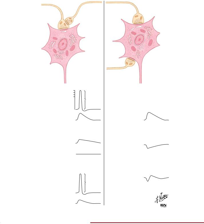
FIGURE 2.7 SYNAPTIC INHIBITORY MECHANISMS•
Inhibitory synapses modulate neuronal activity. Illustrated here is presynaptic inhibition (left panel) and postsynaptic inhibition (right panel) at a motor neuron.
58

Synaptic Transmission: Chemical Synaptic Transmission |
NEUROPHYSIOLOGY |
|
|||||||||||
|
|
Excitatory |
|
|
|
|
Inhibitory |
|
|
||
|
|
|
|
|
Synaptic |
|
|
|
|
|
|
|
|
|
|
|
vesicles |
|
|
|
|
|
|
|
|
|
|
|
in synaptic |
|
|
|
|
|
|
|
|
|
|
|
bouton |
|
|
|
|
|
|
|
|
|
|
|
Presynaptic |
|
|
|
|
|
|
|
|
|
|
|
membrane |
|
|
|
|
|
|
|
|
|
|
|
Transmitter |
|
|
|
|
|
|
|
|
Na |
|
|
substances |
|
|
|
|
||
|
|
Synaptic cleft |
|
|
|
|
|
||||
|
|
|
|||||||||
|
|
Cl |
|
|
|||||||
Postsynaptic |
|
|
|
|
|||||||
|
|
K |
|
|
|
|
|
||||
|
|
|
|
|
|
|
|
|
|||
membrane
When impulse reaches excitatory synaptic bouton, it causes release of a transmitter substance into synaptic cleft. This increases permeability of postsynaptic membrane to Na and K . More Na moves into postsynaptic cell than K moves out, due to greater electrochemical gradient
At inhibitory synapse, transmitter substance released by an impulse increases permeability of the postsynaptic membrane to Cl . K moves out of post-synaptic cell but no net flow of Cl occurs at resting membrane potential
Synaptic bouton
Resultant net ionic current flow is in a direction that tends to depolarize postsynaptic cell. If depolarization reaches firing threshold, an impulse is generated in postsynaptic cell
(mV) |
|
|
Current |
|
|
|
65 |
|
|
Potential |
|
|
|
Potential |
|
|
|
|
|
|
70 |
|
|
|
|
|
|
|
0 |
4 |
8 |
12 |
16 |
|
msec
Current flow and potential change
Resultant ionic current flow is in direction that tends to hyperpolarize postsynaptic cell. This makes depolarization by excitatory synapses more difficult—more depolarization is required to reach threshold
|
|
|
|
|
|
|
msec |
|
|
|
(mV) |
70 |
0 |
4 |
8 |
12 |
16 |
||||
|
|
|
|
|
Potential |
|
|
|
||
|
|
|
|
|
|
|
|
|
||
Potential |
75 |
|
|
|
|
|
|
|
|
|
|
|
|
Current |
|
|
|
||||
|
|
|
|
|
|
|
|
|||
|
|
|
|
|
|
|
|
|
|
|
Current flow and potential change
©
FIGURE 2.8 CHEMICAL SYNAPTIC TRANSMISSION•
Chemical synaptic transmission between neurons may be excitatory or inhibitory. During excitation (left column), a net increase in the inward flow of Na compared with the outward flow of K results in a depolarizing potential change (excitatory postsynaptic potential [EPSP]) that drives the postsynaptic cell closer to its
threshold for an action potential. During inhibition (right column), the opening of K and Cl channels drives the membrane potential away from threshold (hyperpolarization) and decreases the probability that the neuron will reach threshold (inhibitory postsynaptic potential [IPSP]) for an action potential.
59