

NEUROPHYSIOLOGY |
Cerebellum: Afferent Pathways |
Cortical input
Nucleus reticularis tegmenti pontis
Superior cerebellar peduncle
Middle cerebellar peduncle
To contralateral cerebellar cortex
Leg |
Arm |
|
|
|
F |
Primary fissure |
|
|
|
||
|
|
ace |
|
Pontine nuclei (contralateral)
Spinal input
Inferior olive
Upper part of medulla oblongata
Spinal input
Vestibular nerve and ganglion
Lower part of medulla oblongata
Cortical input
Lateral reticular nucleus
Spinal input
Cervical part of spinal cord
Motor interneuron
Rostral spinocerebellar tract
Spinal border cells
Motor interneuron
Lumbar part of spinal cord
Clarke’s column
Ventral spinocerebellar tract
FIGURE 2.21
|
To nodule and flocculus |
|
|
|
||
Vestibular |
Inferior cerebellar peduncle |
|
|
|
||
nuclei |
|
|
|
|
|
|
|
|
Functional Subdivisions of Cerebellum |
||||
Reticulocerebellar |
|
Hemisphere Vermis |
|
|||
|
|
Inter- |
|
|||
tract |
|
|
|
|
||
|
|
Lateral mediate |
|
|||
Cuneocerebellar |
|
part |
part |
Anterior lobe |
||
tract |
|
Leg zone |
|
|
||
|
|
|
Primary |
|||
Gracile nucleus |
Arm zone |
|
|
fissure |
||
Main cuneate |
Face zone |
|
|
|
||
|
|
|
|
|||
nucleus (relay |
|
|
|
Middle |
||
for cutaneous |
2nd spinal |
|
|
(posterior) |
||
information) |
|
|
lobe |
|||
projection |
|
|
||||
External cuneate nucleus |
|
|
|
|||
area (gracile |
|
|
|
|||
(relay for proprioceptive |
lobule) |
|
|
Posterolateral |
||
information) |
|
Archi- |
|
|
fissure |
|
From skin (touch |
Lingula |
Flocculo- |
||||
cerebellum |
||||||
and pressure) |
Flocculus |
|||||
(vestibulo- |
nodular lobe |
|||||
From muscle (spindles |
Nodule |
|||||
cerebellum) |
|
|||||
|
Uvula |
|
||||
and Golgi tendon organs) |
Paleocerebellum |
|
||||
From skin and |
(spinocerebellum) |
Pyramid |
|
|||
deep tissues |
|
|
Vermis |
|
||
Neocerebellum |
Middle vermis |
|||||
(pain and Golgi |
||||||
(pontocerebellum) Hemisphere |
|
|||||
tendon organs) |
Schema of |
|||||
|
|
|
||||
From skin (touch |
|
|
|
|||
|
|
|
theoretical |
|||
and pressure) |
|
|
|
“unfolding” |
||
and from muscle |
|
|
|
of cerebellar |
||
(spindles and |
|
|
|
surface in |
||
Golgi tendon |
|
|
|
derivation of |
||
organs) |
|
|
|
|
above diagram |
|
Dorsal spinocerebellar tract |
|
|
|
|
||
CEREBELLAR AFFERENT PATHWAYS• |
|
|
|
© |
||
|
|
|
|
|||
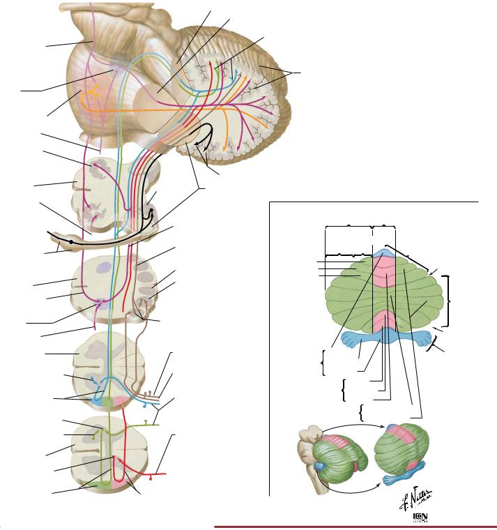
The cerebellum plays an important role in coordinating movement. It receives sensory information and then influences descending motor pathways to produce fine, smooth, and coordinated motion. The cerebellum is divided into three general areas: archicerebellum (also called vestibulocerebellum) paleocerebellum (also called spinocerebellum) and the neocerebellum (also called the cerebrocerebellum). The archicerebellum is primarily involved in controlling posture and balance, as well as the movement of the head and eyes. It receives afferent signals from the vestibular apparatus and then sends efferent fibers to the appropriate descending motor pathways. The paleocere-
bellum primarily controls movement of the proximal portions of the limbs. It receives sensory information on limb position and muscle tone and then modifies and coordinates these movements through efferent pathways to the appropriate descending motor pathways. The neocerebellum is the largest portion of the cerebellum, and it coordinates the movement of the distal portions of the limbs. It receives input from the cerebral cortex and thus helps in the planning of motor activity (e.g., seeing a pencil and then planning and executing the movement of the arm and hand to pick it up).
72
Cerebellum: Efferent Pathways |
NEUROPHYSIOLOGY |
Excitatory endings
Inhibitory endings of Purkinje cells
Ventral anterior and ventral lateral nuclei of thalamus
Mesencephalic reticular formation
Red nucleus
Fastigial nucleus
Globose nuclei
Emboliform nucleus
Dentate nucleus
Cerebellar cortex
Vestibular nuclei
Inferior cerebellar peduncle
Inferior olive
Lateral reticular nucleus
Medulla oblongata
Pontomedullary reticular formation
FIGURE 2.22 CEREBELLAR EFFERENT PATHWAYS•
Motor and premotor cerebral cortex
Internal capsule
Cerebral peduncle
Decussation of superior cerebellar peduncles
Descending fibers from superior cerebellar peduncles
Hook bundle of Russell
Section A–B viewed from below
Section B–C viewed from above
A |
|
|
Planes of |
|
|
section: |
B |
|
red arrows |
|
|
indicate |
C |
|
direction |
||
|
||
of view |
© |
|
|
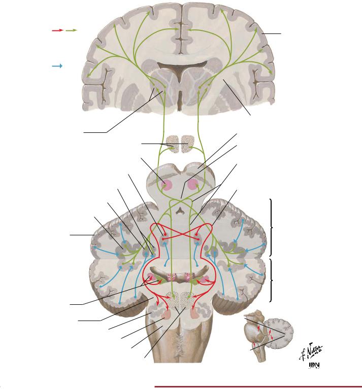
The cerebellum plays an important role in coordinating movement. It influences descending motor pathways to produce fine, smooth, and coordinated motion. The archicerebellum is primarily involved in controlling posture and balance and movement of the head and eyes. It sends efferent fibers to the appropriate descending motor pathways. The paleocerebellum primarily controls movement of
the proximal portions of the limbs. It modifies and coordinates these movements through efferent pathways to the appropriate descending motor pathways. The neocerebellum coordinates the movement of the distal portions of the limbs. It helps in the planning of motor activity (e.g., seeing a pencil and then planning and executing the movement of the arm and hand to pick it up).
73
NEUROPHYSIOLOGY |
Cutaneous Sensory Receptors |
Melanocyte
Arrector muscle of hair
Sebaceous gland
|
Cuticle |
|
follicle |
Internal sheath |
|
External sheath |
||
|
||
Hair |
Glassy |
|
membrane |
||
|
Connective |
|
|
tissue layer |
Hair cuticle
Sweat gland
Hair matrix
Papilla of hair follicle
Pacinian corpuscle
Artery
Detail of Merkel’s disc
Basal epithelial cells
Cytoplasmic protrusion
Mitochondria
Expanded axon terminal
Free nerve endings |
Meissner’s corpuscle |
Stratum corneum |
|
|
||
Hair shaft |
|
Pore of sweat gland |
Stratum |
|
|
|
|
|
|
|
lucidum |
Stratum |
Epidermis |
|
|
|
|
|
||
|
|
|
|
|
granulosum |
|
|
|
|
|
|
|
|
|
|
|
|
Stratum |
|
|
|
|
|
|
spinosum |
|
|
|
|
|
|
|
Stratum |
|
|
|
|
|
|
basale |
|
|
|
|
|
Dermal |
|
|
|
|
|
|
papilla |
|
|
|
|
|
|
(of papillary |
|
|
|
|
|
|
layer) |
|
Dermis |
|
|
|
|
|
|
|
|
|
|
|
Reticular |
|
|
|
|
|
|
layer |
|
|
|
|
|
|
|
|
tissue Subcutaneous |
|
|
|
|
Subcutaneous |
|
|
Vein |
|
|
|
artery and vein |
|
|
Sensory nerves |
|
|
|
|
Basement membrane |
|
Elastic fibers |
|
|
Cutaneous |
|
Axon terminal |
|
Skin ligaments |
|
|
Mitochondrion |
|
||
|
nerve |
|
Schwann cell |
|
||
(retinacula cutis) |
|
|
|
|||
Motor |
|
|
|
|||
|
|
|
|
|
|
|
|
|
(autonomic) |
|
|
|
|
|
|
nerve |
|
Cross section |
|
|
Desmosomes |
|
|
|
|
|
|
|
|
|
|
|
|
|
Merkel |
|
|
|
|
|
|
cell |
|
|
|
|
|
|
Lobulated |
|
|
|
|
|
|
nucleus |
|
|
|
|
|
|
Granulated |
|
|
|
|
|
|
vesicles |
|
|
|
Axon |
|
|
Schwann |
|
|
|
Schwann cells |
|
|
|
|
|
|
|
|
|
cell |
|
|
|
Detail of free nerve ending |
|
|
FIGURE 2.23 SKIN AND CUTANEOUS RECEPTORS•
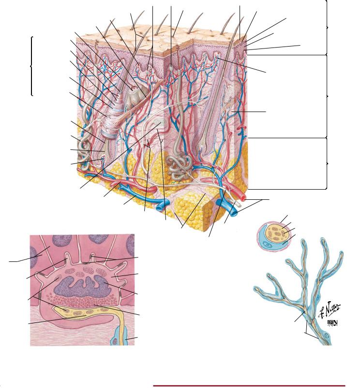
Cutaneous receptors respond to touch (mechanoreceptors), pain (nociceptors), and temperature (thermoreceptors). Several different types of receptors are present in skin. Meissner’s corpuscles have small receptive fields and respond best to stimuli that are applied at low frequency (i.e., flutter). The pacinian corpuscles are located in the subcutaneous tissue and have large receptive fields. They
respond best to high-frequency stimulation (i.e., vibration). Merkel’s discs have small receptive fields and respond to touch and pressure (i.e., indenting the skin). Ruffini’s corpuscles have large receptive fields, and they also respond to touch and pressure. Free nerve endings respond to pain and temperature.
74
Cutaneous Receptors: Pacinian Corpuscle |
NEUROPHYSIOLOGY |
Pacinian Corpuscle |
|
as Pressure Transducer |
Pressure |
|
To amplifier |
|
Generator potential |
1st node
Myelin sheath
Lamellated capsule
Central core
Unmyelinated axon terminal
Action potential
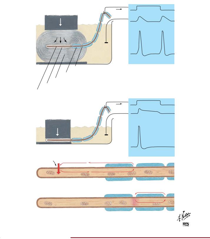
A. Sharp “on and off” changes in pressure at start and end of pulse applied to lamellated capsule are transmitted to central axon and provoke generator potentials, which in turn may trigger action potentials; there is no response to a slow change in pressure gradient. Pressure at central core and, accordingly, generator potentials are rapidly dissipated by viscoelastic properties of capsule (Action potentials may be blocked by pressure at a node or by drugs)
Pressure
To amplifier
Generator potential
B. In absence of capsule, axon responds to slow as well
as to rapid changes in pressure. Generator potential Action potential dissipates slowly, and there is no “off” response
Pressure |
Na+ |
|
|
|
|
|
|
|
|
|
|
|
|
|
|
|
|
|
|
|
|
|
|
|
|
|
|
|
|
|
|
|
|
|
|
|
|
|
|
|
|
|
|
|
|
|
|
|
|
|
|
|
|
|
|
|
|
||||||
|
|
|
|
|
|
|
|
|
|
|
|
|
|
|
|
Pressure applied to axon terminal directly or |
|
|
|
|
|
|
|
|
|
||||||
via capsule causes increased permeability of |
|
|
|
|
|
|
|
|
|
||||||
membrane to Na+, thus setting up ionic |
|
|
|
|
|
|
|
|
|
|
|||||
generator current through 1st node |
|
|
|
|
|
|
|
|
|
|
|
||||
|
|
|
|
|
|
|
|
|
|
|
|
|
|
|
|
|
|
|
|
|
|
|
|
|
|
|
|
|
|
|
|
If resultant depolarization at 1st node is great |
|
enough to reach threshold, an action potential |
|
appears which is propagated along nerve fiber |
© |
|
FIGURE 2.24 PACINIAN CORPUSCLE•
Pacinian corpuscles are mechanoreceptors that transduce mechanical forces (displacement, pressure, vibration) into action potentials that are conveyed centrally by afferent nerve fibers. As the viscoelastic lamellae are displaced, the unmyelinated axon terminal membrane’s ionic permeability is increased until it is capable of
producing a “generator potential.” As demonstrated in the figure, pacinian corpuscles respond to the beginning and end of a mechanical force while the concentric lamellae dissipate slow changes in pressure. In the absence of the capsule, the generator potential decays slowly and yields only a single action potential.
75
NEUROPHYSIOLOGY |
Proprioception and Reflex Pathways: I |
Spinal Effector Mechanisms
Dorsal horn interneuron
From motor neuron
Proprioceptive fibers |
From |
cutaneous |
|
Dorsal horn interneuron |
receptor |
|
From |
|
muscle |
|
spindle |
Dorsal root ganglion
Flexor reflex interneuron |
Ventral root |
To motor neuron |
Dorsal horn interneuron |
|
|
|
|
To motor neuron
motor axon
Schematic representation of motor neurons
In cervical enlargement of spinal cord
|
|
|
|
|
|
In lumbar |
Fl |
|
|
|
|
|
enlargement |
e |
|
s |
|
|
||
|
|
r |
|
|
of spinal cord |
|
|
x o |
|
|
s |
||
|
|
|
|
|
|
|
|
|
|
|
|
r |
|
|
|
|
|
o |
|
|
|
|
|
|
s |
|
|
|
|
|
n |
|
|
|
|
Exte |
|
|
|
||
F
l
|
s |
|
|
r |
|
|
|
o |
|
|
|
ex |
|
|
|
|
|
|
s |
|
|
n |
|
te |
|
||
Ex |
|
|
|
o
r
s
©

FIGURE 2.25 PROPRIOCEPTION: SPINAL EFFECTOR MECHANISM•
Position sense or proprioception involves input from cutaneous mechanoreceptors, Golgi tendon organs, and muscle spindles (middle figure of upper panel). Both monosynaptic reflex pathways (middle figure of upper panel) and polysynaptic pathways involving several spinal cord segments (top and bottom figures of upper
panel) initiate muscle contraction reflexes. The lower panel shows the somatotopic distribution of the motor neuron cell bodies in the ventral horn of the spinal cord that innervate limb muscles (flexor and extensor muscles of upper and lower limbs).
76
Proprioception and Reflex Pathways: II |
NEUROPHYSIOLOGY |
Alpha motor neurons to extrafusal striated muscle end plates
Gamma motor neurons to intrafusal striated muscle end plates
Ia (A ) fibers from annulospiral endings (proprioception)
II (A ) fibers from flower spray endings (proprioception);
from paciniform corpuscles (pressure) and pacinian corpuscles (pressure)
III (A ) fibers from free nerve endings and from some specialized endings (pain and some pressure)
IV (unmyelinated) fibers from free nerve endings (pain)
Ib (A ) fibers from Golgi tendon organs (proprioception)
Alpha motor neuron to extrafusal muscle fiber end plates
Gamma motor neuron to intrafusal muscle fiber and plates
II (A ) fiber from flower spray endings
Ia (A ) fiber from annulospiral endings
Detail of muscle spindle
A fibers from Golgi-type endings
A fibers from paciniform corpuscles and Ruffini terminals
A and C fibers from free nerve endings
Extrafusal muscle fiber
Intrafusal muscle fibers
Sheath
Lymph space
Nuclear bag fiber
Nuclear chain fiber
©
Efferent fibers
Afferent fibers
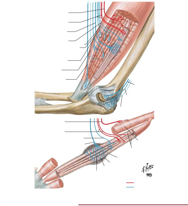
FIGURE 2.26 MUSCLE AND JOINT RECEPTORS•
Muscle spindles and Golgi tendon organs send afferent signals to the brain to convey the position of limbs and help coordinate muscle movement. Muscle spindles convey information on muscle tension and contraction (dynamic forces) and muscle length (static forces). The nuclear bag fibers respond to both dynamic and static
forces, whereas the nuclear chain fibers respond to static forces. Intrafusal fibers maintain appropriate tension on the nuclear bag and nuclear chain fibers. If the muscle tension is too great (e.g., overstretching of muscle or too heavy a load), activation of the Golgi tendon organ causes a reflex relaxation of the muscle.
77
NEUROPHYSIOLOGY |
Proprioception and Reflex Pathways: III |
Golgi tendon organ
Golgi tendon organ
Golgi tendon organ
Ib fibers
Ia fibers 
Extrafusal muscle fiber
Intrafusal muscle fiber
Alpha motor neurons 
Gamma motor neurons
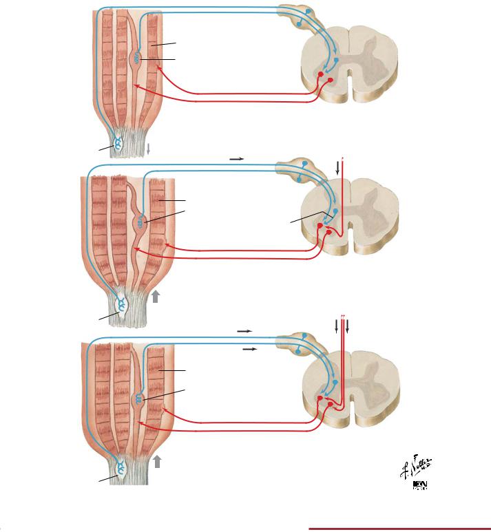
A. Passive stretch. Both intrafusal and extrafusal muscle fibers stretched; spindles activated. Reflex via Ia fibers and alpha motor neurons causes secondary contraction (basis of stretch reflexes, such as knee jerk). Stretch is too weak to activate Golgi tendon organs
Ib fibers
Alpha activation
from brain
Ia fibers
Extrafusal muscle fiber
Intrafusal muscle fiber
Inhibitory interneuron
Alpha motor neurons 
Gamma motor neurons
B. Active contraction. Central excitation of alpha motor neurons only causes contraction of extrafusal muscle fibers with consequent relaxation of intrafusal fibers; spindles not activated. Tension is low; does not adjust to increased resistance. Tendon organ activated, causing relaxation
|
Alpha and |
|
Ib fibers |
gamma |
|
activation |
||
|
||
Ia fibers |
from brain |
|
|
Extrafusal muscle fiber
Intrafusal muscle fiber
Alpha motor neurons 
Gamma motor neurons 
C. Active contraction with gamma coactivation. Intrafusal as well as |
|
extrafusal fibers contract; spindles activated, reinforcing contraction |
|
stimulus via Ia fibers in accord with resistance. Tendon organ |
|
activated, causing relaxation if load is too great |
© |
FIGURE 2.27 PROPRIOCEPTIVE REFLEX CONTROL OF MUSCLE TENSION•
Interaction of the muscle spindle and Golgi tendon organ during passive stretch of a muscle (panel A) and during a contraction (panels B and C).
78
Proprioception and Reflex Pathways: IV |
NEUROPHYSIOLOGY |
|
B. Stretch reflex |
|
A. Afferent inhibition |
(reciprocal inhibition) |
|
From extensor spindle |
From extensor spindle |
|
receptor (Ia, II fibers) |
|
|
From flexor spindle |
receptor (Ia, II fibers) |
|
|
|
|
(Ia, II fibers) |
Axosomatic or |
|
|
axodendritic |
|
|
inhibitory |
|
|
synapse |
|
Axoaxonic presynaptic |
Excitatory |
|
inhibitory synapse |
|
|
synapse |
|
|
|
|
|
To extensors |
To extensors |
|
To flexors |
|
|
|
|
|
C. Recurrent inhibition |
D. Tendon organ reflex |
|
|
From extensor tendon |
|
|
organ (Ib fibers) |
|
|
Inhibitory synapse |
|
Renshaw cells |
Excitatory synapse |
|
Collaterals |
|
|
To synergistic |
To extensors |
|
|
|
|
muscles |
To flexors |
|
|
|
|
E. Flexor withdrawal reflex |
|
|
Nociceptive fibers |
|
|
Ipsilateral |
Contralateral |
|
flexion |
extension |
|
Inhibitory synapse |
Excitatory synapse |
|
Excitatory synapse |
Inhibitory synapse |
|
To extensors |
To extensors |
|
To flexors |
To flexors |
© |
|
||
|
|
|
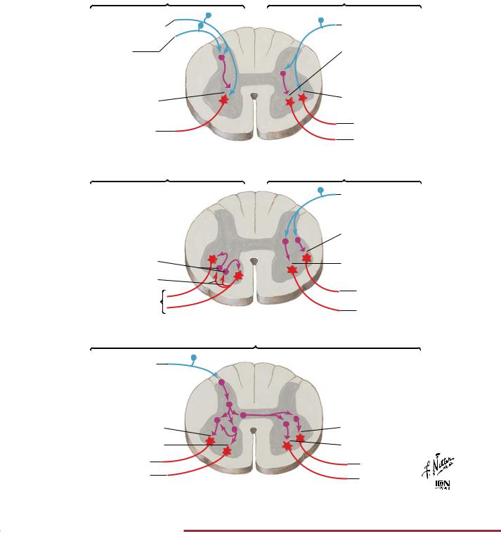
FIGURE 2.28 SPINAL REFLEX PATHWAYS•
Summary of the spinal reflex pathways.
79