

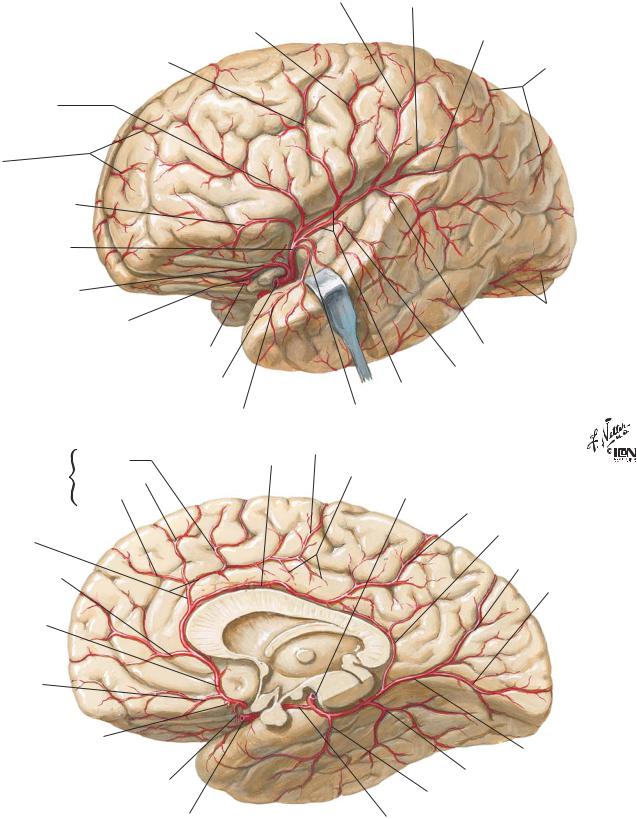
Arteries of Brain: Lateral and Medial Views |
NEUROANATOMY |
Anterior parietal (postcentral sulcal) artery |
Posterior parietal artery |
Central (rolandic) sulcal artery
Precentral (pre-rolandic) sulcal artery
Prefrontal sulcal artery
Terminal branches of anterior cerebral artery
Lateral frontobasal (orbitofrontal) artery
Left middle cerebral artery
Left anterior cerebral artery
Anterior communicating artery
Right anterior cerebral artery
Left internal carotid artery
|
|
Polar temporal artery |
Medial |
Posterior |
Pericallosal artery |
frontal |
Intermediate |
|
branches |
Anterior |
|
|
|
|
Callosomarginal artery
Polar frontal artery
Right anterior cerebral artery
Medial frontobasal (orbitofrontal) artery
Anterior
communicating artery (cut)
Distal medial striate artery (recurrent artery of Heubner)
Right internal carotid artery
Branch to angular gyrus
Terminal branches of posterior cerebral artery
Occipitotemporal branches
Posterior temporal branch
Middle temporal branch
Superior and inferior terminal branches (trunks)
Anterior temporal branch
Paracentral artery
Cingular branches
Right posterior cerebral artery
Precuneal artery
Dorsal branch
to corpus callosum
Parietooccipital branch
Calcarine branch
Medial occipital artery
Posterior temporal branch
Anterior temporal branch
Posterior communicating artery
Note: Anterior parietal (postcentral sulcal) artery also occurs as separate anterior parietal and postcentral sulcal arteries
15
NEUROANATOMY |
Arteries of Posterior Cranial Fossa |
Thalamogeniculate arteries
Anterior choroidal artery
Crura of fornix
Anterolateral central (lenticulostriate) arteries
Heads of caudate nuclei
Septum pellucidum
Corpus callosum
Anterior cerebral arteries
Longitudinal cerebral fissure
Lateral and medial geniculate bodies of left thalamus
Choroid plexuses of lateral ventricles
Pulvinars of left and right thalami
Splenium of corpus callosum
Occipital (posterior) horn of right lateral ventricle
Right dorsal branch to corpus callosum (posterior pericallosal artery)
Parietooccipital |
Branches of |
Calcarine |
right posterior |
cerebral artery |
Optic nerve (II)
IV
Ophthalmic artery |
III |
Anterior |
V |
|
|
cerebral artery |
|
Middle |
VIII |
cerebral artery |
|
Posterior |
VII |
communicating artery |
VI |
IX
Thalamoperforating arteries
X
Left internal carotid artery
XI
Basilar artery
Pontine arteries
Labyrinthine (internal acoustic) artery
Posterior cerebral artery
Superior cerebellar artery
Anterior inferior cerebellar artery
Anterior meningeal branch of vertebral artery
Temporal branches of posterior cerebral artery
Anterior spinal artery
Superior colliculi
Superior vermian branch
Posterior medial choroidal artery to choroid plexus of 3rd ventricle
Posterior lateral choroidal artery
Lateral (marginal) branch
Inferior vermian artery (phantom)
Choroidal branch to 4th ventricle (phantom) and
Cerebellar tonsillar branch
of posterior inferior cerebellar artery
Outline of 4th ventricle (broken line)
Posterior meningeal branch of vertebral artery
Posterior inferior cerebellar artery (PICA)
Left posterior spinal artery
Left vertebral artery
16
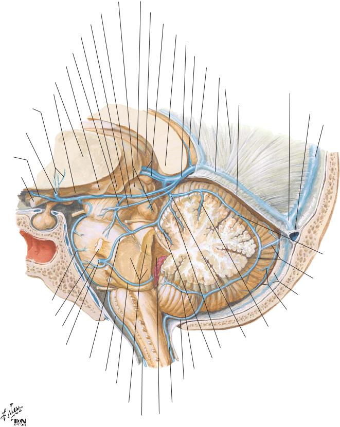
Veins of Posterior Cranial Fossa |
NEUROANATOMY |
Left superior and inferior colliculi
Basal vein (Rosenthal)
Posterior mesencephalic vein
Medial geniculate body
Lateral mesencephalic vein
Lateral geniculate body
Left thalamus (cut surface)
Optic tract
Inferior thalamostriate veins
Deep middle cerebral vein (cut)
Anterior cerebral vein
Optic nerve (II)
Left pulvinar
Right pulvinar
Internal cerebral veins
Splenium of corpus callosum
Great cerebral vein (Galen)
Dorsal vein of corpus callosum
Inferior sagittal sinus
Straight sinus
Falx cerebri
Tentorium cerebelli (cut)
C
|
C |
CL |
D |
|
F
L
TU
N P
U
Confluence of sinuses
Left transverse sinus (cut)
Superior sagittal sinus
Superior vermian vein
Inferior vermian vein
Anterior pontomesencephalic vein
Trigeminal nerve (V)
Transverse pontine vein
Petrosal vein (draining
to superior petrosal sinus)
Lateral pontine vein
Anteromedian medullary vein
Vein of lateral recess of 4th ventricle
Superior, middle and inferior cerebellar peduncles
Anterior spinal vein
T |
|
Falx cerebelli (cut) |
|||
|
and occipital sinus |
||||
|
|
Inferior cerebellar |
|||
|
|
hemispheric veins |
|||
|
Intraculminate vein |
|
|||
Superior cerebellar vein (inconstant) |
|||||
Preculminate vein |
|
|
|
||
Precentral cerebellar vein |
|
|
|||
Superior retrotonsillar vein |
|
|
|
|
|
(Inferior retrotonsillar) |
|
Parts of cerebellum |
|||
vein of cerebellomedullary |
L |
Lingula |
TU Tuber |
||
cistern |
|||||
CL Central lobule |
P |
Pyramid |
|||
|
|||||
Posterior spinal vein |
C |
Culmen |
U Uvula |
||
4th ventricle |
D Declive |
N |
Nodule |
||
F |
Folium |
T |
Tonsil |
||
|
|||||
17
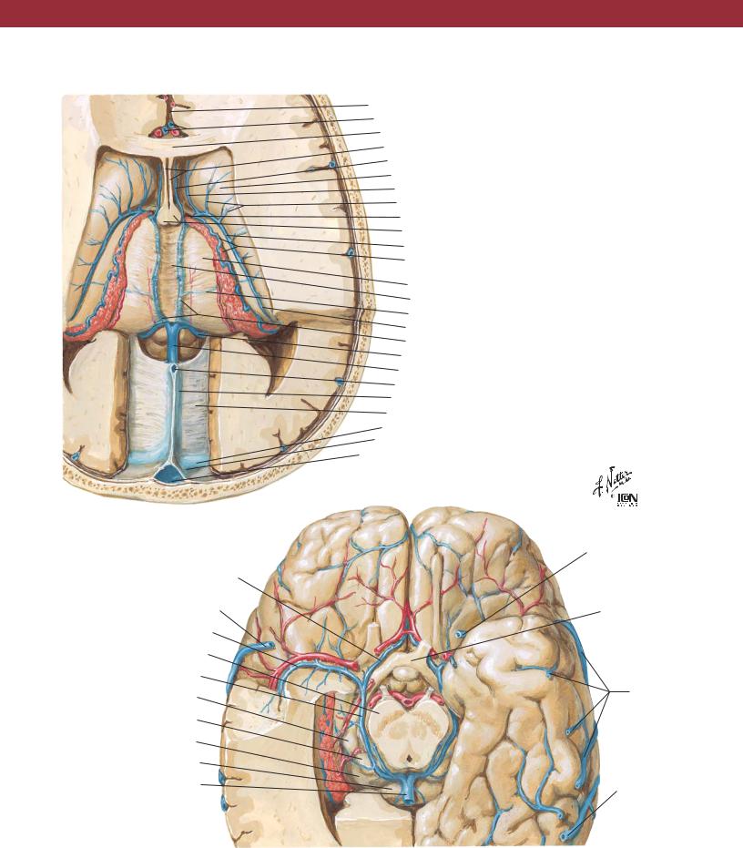
NEUROANATOMY
Dissection: superior view
Anterior cerebral vein
Superficial middle cerebral vein (draining to sphenoparietal sinus)
Deep middle cerebral vein
Cerebral crus
Basal vein (Rosenthal)
Lateral geniculate body
Medial geniculate body
Pulvinar of thalamus
Splenium of corpus callosum
Great cerebral vein (Galen)
Dissection: inferior view
Deep Veins of Brain
Longitudinal cerebral fissure Anterior cerebral veins
Rostrum of corpus callosum Septum pellucidum
Anterior vein of septum pellucidum Head of caudate nucleus
Anterior vein of caudate nucleus Transverse veins of caudate nucleus Interventricular foramen (Monro) Columns of fornix
Superior thalamostriate vein
Superior choroid vein and choroid plexus of lateral ventricle
Thalamus
Tela choroidea of 3rd ventricle Lateral direct vein
Posterior vein of caudate nucleus Internal cerebral veins
Basal vein (Rosenthal) Great cerebral vein (Galen) Inferior sagittal sinus Straight sinus
Tentorium cerebelli Transverse sinus
Confluence of sinuses Superior sagittal sinus
Uncal vein
Optic chiasm
Inferior cerebral veins
Inferior anastomotic vein (Labbé)
18