


NEUROPHYSIOLOGY |
Visual System: Visual Pathway |
G
G
Overlapping
visual fields
A A
BB
HH
R R C |
C |
Projection on |
||
left retina |
||||
|
|
|
||
P |
|
P |
|
|
Choroid |
Choroid |
|
||
Periphery |
Macula |
|
||
Structure of retina (schematic): |
||||
A |
Amacrine cells |
Projection on left |
||
dorsal lateral |
||||
B |
Bipolar cells |
|||
geniculate nucleus |
||||
C |
Cones |
|
||
|
|
|||
G |
Ganglion cells |
|
||
H |
Horizontal cells |
|
||
P |
Pigment cells |
|
||
R |
Rods |
|
|
|
Calcarine fissure
Projection on left occipital lobe
FIGURE 2.33 RETINOGENICULOSTRIATE VISUAL PATHWAY•
Optic (II) nerves Optic chiasm
Optic tracts
Lateral geniculate bodies
Central darker circle represents macular zone
Lightest shades represent monocular fields
Each quadrant a different color
Projection on right retina
Projection on right dorsal lateral geniculate nucleus
Projection |
|
on right |
|
occipital lobe |
© |
The retina has two types of photoreceptors: cones that mediate color vision and rods that mediate light perception but with low acuity. The greatest acuity is found in the region of the macula of the retina, where only cones are found (upper left panel). Visual signals are conveyed by the ganglion cells whose axons course in the optic nerves. Visual signals from the nasal retina cross in the
optic chiasm while information from the temporal retina remains in the ipsilateral optic tract. Fibers synapse in the lateral geniculate nucleus (visual field is topographically represented here and inverted), and signals are conveyed to the visual cortex on the medial surface of the occipital lobe.
84
Auditory System: Cochlea |
NEUROPHYSIOLOGY |
|
Cochlear nerve |
|
|
A. Membranous |
Utricle |
Semicircular |
|
labyrinth within |
canals |
||
|
|||
bony labyrinth |
Saccule |
|
|
(path of sound |
|
|
|
waves) |
|
|
Scala vestibuli |
|
|
||
Cochlear duct |
|
|
|
|
(scala media) |
Scala tympani |
Round window |
Oval window and stapes |
|
|
||||
B. Section |
|
|
|
|
through turn |
|
|
Vestibular (Reissner’s) |
|
of cochlea |
Scala vestibuli |
membrane |
||
|
(perilymph); |
Cochlear duct (scala media; |
||
|
weakly |
|
||
|
+80 mV |
endolymph) |
||
Efferent |
positive |
|||
Tectorial membrane |
||||
nerve fibers |
|
|
||
|
|
|
Spiral ligament |
|
|
|
|
Bone |
|
Afferent |
Scala tympani |
Outer hair cells; 60 mV |
||
nerve fibers |
(perilymph); 0 mV |
Basilar membrane |
||
|
|
|
||
Spiral ganglion |
|
|
Inner hair cell; 60 mV |
|
|
|
|
||
C. Spiral organ |
Hair cells |
|
|
|
of Corti |
|
Tectorial membrane |
||
Inner Outer
Stereocilia
Rods and tunnel of Corti
|
Basilar membrane |
|
|
|
Supporting cells |
|
|
Spiral lamina |
Afferent nerve fibers |
|
|
Spiral ganglion |
Efferent nerve fibers |
|
|
As basilar membrane moves up, hairs are deflected outward, causing |
|
|
|
depolarization of hair cells and increased firing of afferent nerve fibers |
|
© |
|
|
|
|
|
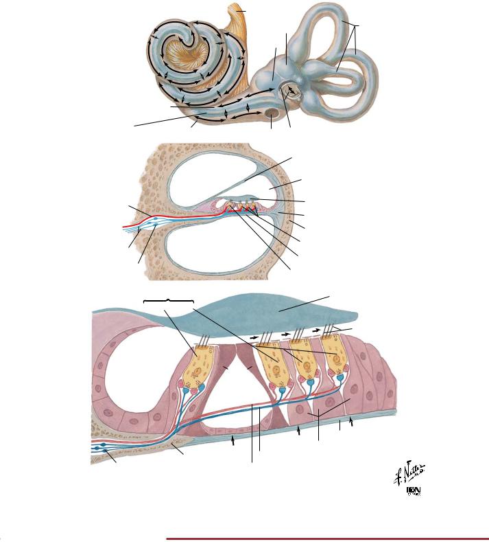
FIGURE 2.34 COCHLEAR RECEPTORS•
The cochlea transduces sound into electrical signals. This is accomplished by the hair cells, which depolarize in response to vibration of the basilar membrane. The basilar membrane moves in
response to pressure changes imparted on the oval window of the cochlea in response to vibrations of the tympanic membrane.
85
NEUROPHYSIOLOGY |
Auditory System: Pathways |
Brachium of inferior colliculus
Inferior colliculus
Midbrain
Lateral lemnisci
Medulla oblongata
Acoustic area of temporal lobe cortex
Medial geniculate body
Correspondence between cochlea and acoustic area of cortex:
 Low tones
 Middle tones
Nuclei of 
 High tones lateral

 lemnisci
Dorsal cochlear nucleus
Inferior cerebellar peduncle
Ventral cochlear nucleus
Cochlear division of vestibulocochlear nerve
Dorsal acoustic stria
Reticular formation  Inner Outer
Trapezoid body |
Hair cells |
Spiral ganglion |
|
Intermediate acoustic stria |
|
Superior olivary complex |
|
©

FIGURE 2.35 AUDITORY PATHWAYS•
The cochlea transduces sound into electrical signals. Axons convey these signals to the dorsal and ventral cochlear nuclei, where it is tonotopically organized. Following a series of integrated relay pathways, the ascending pathway projects to the thalamus (medial
geniculate bodies) and then the acoustic cortex in the transverse gyrus of the temporal lobe, where information is tonotopically represented (low, middle, and high tones).
86
Vestibular System: Receptors |
NEUROPHYSIOLOGY |
A. Membranous labyrinth
Vestibular ganglion
Vestibular and cochlear divisions of
vestibulocochlear n. Maculae Saccule Utricle
Cochlear duct (scala media)
B. Section of crista
Opposite wall of ampulla
Gelatinous cupula Hair tufts
Hair cells
Nerve fibers
Basement membrane
D. Structure and innervation |
|
Excitation |
|
of hair cells |
|
|
|
|
Inhibition |
||
|
|
||
Kinocilium |
|
|
|
Stereocilia |
|
|
|
Cuticle |
Basal body |
||
Cuticle |
|||
|
|||
Hair cell (type I) |
|
|
|
Supporting cells |
|
|
|
Afferent nerve |
|
|
|
calyx |
|
|
|
Efferent nerve |
|
|
|
ending |
|
|
|
Basement |
|
|
|
membrane |
|
|
|
Myelin sheath |
|
|
|
FIGURE 2.36 VESTIBULAR RECEPTORS•
Superior semicircular canal
Cristae within ampullae
Horizontal semicircular canal
Posterior semicircular canal
C. Section of macula
Otoconia Gelatinous otolithic membrane
Hair tuft Hair cells
Supporting cells Basement membrane Nerve fibers
Kinocilium
Stereocilia
Basal body
Hair cell (type II)
Supporting cell
Efferent nerve endings
Afferent nerve endings
Myelin sheath
©

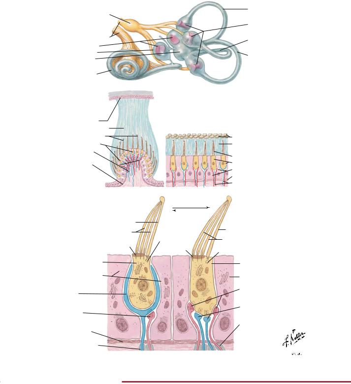
The vestibular apparatus detects movement of the head in the form of linear and angular acceleration. This information is important for the control of eye movements so that the retina can be provided with a stable visual image. It is also important for the control of posture. The utricle and saccule respond to linear acceleration,
such as the pull of gravity. The three semicircular canals are aligned so that the angular movement of the head can be sensed in all planes. The sensory hair cells are located in the maculae of the utricle and saccule and in the cristae within each ampullae.
87