Cardiology / English / Internal_diseases_propedeutics._Part_II._Diagnostics_of_cardiovascular_diseases
.pdf
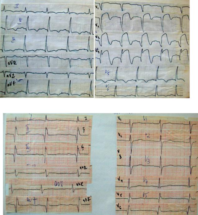
LOCALIZATION OF IM (Fig. 28-29):
1.The FRONT wall of the left ventricle: I, aVL, V1-4; discordant displacement to ST III, aVF;
2.The SIDE wall of the left ventricle: V5-6;
3THE LOWER wall LV: III, aVF; the discordance of the offset ST in I, aVL
Fig.28. MI of the front wall of the left ventricle.
Fig.29. MI of the lower wall of the left ventricle.
81
TEST CONTROL:
1.THE PATIENT COMPLAINS OF PROLONGED (MORE THAN 10 MIN.) CHEST PAIN OF INCREASING CHARACTER, RADIATING TO THE BACK, ARM AND NECK, RESULTING IN A STATE OF REST AND NOT STOPED AFTER TAKING 3 TABLETS OF NITROGLYCERIN. WHAT KIND OF PATHOLOGY YOU CAN THINK OF (GIVE ONE ANSWER)?
a) cardialgia; b) angina;
c) angina at rest;
d) myocardial infarction; e) pericarditis;
f) dissecting aneurysm of the aorta.
2.INDICATE THE 5 CHARACTERISTIC SYMPTOMS OF RIGHT HEART FAILURE:
a) enlargement of the liver;
b) shortness of breath during physical exertion and/or at rest;
c) attacks of breathlessness with difficulty in inhalation and exhalation, forcing to take a half upright position;
d) attacks of breathlessness with difficulty exhaling, forcing to take a sitting position with fixation of the shoulder girdle;
e) presence of free fluid in the abdomen (ascites);
f) the presence of free fluid in the pleural cavity (hydrothorax);
g) the presence of free fluid in the pericardial cavity (hydroperiod); h) swelling of the feet.
3.PERCUSSION BORDERS OF RELATIVE CARDIAC DULLNESS OF THE PATIENT REVEALED THE FOLLOWING DATA:
- right margin – 1 cm outwards from the right edge of the sternum, - left – 4 cm laterally from the left midclavicular line,
- upper – in intercostal space III,
- the width of the vascular bundle – 5 cm.
For what configuration of the heart is typical?
a)for normal;
b)for the mitral;
c)for aortic.
82
4.WHAT ARE 2 FACTORS MOST DETERMINE THE LEVEL OF DIASTOLIC BLOOD PRESSURE (BP)?
a) the tone of resistive vessels (arterioles) – tot al peripheral vascular resistance; b) the volume of the vascular of ejection (stroke volume of the heart);
c) the elasticity of the walls of the aorta;
d) the completeness of closure of the valves of the aorta.
5.DURING THE INSPECTION AND PALPATION OF THE HEART REGION OF THE PATIENT REVEALED AMPLIFIED AND DIFFUSE APEX BEAT. WHAT STATE OF HEART IS THE EVIDENCE?
a) only hypertrophy of the left ventricle; b) only dilatation of the left ventricle; c) only dilatation of the right ventricle
d) hypertrophy of the left ventricle and dilatation of the left ventricle
6.LIST THE FEATURES CHARACTERIZING HEART DISEASES EDEMA:
a)a high density edema ("solid" edema);
b)low density edema (soft swelling);
c)pallor of the skin in the area of edema;
d)cyanotic skin in the area of edema;
e)the presence of trophic skin changes in the area of edema;
f)lack of trophic skin changes in the area of edema.
7.EXPLAIN THE MECHANISM OF OCCURRENCE OF COUGH WITH CLEAR SPUTUM AND HEMOPTYSIS IN PATIENTS WITH LEFT VENTRICULAR HEART FAILURE (GIVE ONE ANSWER):
a) inflammatory process in the alveoli – the exudat ion of blood plasma and red blood cells;
b) high pressure in the blood vessels of the pulmonary circulation – the leakage of blood plasma and erythrocytes in the alveoli;
c) high pressure in the blood vessels of the big circle of blood circulation – the leakage of blood plasma and erythrocytes in the alveoli;
d) high pressure in the blood vessels of the pulmonary circulation – bronchial vessels microreserve – penetration of blood plasma into the lumen of the bronchi.
8.WHAT ARE 3 METHODS OF PHYSICAL AND INSTRUMENTAL EXAMINATION CAN REVEAL DILATATION OF THE LEFT VENTRICLE? a) palpation of the heart;
83
b)percussion of the heart;
c)electrocardiography (ECG);
d)roentgenography of chest organs;
e)echocardiography (EchoCG).
9.HOW TO CHANGE THE LOCALIZATION OF THE APEX BEAT OF THE WOMAN IN THE LAST 2-3 MONTHS OF PREGNANCY?
a) will not change;
b) shift up and to the left; c) will shift to the right.
10.PALPATION OF THE PATIENT IN THE APEX OF THE HEART REVEALED A THRILL THAT DOES NOT COINCIDE WITH THE PULSATION OF THE APEX BEAT. FOR WHAT CONDITION IS THIS TYPICAL?
a) obstruction of blood flow through the mitral valve hole;
b) obstruction of blood flow through the opening of the tricuspid valve; c) obstruction of blood flow through the opening of the aortic valve.
11.SPECIFY 3 MAIN COMPONENTS INVOLVED IN THE FORMATION OF I TONE OF THE HEART:
a) fluctuations of the valves of the atrioventricular valves in the phase of isometric reduction;
b) fluctuations of the valves of the valves of the aorta and pulmonary artery protodiastolic period;
c) fluctuations in the muscular wall of the ventricles in the phase of isometric contraction; d) fluctuations in the muscular wall of the ventricle at protodiastolic period;
e) oscillations of initial segments of the aorta and pulmonary artery in the early phase of exile.
12.SPECIFY THE MAIN 4 MECHANISM OF WEAKENING OF THE II TONE IN THE SECOND INTERCOSTAL SPACE RIGHT OF STERNUM:
a) increase the mobility of the semilunar valves of the aorta; b) reducing the mobility of the semilunar valves of the aorta;
c) non-tight closing of the valves of the aorta in the protodiastolic period; d) lowering the pressure in the aorta;
e) the increase in the rate of relaxation of ventricular myocardium; f) decrease in the rate of relaxation of the ventricular myocardium.
84
13.WHAT ARE 3 OF THE LISTED SIGNS CAN BE USED TO IDENTIFY THE I HEART TONE?
a) the tone coincides with the apex beat and carotid pulsation;
b) a tone does not coincide with the apex beat and carotid pulsation; c) the tone is listened after a long pause;
d) the tone is listened after a short pause; e) a low frequency tone and long lasting.
14.HOW TO CHANGE THE VOLUME OF I TONE AT THE APEX IN MITRAL VALVE INSUFFICIENCE?
a) increase; b) decrease;
in) will not change.
15.WHAT 2 CHANGES II TONE OF THE HEART CAN REVEAL IN THE 2ND INTERCOSTAL SPACE LEFT OF STERNUM IN PATIENTS PRESENTING COMPLAINTS OF SIGNIFICANT DYSPNEA AND A COUGH WITH STREAKS OF BLOOD, INCREASING IN A HORIZONTAL POSITION AND DECREASING IN ORTHOPNEA POSITION?
a) gain (emphasis) II tone; b) weakining II tone;
c) no change II tone.
16.SPECIFY THE 2 CHARACTERISTIC AUSCULTATION SIGNS OF ATRIAL FIBRILLATION:
a) correct (regular) rhythm of the heart;
b) isolated premature occurring I and II heart tones followed by a pause on the background of the right rhythm;
c) completely irregular (chaotic) rhythm of the heart; d) the same volume I of tones in each cardiac cycle;
e) the constant change of volume I of the tone ( from one cardiac cycle to another).
17.WHEN REGISTERING PHONOCARDIOGRAM (FKG) AT THE APEX OF THE HEART, THE PATIENT RECORDED HIGH-FREQUENCY DIASTOLIC EXTRATONE ARISING FROM CLOSE II TONE (EVERY 0.1 S). NAME IT:
a) III the tone of the heart; b) IV is the heart;
c) the tone of the opening of the mitral valve.
85
18.2 SPECIFY THE SYMPTOMS OF THE PHYSIOLOGICAL SPLITTING OF II HEART SOUND:
a) the constancy of this phenomenon;
b) the impermanence of this phenomenon; c) the appearance during inhalation.
19.THE PATIENT HAS A PRONOUNCED ATHEROSCLEROTIC DISEASE OF THE ASCENDING AORTA AND AORTIC VALVE. MYOCARDIAL CONTRACTILITY OF THE LEFT VENTRICLE SATISFACTORY, THE AMPLITUDE OF THE DISCLOSURE OF THE AORTIC VALVE IS SUFFICIENT. WHAT IS THE LIKELY CHANGE IN II TONE IN THE SECOND INTERCOSTAL SPACE TO THE RIGHT OF THE STERNUM?
a) enhanced II tone (accent II tone on the aorta); b) weakened II tone;
in) II unmodified tone.
20.THE PATIENT HAS LARGE FOCUS OF POSTINFARCTION CARDIOSCLEROSIS OF THE LEFT VENTRICLE AND OBJECTIVE SIGNS OF MYOGENIC DILATATION (THE WEAKENING AND SHIFT TO THE LEFT OF THE DIFFUSE APEX BEAT, OFFSET TO THE LEFT RELATIVE DULLNESS OF THE HEART). WHAT 3 CHANGES OF HEART TONES WE CAN IDENTIFY IN THIS SITUATION?
a) the strengthening of tone I on the top; b) I weakening tone at the apex;
c) increased the I tone at the base of the xiphoid process;
d) the appearance of the pathological III tone heart at the apex; f) the appearance of the pathological IV tone heart at the apex; g) appearance at the top of the tone of mitral valve opening.
21.GIVE THE DEFINITION OF "VALVULAR REGURGITATION" (CHOOSE ONE ANSWER):
a) any turbulent blood flow through the valve opening;
b) turbulent blood flow through the sash of the diseased valve during systole; c) turbulent blood flow through the sash of the diseased valve in diastole;
d) blood flow through the affected valve flaps, incapable of full closure; e) blood flow through the affected valve flaps, are not able to fully open.
86
22.SPECIFY THE NATURE AND AREA OF THE BEST LISTENING OF MURMURS, IF THE PATIENT HAS THE MITRAL STENOSIS:
a) systolic murmur at the apex; b) diastolic murmur at the apex;
c) systolic murmur in the second intercostal space to the right of the sternum; d) diastolic murmur in the second intercostal space to the right of the sternum; e) systolic murmur in the second intercostal space left of the sternum;
23.SPECIFY THE NATURE AND AREA OF THE BEST LISTENING OF MURMURS, IF THE PATIENT HAS PULMONARY ARTERY VALVE INSUFFICIENCE
a) systolic murmur at the apex; b) diastolic murmur at the apex;
c) systolic murmur in the second intercostal space to the right of the sternum; d) diastolic murmur in the second intercostal space to the right of the sternum; e) systolic murmur in the second intercostal space left of the sternum;
f) diastolic murmur in the second intercostal space left of the sternum; g) systolic murmur at the base of the xiphoid process.
24.SPECIFY THE NATURE AND AREA OF THE BEST LISTENING OF MURMURS, IF THE PATIENT HAS SWELLING AND PULSATION OF THE JUGULAR VEINS OF THE NECK, COINCIDING WITH THE PULSATION OF THE APEX BEAT?
a) systolic murmur at the apex; b) diastolic murmur at the apex;
c) systolic murmur in the second intercostal space to the right of the sternum; d) diastolic murmur in the second intercostal space to the right of the sternum; e) systolic murmur in the second intercostal space left of the sternum;
f) diastolic murmur in the second intercostal space left of the sternum; g) systolic murmur at the base of the xiphoid process.
25.WHAT 2 GROUPS OF HEART MURMURS ARE FUNCTIONAL?
a)murmurs resulting from lesions of the heart valves and great vessels;
b)murmur occurring due to a decrease in blood viscosity or increased blood flow velocity;
c)murmur arising from the relative insufficiency or valve stenosis holes;
87
d)pericardial RUB and pleuropericardial murmur;
e)the murmur arising from congenital heart defects.
26.ON SOME OF THESE HEART LESIONS CAN BE CONSIDER IF THE PATIENT STUDY REVEALED: INCREASED I TONE AND MESOCESTOIDES LOW FREQUENCY RUMBLING MURMUR AT THE APEX OCCURRING AFTER AN AUDIBLE TONE BY OPENING OF THE MITRAL VALVE?
a) mitral valve insufficiency; b) mitral stenosis;
c) the aortic insufficiency; d) aortic stenosis.
27.HOW TO CHANGE THE LOUDNESS OF THE SYSTOLIC MURMUR OF MITRAL REGURGITATION WITH INCREASING THE CONTRACTILITY OF THE LEFT VENTRICLE?
a) decreases;
b) will increase; in) will not change.
28.GIVE A DETAILED CHARACTERIZATION OF THE MURMUR ORGANIC MITRAL REGURGITATION (GIVE ANSWER 4):
a) auscultated at the apex; b) is the systolic;
c) is the diastolic;
d) starts immediately after the I tones; e) starts some time after I tone;
f) is held in the left axillary region;
g) is carried out on the carotid and subclavian arteries.
29.GIVE A DETAILED CHARACTERIZATION OF THE ORGANIC MURMUR OF AORTIC VALVE INSUFFICIENCY (GIVE 5 ANSWERS):
a) auscultated in the 2nd intercostal space right of the sternum; b) auscultated in the 2nd left intercostal space from the sternum;
c) auscultated left of the sternum, in the area of attachment of III – IV ribs; d) is the systolic;
e) is diastolic;
f) starts immediately after the II tone;
88
g)starts some time after II tone;
h)is held in the left axillary region;
i)is carried out according to the flow of blood from the aorta into the left ventricle.
30.SPECIFY 2 PERCUS AND RADIOGRAPHIC SYMPTOM OF MITRAL CONFIGURATION OF THE HEART:
a) displacement of right border of relative dullness of heart to the right; b) the offset of the left border of relative dullness of heart to the left;
c) the displacement of the upper borders of relative dullness of the heart upward; d) accentuated waist of heart;
e) waist smoothed heart.
31.DESCRIBE THE APEX BEAT IN A PATIENT WITH MITRAL VALVE INSUFFICIENCY IN THE STAGE OF COMPENSATION DEFECT (REPLY 3): a) enhanced apex beat;
b) weakened apex beat;
c) the displacement of the apex beat to the left only;
d) the displacement of the apex beat to the left and down (6th – 7th intercostal space); e) apex beat advanced;
f) apex beat concentric.
32.CHECK THE 4 TYPICAL SYMPTOMS OF DECOMPENSATION OF MITRAL HEART DEFECTS:
a) acrocyanosis;
b) dense bluish swelling of legs and feet; c) fluid effusion in the pleural cavity;
d) shortness of breath on exertion;
e) pain in the heart area character (removed nitroglycerin); f) enlargement of the liver.
33.HOW TO CHANGE THE PERCUS SHAPE OF THE HEART IN A PATIENT WITH MITRAL VALVE STENOSIS AT THE STAGE OF COMPENSATION (ONE ANSWER)
a) displacement of the upper border of relative dullness of heart - up, and the waist smoothed heart.;
b) significant shift of the left border of relative dullness of heart to the left.
89
34.WHICH 3 OF THE FOLLOWING COMPLAINTS ARE CHARACTERISTIC FOR A MITRAL VALVE INSUFFICIENCY PATIENT?
a) shortness of breath during physical exertion and in the supine position;
b) pain in the heart area and behind the breastbone radiating to the left arm and under the shoulder blade;
c) sensations of faults in work of heart and heartbeat ("chaotic rhythm hearts");
d) swelling of the feet (more towards evening);
e) dizziness and (sometimes) momentary fainting during physical load;
f) dyspnea and cough with blood streaks at night.
35.WHAT ARE 2 VARIATIONS OF THE PULSE AT THE RADIAL ARTERIES ARE TYPICAL OF PATIENTS WITH MITRAL VALVE STENOSIS WITH ATRIAL FIBRILLATION?
a) high and imminent ("galloping" - pulse Corrigan); b) low and slow;
c) pulsus deficience;
d) different (pulsus difference); e) pulsus paradoxus.
36.SPECIFY 2 AUSCULTATION SIGN OF ATRIAL FIBRILLATION :
a)correct the heart rhythm;
b)single failures in the activities of the heart on the background of the right rhythm;
c)"chaotic" (completely irregular) heart rhythm;
d)strengthening of tone I at the apex;
e)changing the volume I tone at the apex.
37.SPECIFY 3 MAIN DIAGNOSTIC AUSCULTATION SIGNS OF STENOSIS THE MITRAL VALVE (RHEUMATIC ETIOLOGY):
a) the strengthening of tone I at the apex; b) I weakening tone at the apex;
c) the presence of pathological III tone at the apex;
d) the presence of the tone of mitral valve opening at the apex;
e) low-frequency diastolic murmur at the apex without holding in other areas.
38.SPECIFY 2 PERCUSSION AND RADIOGRAPHIC CHARACTERISTIC AORTIC CONFIGURATION OF THE HEART:
90
