

Cardiology / English / Internal_diseases_propedeutics._Part_II._Diagnostics_of_cardiovascular_diseases
.pdfNo definition of heart failure (HF) is entirely satisfactory. Congestive heart failure (CHF) develops when plasma volume increases and fluid accumulates in the lungs, abdominal organs (especially the liver), and peripheral tissues.
Physiology At rest and during exercise, cardiac output (CO), venous return, and distribution of blood flow with O2 delivery to the tissues are balanced by neurohumoral and intrinsic cardiac factors. Preload, the contractile state, afterload, the rate of contraction, substrate availability, and the extent of myocardial damage determine left ventricular (LV) performance and myocardial O2 requirements. The Frank-Starling principle, cardiac reserve, and the oxyhemoglobin dissociation curve play a role.
Preload (the degree of end-diastolic fiber stretch) reflects the end-diastolic volume, which is influenced by diastolic pressure and the composition of the myocardial wall. For clinical purposes, the end-diastolic pressure, especially if above normal, is a reasonable measure of preload in many conditions. LV dilatation, hypertrophy, and changes in myocardial distensibility or compliance modify preload.
The contractile state in isolated cardiac muscle is characterized by the force and velocity of contraction, which are difficult to measure in the intact heart. Clinically, the contractile state is often expressed as the ejection fraction (LV stroke volume/enddiastolic volume).
Afterload (the force resisting myocardial fiber shortening after stimulation from the relaxed state) is determined by the chamber pressure, volume, and wall thickness at the time of aortic valve opening. Clinically, afterload approximates systemic BP at or shortly after aortic valve opening and represents peak systolic wall stress. The heart rate and rhythm also influence cardiac performance.
Reduced substrate availability (eg, of fatty acid or glucose), particularly if O2 availability is reduced, can impair the vigour of cardiac contraction and myocardial performance.
Tissue damage (acute with myocardial infarction or chronic with fibrosis due to various diseases) impairs local myocardial performance and imposes an additional load on viable myocardium.
51
The Frank-Starling principle states that the degree of end-diastolic fiber stretch (preload) within a physiologic range is proportional to the systolic performance of the subsequent ventricular contraction
This mechanism operates in HF, but, because ventricular function is abnormal, the response is inadequate. If the Frank-Starling curve is depressed, fluid retention, vasoconstriction, and a cascade of neurohumoral responses lead to the syndrome of CHF. Over time, LV remodeling (change from the normal ovoid shape) with dilatation and hypertrophy further compromises cardiac performance, especially during physical stress. Dilatation and hypertrophy may be accompanied by increased diastolic stiffness.
Classification and Etiology
In many forms of heart disease, the clinical manifestations of HF may reflect impairment of the left or right ventricle.
Left ventricular (LV) failure characteristically develops in coronary artery disease, hypertension, and most forms of cardiomyopathy and with congenital defects (eg, ventricular septal defect, patent ductus arteriosus with large shunts).
Right ventricular (RV) failure is most commonly caused by prior LV failure (which increases pulmonary venous pressure and leads to pulmonary arterial hypertension) and tricuspid regurgitation. Causes are also mitral stenosis, primary pulmonary hypertension, multiple pulmonary emboli, pulmonary artery or valve stenosis, and RV infarction.
One distinguishes chrionic and acute heart failure. In Russian Federation clinicians use classification of chronic heart failure, stipulated by National Scientific Society of Cardiologists in 2002. It joins two classifications: one – after N.D. Strazhesko and V.H.Vasilenko (1935) and New York Heart Association functional classification (NYHA) (1964).
According to this classification stages and functional classes of chronic heart failure are distinguished (Table 1 &6).
|
|
Table 6 |
|
Stages of chronic heart failure (may worsen despite on treatment) |
|
|
|
|
I stage |
|
Initial stage of heart disease (damage). Hemodynamics |
|
|
|
|
52 |
|
|
isn't altered. Latent heart failure. Asymtomatic left |
|
ventricular dysfunction. |
|
|
IIA stage |
Clinically pronounced stage of heart disease (damage). |
|
Hemodynamics is altered in one of circulation circles, |
|
moderately pronounced. Adaptive remodeling of the heart |
|
and vessels. |
|
|
IIB stage |
Grave stage of heart disease (damage). Pronounced |
|
hemodynamics alterations in both circulation circle. |
|
Disadaptive remodeling of the heart and vessels. |
|
|
III stage |
Terminal stage of the heart damage. Pronounced |
|
hemodynamics alterations and severe (irreversible) |
|
structural changes of target organs (heart, lungs, vessels, |
|
brain, kidneys). Final stage of organs remodeling. |
|
|
HF is manifested by systolic or diastolic dysfunction or both. Combined systolic and diastolic abnormalities are common.
In systolic dysfunction (primarily a problem of ventricular contractile dysfunction), the heart fails to provide tissues with adequate circulatory output. A wide variety of defects in energy utilization, energy supply, electrophysiologic functions, and contractile element interaction occur, which appear to reflect abnormalities in intracellular Ca++ modulation and cyclic adenosine monophosphate (cAMP) production.
Systolic dysfunction has numerous causes; the most common are coronary artery disease, hypertension, and dilated congestive cardiomyopathy. There are many known and probably many unidentified causes for dilated myocardiopathy. More than 20 viruses have been identified as causal. Toxic substances damaging the heart include alcohol, a variety of organic solvents, certain chemotherapeutic drugs (eg, doxorubicin), β-blockers, Ca blockers, and antiarrhythmic drugs.
Diastolic dysfunction (resistance to ventricular filling not readily measurable at the bedside) accounts for 20 to 40% of cases of HF. It is generally associated with prolonged ventricular relaxation time, as measured during isovolumic relaxation (the time between
53
aortic valve closure and mitral valve opening when ventricular pressure falls rapidly). Resistance to filling (ventricular stiffness) directly relates to ventricular diastolic pressure; this resistance increases with age, probably reflecting myocyte loss and increased interstitial collagen deposition. Diastolic dysfunction is presumed to be dominant in hypertrophic cardiomyopathy, circumstances with marked ventricular hypertrophy (eg, hypertension, advanced aortic stenosis), and amyloid infiltration of the myocardium.
Pathophysiology
In LV failure, CO declines and pulmonary venous pressure increases. Elevated pulmonary capillary pressure to the levels that exceed the oncotic pressure of the plasma proteins (about 24 mm Hg) leads to increased lung water, reduced pulmonary compliance, and a rise in the O2 cost of the work of breathing. Pulmonary venous hypertension and edema resulting from LV failure significantly alter pulmonary mechanics and, thereby, ventilation/perfusion relationships. Dyspnea correlates with elevated pulmonary venous pressure and the resultant increased work of breathing, although the precise cause is debatable. When pulmonary venous hydrostatic pressure exceeds plasma protein oncotic pressure, fluid extravasates into the capillaries, the interstitial space, and the alveoli. Pleural effusions characteristically accumulate in the right hemithorax and later bilaterally. Lymphatic drainage is greatly enhanced but cannot overcome the increase in lung water. Unoxygenated pulmonary arterial blood is shunted past nonaerated alveoli, decreasing mixed pulmonary capillary PaO2. A combination of alveolar hyperventilation due to increased lung stiffness and reduced PaO2 is characteristic of LV failure. Thus, arterial blood gas analysis reveals an increased pH and a reduced PaO2 (respiratory alkalosis) with decreased saturation reflecting increased intrapulmonary shunting. Typically, PaCO2 is reduced too. A PaCO2 above normal signifies alveolar hypoventilation possibly due to respiratory muscle failure and requires urgent ventilatory support.
In RV failure, systemic venous congestive symptoms develop. Moderate hepatic dysfunction commonly occurs in CHF secondary to RV failure, with usually moderate increases in conjugated and unconjugated bilirubin, prothrombin time, and hepatic enzymes (eg, alkaline phosphatase, AST, ALT). However, in severely compromised
54
circulatory states with markedly reduced splanchnic blood flow and hypotension, increases due to central necrosis around the hepatic veins may be severe enough to suggest hepatitis with acute liver failure. Reduced aldosterone breakdown by the impaired liver further contributes to fluid retention.
In systolic dysfunction, inadequate ventricular emptying leads to increased preload, diastolic volume, and pressure. Sudden (as in MI) and progressive (as in dilated cardiomyopathy) myocyte loss induces ventricular remodeling, resulting in increased wall stress accompanied by apoptosis (accelerated myocardial cell death) and inappropriate ventricular hypertrophy. Later, the ejection fraction falls, resulting in progressive pump failure. Systolic HF may primarily affect the LV or the RV (see above), although failure of one ventricle tends to lead to failure of the other.
In diastolic dysfunction, increased resistance to LV filling as a consequence of reduced ventricular compliance (increased stiffness) results in prolonged ventricular relaxation (an active state following contraction) and alters the pattern of ventricular filling. Ejection fraction may be normal or increased. Normally, about 80% of the stroke volume enters the ventricle passively in early diastole, reflected in a large e wave and smaller a wave on pulsed-wave Doppler echocardiography. Generally, in diastolic LV dysfunction the pattern is reversed, accompanied by increased ventricular filling pressure and a-wave amplitude.
Whether the failure is primarily systolic or diastolic and regardless of which ventricle is affected, various hemodynamic, renal, and neurohumoral responses may occur.
Hemodynamic responses: With reduced CO, tissue O2 delivery is maintained by increasing A-VO2. Measurement of A-VO2 with systemic arterial and pulmonary artery blood samples is a sensitive index of cardiac performance and reflects, via the Fick equation (VO2 = CO . A-VO2), CO (inversely related) and the body's O2 consumption (VO2--directly related).
Increased heart rate and myocardial contractility, arteriolar constriction in selected vascular beds, venoconstriction, and Na and water retention compensate in the early stages for reduced ventricular performance. Adverse effects of these compensatory efforts
55
include increased cardiac work, reduced coronary perfusion, increased cardiac preload and afterload, fluid retention resulting in congestion, myocyte loss, increased K excretion, and cardiac arrhythmia.
Renal responses: The mechanism by which an asymptomatic patient with cardiac dysfunction develops overt CHF is unknown, but it begins with renal retention of Na and water, secondary to decreased renal perfusion. Thus, as cardiac function deteriorates, renal blood flow decreases in proportion to the reduced CO, the GFR falls, and blood flow within the kidney is redistributed. The filtration fraction and filtered Na decrease, but tubular resorption increases.
Neurohumoral responses: Increased activity of the renin-angiotensin-aldosterone system influences renal and peripheral vascular response in HF. The intense sympathetic activation accompanying HF stimulates the release of renin from the juxtaglomerular apparatus near the descending loop of Henle in the kidney. Probably, decreased arterial systolic stretch secondary to declining ventricular function also stimulates renin secretion. Reflex and adrenergic stimulation of the renin-angiotensin-aldosterone system produces a cascade of potentially deleterious effects: Increased aldosterone levels enhance Na reabsorption in the distal nephron, contributing to fluid retention. Renin produced by the kidney interacts with angiotensinogen, producing angiotensin I from which is cleaved the octapeptide angiotensin II by ACE. Angiotensin II has various effects believed to enhance the syndrome of CHF, including stimulation of the release of arginine vasopressin (AVP), which is antidiuretic hormone (ADH); vasoconstriction; enhanced aldosterone output; efferent renal vasoconstriction; renal Na retention; and increased norepinephrine release. Angiotensin II is also believed to be involved in vascular and myocardial hypertrophy, thus contributing to the remodeling of the heart and peripheral vasculature, which contributes to HF in various myocardial and other heart diseases.
Plasma norepinephrine levels are markedly increased, largely reflecting intense sympathetic nerve stimulation, because plasma epinephrine levels are not increased. High plasma norepinephrine levels in patients with CHF are associated with a poor prognosis.
The heart contains many neurohormonal receptors (α1, β1, β2, β3, adrenergic, muscarinic, endothelin, serotonin, adenosine, angiotensin II). In patients with HF, β1
56
receptors (which constitute 70% of cardiac β receptors), but not the other adrenergic receptors, are down-regulated, potentially adversely affecting myocardial function. This down-regulation, which is probably a response to intense sympathetic overdrive, has been detected even in asymptomatic patients with the early stages of HF. Altered myocardial stimulator or receptor functions for various other neurohormonal factors may adversely influence myocyte performance in HF.
Serum levels of atrial natriuretic peptide (released in response to increased atrial volume and pressure load) and brain natriuretic peptide (released from the ventricle in response to ventricular stretch) are markedly increased in patients with CHF. These peptides enhance renal excretion of Na, but, in patients with CHF, the effect is blunted by decreased renal perfusion pressure, receptor down-regulation, and perhaps enhanced enzymatic degradation. Serum, brain (B-type) natriuretic peptide appears to be important for diagnosis in CHF and correlates well with functional impairment.
AVP is released in response to a fall in BP or ECF volume and by the effects of various neurohormonal stimuli. An increase in plasma AVP diminishes excretion of free water by the kidney and may contribute to the hyponatremia of HF. AVP levels in CHF vary, but experimental AVP blockers have increased water excretion and serum Na levels.
Other consequences: Protein-losing enteropathy characterized by marked hypoalbuminemia, ischemic bowel infarction, acute and chronic GI hemorrhage, and malabsorption may result from severe chronic venous hypertension. Peripheral gangrene in the absence of large vessel occlusion or chronic irritability and decreased mental performance may result from chronic markedly reduced PO2, reflecting severely reduced cerebral blood flow and hypoxemia.
Cardiac cachexia (loss of lean tissue >= 10%) may accompany severely symptomatic HF. The failing heart produces tumor necrosis factor-, which is a key cytokine in the development of catabolism and possibly of cardiac cachexia. Marked anorexia is characteristic of this syndrome. Restoring cardiac function to normal can reverse cardiac cachexia.
Symptoms and Signs
57
HF may be predominantly right-sided or left-sided and may develop gradually or suddenly (as with acute pulmonary edema).
Cyanosis may occur with any form of HF. The cause may be central and may reflect hypoxemia. A peripheral component due to capillary stasis with increased A-VO2 and resultant marked venous oxyhemoglobin unsaturation may also be present. Improved color of the nail bed with vigorous massage suggests peripheral cyanosis. Central cyanosis cannot be altered by increasing local blood flow.
LV failure: Pulmonary venous hypertension may become apparent with tachycardia, fatigue on exertion, dyspnea on mild exercise, and intolerance to cold. Paroxysmal nocturnal dyspnea and nocturnal cough reflect the redistribution of excess fluid into the lung with the recumbent position. Occasionally, pulmonary venous hypertension and increased pulmonary fluid manifest as bronchospasm and wheezing. Cough may be prominent, and pink-tinged or brownish sputum due to blood and the presence of HF cells is common. Frank hemoptysis due to ruptured pulmonary varices with massive blood loss is uncommon but may occur. Signs of chronic LV failure include diffuse and laterally displaced apical impulse, palpable and audible ventricular (S3) and atrial gallops (S4), accentuated pulmonic second sound, and inspiratory basilar rales (crepitation). Rightsided pleural effusion is common.
Acute pulmonary edema is a life-threatening manifestation of acute LV failure secondary to sudden onset of pulmonary venous hypertension. A sudden rise in LV filling pressure results in rapid movement of plasma fluid through pulmonary capillaries into the interstitial spaces and alveoli. The patient presents with extreme dyspnea, deep cyanosis, tachypnea, hyperpnea, restlessness, and anxiety with a sense of suffocation. Pallor and diaphoresis are common. The pulse may be thready (pulsus filiformis), and BP may be difficult to obtain. Respirations are labored, and moist bubbling rales are widely dispersed over both lung fields anteriorly and posteriorly. Some patients manifest marked bronchospasm or wheezing (cardiac asthma). Noisy respiratory efforts often render cardiac auscultation difficult, but a summation gallop, merger of S3 and S4, may be heard. Hypoxemia is severe. CO2 retention is a late, ominous manifestation of secondary hypoventilation and requires immediate attention.
58
RV failure: The principal symptoms include fatigue; awareness of fullness in the neck; fullness in the abdomen, with occasional tenderness in the right upper quadrant (over the liver); ankle swelling; and, in advanced stages, abdominal swelling due to ascites. Edema over the sacrum is likely in supine patients. Signs include evidence of systemic venous hypertension, abnormally large a or v waves in the external jugular pulse, an enlarged and tender liver, a murmur of tricuspid regurgitation along the left sternal border, RV S3 and S4, and pitting edema of the lowest parts of the body Diagnosis
Although symptoms and signs (eg, exertional dyspnea, orthopnea, edema, tachycardia, pulmonary rales, a third heart sound, jugular venous distention) have a diagnostic specificity of 70 to 90%, the sensitivity and predictive accuracy are low.
Elevated levels of B-type natriuretic peptide are diagnostic. Adjunctive tests include CBC, blood creatinine, electrolytes (eg, Mg, Ca), glucose, albumin, and liver function tests. Thyroid function test results should be assessed in patients with atrial fibrillation and in selected, especially older, persons. In patients with suspected coronary artery disease, stress testing with radionuclide or ultrasound imaging or coronary angiography may be indicated. Endocardial biopsy is of limited usefulness.
ECG should be performed in all patients with HF, although findings are not specific; ambulatory ECG is not generally useful. Various abnormalities (eg, of ventricular hypertrophy, MI, or bundle branch block) may provide etiologic clues. Recent onset of rapid atrial fibrillation may precipitate acute LV or RV failure. Frequent premature ventricular contractions may be secondary and may subside when the HF is treated.
Chest x-ray should be performed in all patients (Fig.25 &26). Pulmonary venous congestion and interstitial or alveolar edema are characteristics of pulmonary edema. Kerley B lines reflect chronic elevation of left atrial pressure and chronic thickening of the intralobular septa from edema.
59
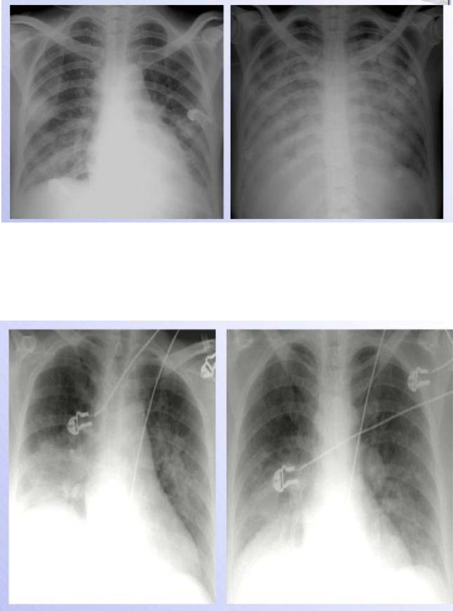
Fig.25. Heart failure
The upper zone bloo d vessels are distended and there are li near densities in the periphery of the lower z ones (interstitial or Kerley‘s B lines) a nd there are some areas of apparent co nsolidation, indicating alveolar pulmonary edema.
Fig.26 H eart failure following myocardial infarction.
60