

книги студ / Color Atlas of Pathophysiology (S Silbernagl et al, Thieme 2000)
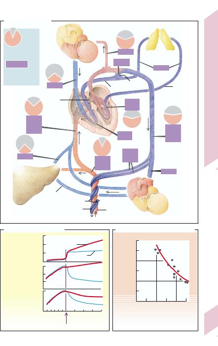
.pdfA. Causes and Consequences of Aortic Regurgitation |
|
|
|
|
|
|||||||||||
Congenital |
Rheumatic |
Bakterial |
Marfan’s |
Syphilis |
Arthritis |
etc. |
|
|
||||||||
fever |
|
|
endocarditis |
syndrome |
|
|
||||||||||
|
|
|
|
|
|
|
|
|
|
|
|
|||||
Aortic valve |
|
|
|
|
Systole |
|
|
Diastole |
|
|
|
|||||
|
|
|
|
|
|
|
|
|
|
|
|
|
|
|||
Thickened, |
|
|
|
|
|
|
|
Regurgitant volume |
|
Regurgitation |
||||||
stiffed, |
|
|
|
|
|
|
|
|
|
|
||||||
perforated |
Aortic regurgitation |
|
|
|
|
|
|
|
|
|||||||
|
|
|
|
|
|
|
|
|
|
|
||||||
|
|
|
|
|
|
|
Diastolic |
|
|
|
|
Effective |
|
|||
|
|
|
|
|
|
|
|
|
|
|
|
Aortic |
||||
|
|
|
1s |
|
|
|
blood pressure |
|
|
|
stroke volume |
|||||
|
|
|
|
|
|
|
|
|
|
|
|
|
|
|
||
|
|
ECG |
|
|
|
|
|
|
|
|
|
|
|
Compensation |
||
mmHg |
|
|
|
|
|
|
|
|
Ventricular dilation |
|
End-diastolic |
7.14 |
||||
|
|
|
|
|
|
|
|
|
|
Laplace’s |
|
volume |
|
|||
150 |
|
|
|
|
|
|
|
|
|
|
|
|
||||
|
|
|
|
|
|
|
|
|
|
|
|
|
||||
|
|
|
|
|
|
|
|
|
|
|
law |
|
|
|
|
Plate |
100 |
|
PAo |
|
|
|
|
|
|
|
Wall tension |
|
Total |
|
|||
|
|
|
|
|
|
|
|
|
|
|
|
stroke volume |
|
|||
|
|
|
|
|
|
|
|
|
|
|
|
|
|
|||
50 |
|
PLV |
|
|
|
|
|
|
LV hypertrophy |
|
Effective |
|
||||
|
|
|
|
|
|
|
|
|
|
|
|
|
||||
|
|
PLA |
|
|
|
|
|
|
|
|
|
|
|
stroke volume |
|
|
|
|
|
|
|
|
|
|
O2 consumption |
|
normalized |
|
|||||
|
|
|
|
|
|
|
|
|
|
|
||||||
|
|
|
|
|
|
|
|
|
|
|
|
|
||||
0 |
|
|
|
|
|
|
|
|
|
|
|
|
|
Years to |
|
|
|
|
SM |
EDM |
|
|
|
|
|
|
|
|
|
|
|||
Basis |
|
|
|
|
|
|
Myocardial hypoxia |
|
|
|
decades |
|
||||
|
|
|
MDM |
|
|
|
|
|
|
|
|
|||||
Apex |
|
|
|
|
(angina pectoris) |
|
|
Decompensation |
|
|
||||||
|
|
|
|
|
|
|
|
|
|
|||||||
Heart |
|
|
II |
I |
Click |
|
|
|
|
|
|
Left heart failure |
|
|
||
sounds |
|
|
|
|
|
|
|
|
|
|||||||
1 |
|
|
|
(after Criley) |
|
|
|
|
|
|
|
|
|
|
||
|
|
|
|
|
|
|
|
|
|
|
|
|
|
Effective |
|
|
|
|
|
|
|
|
|
|
|
|
|
|
|
|
stroke volume |
|
|
|
|
Normal |
Compensated |
Decompensated |
|
|
|
|
Ventricular |
|
|
|||||
|
|
EDV |
|
|
|
|
|
|
|
|
|
|
|
|
||
|
|
|
|
|
|
|
|
|
|
|
|
dilation |
|
|
||
mmHg ESV |
SV |
|
|
|
|
|
|
al.) |
|
|
|
|
|
|
||
100 |
Diastole |
|
|
|
|
|
|
|
(after Rackley et |
Functional |
Ventricular |
|
|
|||
LV |
|
|
|
|
|
|
|
|
|
|
||||||
|
|
|
|
|
|
|
|
mitral |
|
|
|
|||||
Leftventricular pressure = P |
|
|
|
|
|
|
|
|
|
compliance |
|
|
||||
50 |
|
|
|
|
|
|
|
regurgitation |
|
|
||||||
Systole |
|
|
|
|
|
|
|
|
||||||||
|
|
|
|
|
|
|
|
|
|
|
||||||
0 |
|
0.2 |
0.3 |
0.4 |
|
|
Left atrial pressure |
|
|
|||||||
|
|
0 |
0.1 |
|
|
0.5 |
|
|
|
|
|
|
||||
2 |
|
|
|
|
LV volume (L) |
|
|
|
|
|
|
|
|
|
||
|
|
|
|
|
|
|
|
|
|
|
|
Pulmonary |
|
|
||
|
|
|
|
|
|
|
|
|
|
|
|
|
Left heart |
201 |
||
|
|
|
|
|
|
|
|
|
|
|
|
|
edema |
failure |
||
Silbernagl/Lang, Color Atlas of Pathophysiology © 2000 Thieme
All rights reserved. Usage subject to terms and conditions of license.

7 Heart and Circulation
202
Defects of the Tricuspid
and Pulmonary Valves
In principle the consequences of stenotic or regurgitant valves of the right heart resemble those of the left one (→ p.194 – 201). Differences are largely due to the properties of the downstream and upstream circulations (pulmonary arteries and venae cavae, respectively).
The cause of the rare tricuspid stenosis (TS) is usually rheumatic feverinwhich, as in tricuspid regurgitation (TR) of the same etiology, mitral valve involvement usually coexists. TR may also be congenital, for example, Ebstein’s anomaly, in which the septal leaflet of the tricuspid valve is attached too far into the right ventricle (atrialization of the RV). However, most often TR has a functional cause (dilation and failure of the right ventricle). Pulmonary valve defects are also uncommon. Pulmonary stenosis (PS) is usually congenital and often combined with a shunt (→ p. 204), while pulmonary regurgitation (PR) is most often functional (e.g., in advanced pulmonary hypertension).
Consequences. In TS the pressure in the right atrium (PRA) is raised and the diastolic flow through the valve is diminished. As a result, cardiac output falls (valve opening area, normally ca. 7 cm2, reduced to < 1.5 – 2.0 cm2). The low cardiac output limits physical activity. A rise in mean PRA to more than 10 mmHg leads to increased venous pressure (high a wave in the central venous pulse; → p.179), peripheral edema, and possibly atrial fibrillation. The latter increases the mean PRA, and thus the tendency toward edema. Edemas can also occur in TR, because the PRA is raised by the systolic regurgitation (high v wave in the central venous pulse). Apart from the situation in Ebstein’s anomaly, serious symptoms of TR occur only when there is also pulmonary hypertension or right heart failure (→ p. 214). PR increases the volume load on the right ventricle. As PR is almost always of a functional nature, the effect on the patient is mainly determined by the consequences of the underlying pulmonary hypertension (→ p. 214). Although, PS, similar to AS, can be compensated by concentric ventricular hypertrophy, physical activity will be limited (cardiac output↓), and fatigue and syncope may occur.
At auscultation the changes due to valvar defects of the right heart are usually louder during inspiration (venous return increased).
–TS: First heart sound split, early diastolic tricuspid opening sound followed by diastolic murmur (tricuspid flow murmur) that increases in presystole during sinus rhythm (atrial contraction);
–TR: Holosystolic murmur of regurgitant flow; presence (in adults) or accentuation (in children) of third heart sound (due to increased diastolic filling) and of the fourth heart sound (forceful atrial contraction);
–PS: Occurrence or accentuation of fourth heart sound, ejection click (not in subvalvar or supravalvar stenosis); systolic flow murmur;
–PR: Early diastolic regurgitation murmur (Graham–Steell murmur).
Circulatory Shunts
A left-to-right shunt occurs when arterialized blood flows back into the venous system without having first passed through the peripheral capillaries. In right-to-left shunts systemic venous (partially deoxygenated) blood flows directly into the arterial system without first passing through the pulmonary capillaries.
In the fetal circulation (→ A) there is
–low resistance in the systemic circulation (placenta!),
–high pressure in the pulmonary circulation (→ B2),
–high resistance in the pulmonary circulation (lungs unexpanded and hypoxic vasoconstriction; → C),
–right-to-left shunt through the foramen ovale (FO) and ductus arteriosus Botalli (DA).
At birth the following important changes occur:
1.Clamping or spontaneous constriction of the umbilical arteries to the placenta increases the peripheral resistance so that the systemic pressure rises.
2.Expansion of the lungs and rise in the alveo-
lar PO lower the pulmonary vascular resistance2 (→ C), resulting in an increase in
blood flow through the lungs and a drop in the pressure in the pulmonary arteries
(→ B1,2).
!
Silbernagl/Lang, Color Atlas of Pathophysiology © 2000 Thieme
All rights reserved. Usage subject to terms and conditions of license.
A. Fetal Circulation
Upper half of body
O2 |
|
|
0,37 |
|
|
|
|
|
|
O2 saturation |
|
|
78 |
Lung |
(full saturation = 1.0) |
|
|
156 |
(not yet |
(ml/min) |
|
|
expanded) |
|
|
|
|
13 |
|
approx. flow/kg |
|
|
|
|
|
|
|
|
|
body weight |
0,16 |
|
|
|
|
|
Ductus arteriosus |
|
|
|
78 |
|
Pulmonary |
|
|
|
Pulmonary artery |
||
|
|
|
vein |
|
|
Foramen |
|
|
|
|
|
|
|
|
|
ovale |
|
104 |
|
|
|
|
|
|
0,40 |
|
|
|
|
182 |
|
|
0,37 |
0,30 |
|
|
|
||
|
|
|
169 |
182 |
|
|
|
|
|
0,36 |
|
0,6 |
130 |
|
78 |
|
|
||
|
|
|
||
|
|
130 |
|
|
|
|
|
|
52 |
Lower half of body
Portal vein Umbilical cord
|
|
Umbilical arteries |
|
|
|
Umbilical vein |
|
|
|
|
|
||
|
|
|
|
|
Placenta |
|
|
|
|
|
|
|
|
B. Pulmonary Circulation |
|
|
|
|
C. Fetal Hypoxic Vasoconstriction |
||||||||
Pulmonary artery: |
3 |
Ventricular |
|
|
|
|
|
|
|
|
|
|
|
|
|
|
|
|
|
|
|
|
|
|
|
||
1 Blood flow |
2 |
septal defect |
|
|
|
resistance · min) |
|
|
|
|
|
|
|
(L/min) |
1 |
|
Normal |
|
1.0 |
|
|
|
|
|
|||
|
|
|
|
|
|
|
|
|
|||||
|
|
|
|
|
|
|
|
|
|
||||
|
0 |
|
|
|
|
|
0.8 |
|
|
|
|
|
|
|
75 |
|
|
|
|
|
–1 |
|
|
|
|
|
|
2 Systolic |
|
|
|
|
|
Pulmonaryvascular (mmHg · mL |
0.6 |
|
|
|
|
|
|
50 |
|
|
|
|
|
|
|
|
|
|
|||
pressure |
|
|
|
|
|
|
|
|
|
|
|
||
(mmHg) |
25 |
|
|
|
|
|
0.4 |
|
|
|
|
|
|
|
|
|
|
|
|
|
|
|
|
|
|||
|
0 |
|
|
|
|
|
0.2 |
|
|
|
|
|
|
|
|
|
|
|
|
|
|
|
|
|
|
||
3 Muscle thickness |
|
|
|
|
|
00 |
|
|
|
|
|
||
|
|
|
|
|
|
|
|
|
|
|
|||
in vessel wall |
|
|
|
|
|
|
|
5 |
10 |
15 |
20 |
25 |
|
|
20 |
28 36 |
1 |
2 |
3 |
4 |
|
O2 |
pressure in the pulmonary |
||||
|
|
|
|
artery (mmHg) |
|
||||||||
|
Week of |
Week after birth |
|
|
|
Data from fetal lamb |
|||||||
|
pregnancy |
|
|
|
|
|
|
|
|||||
(after Rudolph) |
|
|
|
|
(after Levine) |
|
|
|
|
|
|||
|
Birth |
|
|
|
|
|
|
|
|
|
|||
Plate 7.15 Circulatory Shunts I
203
Silbernagl/Lang, Color Atlas of Pathophysiology © 2000 Thieme
All rights reserved. Usage subject to terms and conditions of license.

7 Heart and Circulation
204
!
3.As a result, there is physiological reversal of the shunt through the foramen ovale (FO) and ductus arteriosus (DA), from right-to- left to left-to-right (left atrium to right atrium and aorta to pulmonary artery).
4.These shunts normally close at or soon after birth, so that systemic and pulmonary circulations are now in series.
Abnormal shunts can be caused by patency of the duct (patent or persisting DA [PDA]; → E) or of the FO (PFO), by defects in the atrial or ventricular septum (ASD or VSD), or by arteriovenous fistulae, etc. Size and direction of the shunt in principle depend on: 1) the cross-sec- tional area of the shunt opening; and 2) the pressure difference between the connected vessels or chambers (→ D). If the opening is relatively small, 1) and 2) are the principal determining factors (→ D1). However, if the shunt between functionally similar vascular spaces (e.g., aorta and pulmonary artery; atrium and atrium, ventricle and ventricle) is across a large cross-sectional area, pressures in the two vessels or chambers become (nearly) equalized. In this case the direction and volume of the shunt is determined by 3) outflow resistance from the shunt-connected vessels or chambers (→ D2; e.g., PDA), as well as 4) their compliance (= volume distensibility; e.g., of the ventricular walls in VSD; → D3).
The ductus arteriosus (DA) normally closes within hours, at most two weeks, of birth due to the lowered concentration of the vasodilating prostaglandins. If it remains patent (PDA), the fetal right-to-left shunt turns into a left- to-right shunt (→ E, top), because the resistances in the systemic and pulmonary circuits have changed in opposite directions. At auscultation a characteristic flow murmur can be heard, louder in systole than diastole (“machinery murmur”). If the cross-sectional area of the shunt connection is small, the aortic pressure is and remains much higher than that in the pulmonary artery (→ D1, P), the shunt volume will be small and the pulmonary artery pressure nearly normal. If the cross-sec- tional area of the shunt connection is large, the shunt volume will also be large and be added to the normal ejection volume of the right ventricle, with the result that pulmonary blood flow and inflow into the left heart chambers are much increased (→ E, left). In compensa-
tion the left ventricle ejection volume is increased (Frank–Starling mechanism; possibly ventricular hypertrophy), and there will be a lasting increased volume load on the left ventricle (→ E, left), especially when the pulmonary vascular resistance is very low postnatally (e.g., in preterm infants). As the ability of the neonate’s heart to hypertrophy is limited, the high volume load can often lead to left ventricular failure in the first month of life.
If, on the other hand, the pulmonary vascular resistance (Rpulm) remains relatively high postnatally (→ E, right), and therefore the shunt volume through the ductus is relatively small despite a large cross-sectional area, a moderately increased left ventricular load can be compensated for a long time. However, in these circumstances the level of pulmonary artery pressure will become similar to that of the aorta. Pulmonary (arterial) hypertension occurs (→ E, right and p. 214). This, if prolonged, will lead to damage and hypertrophy of the pulmonary vessel walls and thus to a further rise in pressure and resistance. Ultimately, a shunt reversal may occur with a right-to-left shunt through the ductus (→ E, bottom left). Aortic blood distal to the PDA will now contain an admixture of pulmonary arterial (i.e., hypoxic) blood (cyanosis of the lower half of the body; clubbed toes but not fingers). The pressure load on the right heart will after a period of compensating right ventricular hypertrophy ultimately lead to right ventricular failure. If functional pulmonary valve regurgitation occurs (caused by the pulmonary hypertension), it may accelerate this development because of the additional right ventricular volume load. Early closure of the PDA, whether by pharmacological inhibition of prostaglandin synthesis, by surgical ligation or by transcatheter closure, will prevent pulmonary hypertension. However, closure of the ductus after shunt reversal will aggravate the hypertension.
A large atrial septal defect initially causes a left-to-right shunt, because the right ventricle being more distensible than the left ventricle offers less resistance to filling during diastole and can thus accommodate a larger volume than the left ventricle. However, when this volume load causes hypertrophy of the right ventricle its compliance is decreased, right atrial pressure rises and shunt reversal may occur.
Silbernagl/Lang, Color Atlas of Pathophysiology © 2000 Thieme
All rights reserved. Usage subject to terms and conditions of license.
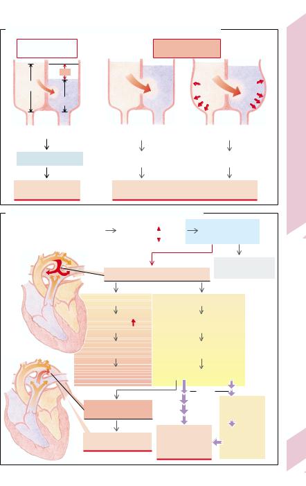
D. Determining Factors for Direction and Size of Circulatory Shunts
Septal defect |
|
Septal defect |
|
small |
|
large |
|
P |
|
|
|
Plt |
|
Clt |
Crt |
Prt |
|
|
|
1 |
2 |
3 |
|
left |
right |
|
|
Rlt |
Rrt |
Plt > Prt remains
P determines shunt volume
Rlt > Rrt |
|
Clt < Crt |
||
|
|
|
|
|
|
Plt |
≈ |
Prt |
|
Outflow resistance R or compliance C determine shunt volume
(after Levine)
E. Consequences of Postnatal Patent Ductus Arteriosus (PDA)
Prenatal |
|
Birth |
|
Vascular resistance: |
|
|
ductus arteriosus: |
|
|
peripheral |
|
|
|
right-to-left shunt |
|
|
|
pulmonary |
|
|
|
|
|
|
|
Persisting |
|
Left-to-right shunt
Postnatally: left-to-right shunt
Spontaneous closure after birth
Rpulm small |
Rpulm large |
|
|
Pulmonary |
Pulmonary artery: |
|
|
blood flow |
pressure load |
|
|
Left heart: |
Damage, |
|
|
volume load |
hypertrophy |
|
|
(Left ventr. hypertrophy) |
Pulmonary hypertension |
||
Left ventricular failure |
|||
|
|
||
|
Years |
|
|
|
to |
Functional |
|
Shunt reversal: |
decades |
||
|
pulmonary |
||
right-to-left shunt |
|
regurgitation |
|
Cyanosis of |
Right heart: |
Volume |
|
hypertrophy, |
load on |
||
lower half of body |
right ventricle |
||
failure |
|||
|
|
||
Silbernagl/Lang, Color Atlas of Pathophysiology © 2000 Thieme
All rights reserved. Usage subject to terms and conditions of license.
Plate 7.16 Circulatory Shunts II
205

Arterial Blood Pressure and its Measurement
|
The systemic arterial blood pressure rises to a |
||||||||
|
maximum (the systolic pressure [PS]), during |
||||||||
|
the ejection period, while it falls to a minimum |
||||||||
|
(the diastolic pressure [PD]) during diastole |
||||||||
|
and the iso(volu)metric period of systole (aor- |
||||||||
|
tic valve closed) (→ A). Up to about 45 years of |
||||||||
|
age the resting (sitting or recumbent) PD |
||||||||
|
ranges |
from |
60– 90 mmHg (8 – 12 kPa); |
PS |
|||||
|
ranges |
from |
100– 140 mmHg |
(13 – 19 kPa) |
|||||
|
(→ p. 208). The difference between PD and PS |
||||||||
|
is the blood pressure amplitude or pulse pres- |
||||||||
Circulation |
sure. |
|
|
|
|
|
|
|
|
The mean blood pressure is decisive for pe- |
|||||||||
|
|||||||||
|
ripheral arterial perfusion. It can be deter- |
||||||||
|
mined graphically (→ A) from the invasively |
||||||||
and |
measured blood pressure curve (e.g., arterial |
||||||||
catheter), or while recording such a curve by |
|||||||||
|
|||||||||
Heart |
dampening down the oscillations until only |
||||||||
the mean pressure is recorded. |
|
|
|
||||||
In the vascular system the flow fluctuations |
|||||||||
7 |
in the great arteries are dampened through the |
||||||||
|
“windkessel” (compression chamber) effect to |
||||||||
|
an extent that precapillary blood no longer |
||||||||
|
flows in spurts but continuously. Such a sys- |
||||||||
|
tem consisting of highly compliant conduits |
||||||||
|
and high-resistance terminals, is called a hy- |
||||||||
|
draulic filter. The arteries become more rigid |
||||||||
|
with age, so that the PS rise per volume in- |
||||||||
|
crease |
( P/ V = elastance) |
becomes |
greater |
|||||
|
and compliance decreases. This mainly in- |
||||||||
|
creases PS (→ C), without necessarily increas- |
||||||||
|
ing the mean pressure (the shape of the pres- |
||||||||
|
sure curve is changed). Thoughtless pharma- |
||||||||
|
cological lowering of an elevated PS in the el- |
||||||||
|
derly can thus result in dangerous underperfu- |
||||||||
|
sion (e.g., of the brain). |
|
|
|
|
||||
|
Measuring blood pressure. Blood pressure |
||||||||
|
(at the level of the heart) is routinely measured |
||||||||
|
according to the Riva-Rocci method, by sphyg- |
||||||||
|
momanometer (→ B). An inflatable cuff is fit- |
||||||||
|
ted snugly around the upper arm (its width at |
||||||||
|
least 40% of the arm’s circumference) and un- |
||||||||
|
der manometric |
control |
inflated |
to |
ca. |
||||
|
30 mmHg (4 kPa) above the value at which |
||||||||
|
the palpated radial pulse disappears. A stetho- |
||||||||
|
scope having been placed over the brachial ar- |
||||||||
|
tery near the elbow, at the lower edge of the |
||||||||
206 |
cuff, the cuff pressure is then slowly lowered |
||||||||
(2 – 4 mmHg/s). The occurrence |
of |
the first |
|||||||
|
pulse-synchronous |
sound |
(clear, |
tapping |
|||||
sound; phase 1 of Korotkoff) represents PS and is recorded. Normally this sound at first becomes softer (phase 2) before getting louder (phase 3), then becomes muffled in phase 4 and disappears completely (phase 5). The latter is nowadays taken to represent PD and is recorded as such.
Sources of error when measuring blood pressure. Complete disappearance of the sound sometimes occurs at a very low pressure. The difference between phases 4 and 5 (normally about 10 mmHg) is increased by conditions and diseases that favor flow turbulence (physical activity, fever, anemia, thyrotoxicosis, pregnancy, aortic regurgitation, AV fistula). If blood pressure is measured again, the cuff pressure must be left at zero for one to two minutes, because venous congestion may give a falsely high diastolic reading. The cuff should be 20% broader than the diameter of the upper arm. A cuff that is too small (e.g., in the obese, in athletes or if measurement has to be made at the thigh) also gives falsely high diastolic values, as does a too loosely applied cuff. A false reading can also be obtained when the auscultatory sounds are sometimes not audible in the range of higher amplitudes (auscultatory gap). In this case the true PS is obtained only if the cuff pressure is high enough to begin with (see above).
It is sufficient in follow-up monitoring of systemic hypertension (e.g., in labile hypertension from which fixed hypertension can often develop; → D and p. 208) to measure blood pressure in one arm only (the same one every time, if possible). Nevertheless, in cases of stenosis in one of the great vessels there can be considerable, diagnostically important, differences in blood pressure between left and right arm (pressure on the right > left, except in dextrocardia). This occurs in supravalvar aortic stenosis (mostly in children) and the subclavian steal syndrome, caused by narrowing in the proximal subclavian artery, usually of atherosclerotic etiology (ipsilateral blood pressure reduced). Blood pressure differences between arms and legs can occur in congenital or acquired (usually atherosclerotic) stenoses of the aorta distal to the origin of the arteries to the arms.
Silbernagl/Lang, Color Atlas of Pathophysiology © 2000 Thieme
All rights reserved. Usage subject to terms and conditions of license.
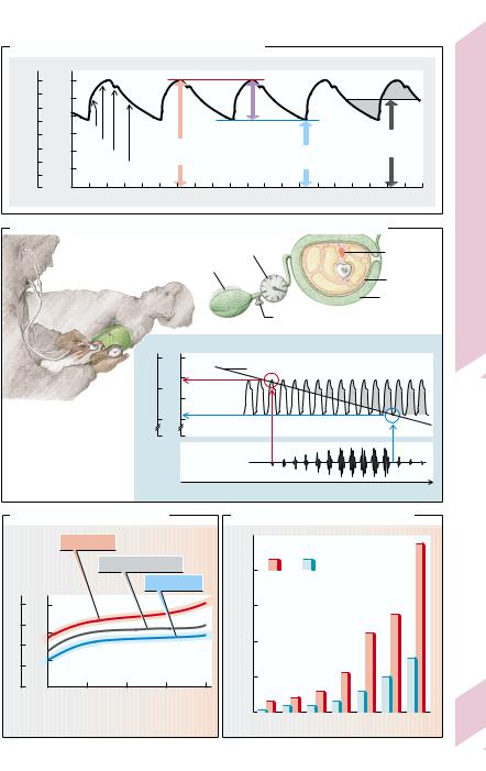
A. Aortic Pressure Curve (Invasive Measurement)
|
16 |
120 |
|
|
|
|
|
|
F2 |
|
|
|
|
|
|
|
|
|
|
|
|
|
12 |
|
80 |
|
|
|
|
|
|
F1 |
|
|
mmHg |
|
|
|
Pressure |
|
|
||
kPa |
8 |
|
Steep |
|
|
|
Mean pressure |
|||
|
|
|
amplitude |
|
||||||
|
40 |
Flat |
|
Systolic |
Diastolic |
if F1 = F2 |
||||
Pressure |
|
|
Incisura |
|
|
|
||||
4 |
|
|
pressure |
|
pressure |
|
||||
|
|
|
Exponential |
|
|
|
|
|
||
|
|
|
|
|
|
|
|
|
|
|
|
0 |
|
0 |
decrease |
|
|
|
|
|
|
|
|
0 |
1 |
|
Time (s) |
2 |
3 |
4 |
||
|
|
|
|
|
|
|
|
|
|
|
B. Measuring Blood Pressure with Sphygmomanometer (after Riva-Rocci)
Sphygmo- |
|
manometer |
Brachial a. |
Pump
Upper arm
Cuff
Release valve
kPa mmHg |
Pressure (brachial a.) |
||
|
|||
20 |
150 |
|
Cuff pressure |
|
125 |
|
|
15 |
Systolic |
|
|
|
|
||
|
100 |
level |
|
10 |
75 |
Diastolic |
|
0 |
0 |
level |
|
|
|
||
|
|
Korotkoff |
|
|
|
sounds |
|
|
|
|
Time |
Plate 7.17 Measuring Arterial Blood Pressure
C. Age-related Blood Pressure
|
|
Systolic |
|
|
|
Blood |
|
Mean pressure |
|
||
pressure |
|
|
Diastolic |
|
|
kPa |
mmHg |
|
|
|
|
20 |
150 |
|
|
|
|
15 |
125 |
|
|
|
|
|
|
|
|
|
|
10 |
|
|
|
|
|
5 |
75 |
|
|
|
|
|
|
|
|
|
|
0 |
00 |
20 |
40 |
60 |
80 |
|
|
|
Age (years) |
|
|
|
|
|
|
(after Guyton) |
|
D. Incidence of Fixed Hypertension
|
50 |
|
|
|
1000 |
|
People |
|
|
40 |
with |
without |
|
|
per |
|
|||
previously labile hypertension |
|
|||
|
|
|||
incidence |
30 |
|
|
|
|
|
|
|
|
Fixied hypertension: |
20 |
|
|
(after Julius and Esler) |
10 |
|
|
||
0 |
|
|
||
|
25–29 30–34 35–39 40–44 45–49 50–54 55–59 |
207 |
||
Age (years)
Silbernagl/Lang, Color Atlas of Pathophysiology © 2000 Thieme
All rights reserved. Usage subject to terms and conditions of license.

7 Heart and Circulation
208
Hypertension
Hypertension (H.), used as a term by itself, refers to an abnormally high arterial pressure in the systemic circulation (for pulmonary hypertension, → p. 214). In the industrialized countries it affects about 20% of the population. As H. almost always begins insidiously, yet can be treated effectively, the upper limit of normal blood pressure needs to be determined. The World Health Organisation (WHO) has proposed the following values for all age groups (mmHg/7.5 = kPa):
|
normal |
Threshold |
Hyper- |
|
|
|
hyper- |
ten- |
|
|
|
tension |
sion |
|
|
|
|
|
|
diastolic pressure |
< 90 |
90 |
– 95 |
> 95 |
(PD [mmHg]) |
|
|
|
|
systolic pressure |
< 140 |
140 |
– 160 |
> 160 |
(PS [mmHg]) |
|
|
|
|
Cases of alternating normal and elevated levels (labile H.) are included in the column ‘Threshold hypertension’. Patients with a labile H. often develop fixed H. later (→ p. 207 D). As PS regularly rises with age (→ p. 207 C), the upper limit of PS in adults has been widely set at 150 mmHg for those aged 40 – 60 years and at 160 mmHg for those aged over 60 years (PD at 90 mmHg for both adult groups). Lower values have been set for children. Assessment of blood pressure should be based on the mean values of at least 3 readings on two days (see also p. 206).
The product of cardiac output (= stroke volume [SV] · heart rate) and total peripheral resistance (TPR) determines blood pressure (Ohm’s law). H. thus develops after an increase in cardiac output or TPR, or both (→ A). In the former case one speaks of hyperdynamic H. or cardiac output H., with the increase in PS being much greater than that in PD. In resistance H., PS and PD are either both increased by the same amount or (more frequently) PD more than PS. The latter is the case when the increased TPR delays ejection of the stroke volume.
The increase of cardiac output in hyperdynamic hypertension is due to an increase in either heart rate or extracellular volume, leading to an increased venous return and thus an increased stroke volume (Frank–Starling mecha-
nism). Similarly, an increase in sympathetic activity of central nervous system origin and/or raised responsiveness to catecholamines (e.g., caused by cortisol or thyroid hormone) can cause an increase in cardiac output (→ A, left).
Resistance hypertension is caused mainly by abnormally high peripheral vasoconstriction
(arterioles) or some other narrowing of peripheral vessels (→ A, right), but may also be due to an increased blood viscosity (increased hematocrit). Vasoconstriction mainly results from increased sympathetic activity (of nervous or adrenal medullary origin), raised responsiveness to catecholamines (see above), or an increased concentration of angiotensin II. Autoregulatory mechanisms also include vasoconstriction. If, for example, blood pressure is increased by a rise in cardiac output (see above), various organs (e.g., kidneys, gastrointestinal tract) “protect” themselves against this high pressure (→ A, middle). This is responsible for the frequently present vasoconstrictor component in hyperdynamic H. that may then be transformed into resistance H. (→ A). Additionally, there will be hypertrophy of the vasoconstrictor musculature. Finally, H. will cause vascular damage that will increase TPR (fixation of the H.).
Some of the causes of hypertension are known (e.g., renal or hormonal abnormalities; → B2,3), but these forms make up only about 5 – 10% of all cases. In all others the diagnosis by exclusion is primary or essential hypertension (→ B1). Apart from a genetic component, more women than men and more urbanites than country dwellers are affected by primary H. In addition, chronic psychological stress, be it job-related (pilot, bus driver) or personal- ity-based (e.g., “frustrated fighter” type), can induce hypertension. Especially in “salt-sensi- tive” people (ca. 1⁄3 of patients with primary H.; increased incidence when there is a family history) the high NaCl intake (ca. 10–15 g/ d = 170–250 mmol/d) in the western industrialized countries might play an important role. While the organism is well protected against Na+ loss (or diminished extracellular volume) through an increase in aldosterone, those with an increased salt sensitivity are apparently relatively unprotected against a high
!
Silbernagl/Lang, Color Atlas of Pathophysiology © 2000 Thieme
All rights reserved. Usage subject to terms and conditions of license.
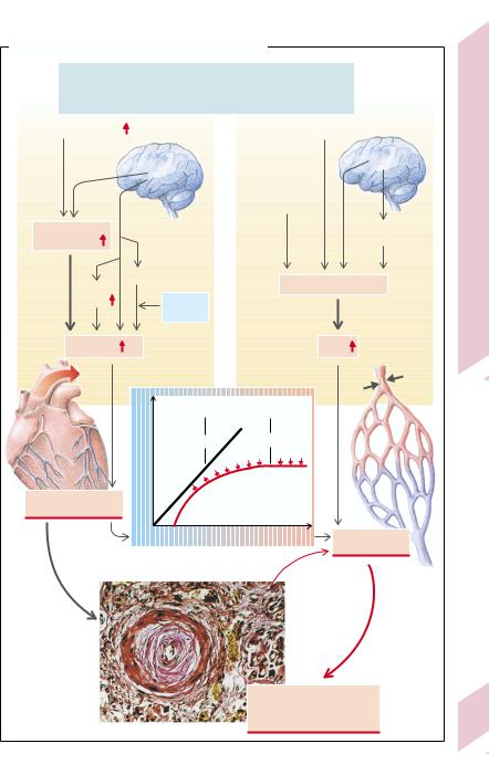
A. Principles of the Development of Hypertension |
|
|
||||
|
Arterial blood pressure = |
|
|
|||
Cardiac output |
x |
Total peripheral resistance |
|
|
||
(CO) |
|
|
(TPR) |
|
|
|
Extracellular volume |
|
|
|
Angiotensin II |
|
|
|
CNS |
|
|
CNS |
|
|
|
|
|
|
|
||
|
|
|
|
Vascular |
|
|
|
|
|
|
hyperreactivity |
|
I |
Central |
|
|
|
|
Adrenal |
Hypertension |
blood volume |
|
|
|
|
medulla |
|
Venous |
Catecholamines |
|
Vasoconstriction |
|||
|
|
|
||||
|
|
|
|
|||
|
|
|
|
|
|
|
tone |
|
T3, T4, |
|
|
|
7.18 |
|
|
cortisol |
|
|
|
|
CO |
|
|
|
TPR |
Plate |
|
|
|
|
|
|||
|
|
Vascular resistance (radius) |
|
|
||
|
perfusion |
constant |
pressure |
|
|
|
|
dependent |
|
|
|||
|
|
|
|
|
||
|
|
|
|
|
|
|
|
Organ |
|
|
Autoregulation |
|
|
Hyperdynamic |
|
|
|
|
|
|
hypertension |
|
|
|
|
|
|
|
|
|
|
|
|
|
|
00 |
Systemic blood pressure |
Resistance |
|
||
|
|
|
|
|
hypertension |
|
|
|
|
|
Vicious |
|
|
|
|
|
|
circle |
|
|
Pfeifer |
|
|
|
Hypertrophy of |
|
|
U. |
|
|
|
vessel musculature and |
|
|
Photo: |
|
|
|
|
||
|
|
|
vascular damage: |
|
209 |
|
|
|
|
fixation of hypertension |
|||
|
|
|
|
|||
Silbernagl/Lang, Color Atlas of Pathophysiology © 2000 Thieme
All rights reserved. Usage subject to terms and conditions of license.

7 Heart and Circulation
210
!
NaCl intake. In these patients, aldosterone release is so strongly inhibited even at “normal” Na+ intake (> 100 mmol/d) that it cannot be lowered any further. A diet with low NaCl intake would in this case bring NaCl balance into the aldosterone regulatory range.
The actual connection between NaCl sensitivity and primary H. has not been fully elucidated, but the possibility is being considered that responsiveness to catecholamines is raised in people sensitive to NaCl. This results, for example, on psychological stress, in a greater than normal rise in blood pressure, on the one hand, due directly to the effect of increased cardiac stimulation (→ B, upper right) and, on the other hand, indirectly as a result of increased renal absorption and thus retention of Na+ (rise in extracellular volume leads to hyperdynamic H.). The increased blood pressure leads to pressure diuresis with increased Na+ excretion, restoring Na+ balance (Guyton). This mechanism also exists in healthy people, but the pressure increase required for excretion of large amounts of NaCl is much lower (→ C, a b). In primary H. (as in disorders of renal function) the NaCl-dependent increase in blood pressure is greater than normal (→ C, c d). A diet that is low in Na+ can thus lower (not yet fixed) H. in these cases (C, c e). A simultaneously elevated K+ supply accentuates this effect for unknown reasons. The cellular mechanism of salt sensitivity still awaits clarification. It is possible that changes in cellular Na+ transport are important. In fact cellular Na+ concentration is raised in primary H., which decreases the driving force for the 3 Na+/Ca2+ exchange carrier in the cell membrane, as a result of which the intracellular Ca2+ concentration rises, which in turn increases the tone of the vasoconstrictor muscles (Blaustein). It is possible that digitalislike inhibitors of Na+-K+-ATPase are involved (ouabain?). They may be present in larger amounts, or there may be a special sensitivity to them in primary H. Atriopeptin (= atrial natriuretic peptide [ANP]), which has vasodilator and natriuretic effects, is probably not involved in the development of primary H. Although the concentration of renin is not elevated in primary H., blood pressure can be reduced even in primary H. by inhibiting the angiotensin-converting enzyme (ACE inhibi-
tors; see below) or angiotensin receptor antagonists.
The various forms of secondary hypertension make up only 5 – 10% of all hypertensive cases (→ B2,3,4), but contrary to primary H. their cause can usually be treated. Because of the late consequences of H. (→ E), such treatment must be initiated as early as possible. Renal hypertension, the most common form of secondary H., can have the following, often partly overlapping, causes (→ B2, see also p.114): Every renal ischemia, for example, resulting from aortic coarctation or renal artery stenosis, but also from narrowing of the renal arterioles and capillaries (glomerulonephritis, hypertension-induced atherosclerosis), leads to the release of renin in the kidneys. It splits the dekapeptide angiotensin I from angiotensinogen in plasma. A peptidase (angiotensin– converting enzyme, ACE), highly concentrated especially in the lungs, removes two amino acids to form angiotensin II. This octapeptide has a strong vasoconstrictor action (TPR rises) and also releases aldosterone from the adrenal cortex (Na+ retention and increase in cardiac output), both these actions raising the blood pressure (→ B2). In kidney disease with a significant reduction of the functioning renal mass, Na+ retention can therefore occur even during normal Na+ supply. The renal function curve is steeper than normal, so that Na+ balance is restored only at hypertensive blood pressure levels (→ C, c d). Glomerulonephritis, renal failure, and nephropathy of pregnancy are some of the causes of the primarily hypervolemic form of renal H. Renal H. can also be caused by a renin-producing tumor or (for unknown reasons) by a polycystic kidney. The kidney is also central to other forms of hypertension that do not primarily originate from it (primary H., hyperaldosteronism, adrenogenital syndrome, Cushing’s syndrome). Furthermore, in every case of chronic H. secondary changes will occur sooner or later (vascular wall hypertrophy, atherosclerosis): they fix the H. even with effective treatment of the primary cause. If unilateral renal artery stenosis is repaired surgically rather late, for example, the other kidney, damaged in the meantime by the hypertension, will maintain the H.
Hormonal hypertension can have several causes (→ B3):
!
Silbernagl/Lang, Color Atlas of Pathophysiology © 2000 Thieme
All rights reserved. Usage subject to terms and conditions of license.