Добавил:
Опубликованный материал нарушает ваши авторские права? Сообщите нам.
Вуз: Предмет: Файл:

книги студ / Color Atlas of Pathophysiology (S Silbernagl et al, Thieme 2000)

.pdf
Скачиваний:
953
Добавлен:
03.09.2020
Размер:
74.89 Mб
Скачать

A. Causes and Consequences of Chronic Pancreatitis


Tumor, stricture, papillary stenosis


Pancreas


Alcohol abuse


Occlusion of

Pancreatitis


main pancreas duct

Pancreatic juice:


Chronic

Secretion of

Citrate concentration


HCO3and fluid

Lithostatin concentration


Proenzyme concentration


Calcium salt


6.14


precipitation


Plate


Normal ERP


Protein


plug


Duct


distension


Calcium


deposition


Epithelial lesions


Enzyme activation


Chronic inflammation and fibrosis of exocrine


and endocrine pancreas, damage to neighboring organs


Tissue atrophy

Pain


Diabetes mellitus

Obstructive


Ductal stenosis

Malabsorption

Pancreatic ascites

jaundice


Diarrhea


Periductal fibrosis

Weight loss

Thrombosis of portal


Pseudocysts


and splenic veins

161


Photo of dilated duct from: Thurn P. et al. Einführung in die radiologische Diagnostik. 10th ed. Stuttgart: Thieme; 1998


Silbernagl/Lang, Color Atlas of Pathophysiology © 2000 Thieme

All rights reserved. Usage subject to terms and conditions of license.

Cystic Fibrosis

6 Stomach, Intestines, Liver

Cystic fibrosis (CF) is a genetic syndrome in which the epithelial secretion, for example, in the lungs, pancreas, liver, genital tract, intestine, nasal mucosa, and sweat glands are affected. Among Caucasians CF is the most frequent lethal (after a mean of 40 years) gene defect (1 per 2 500 births).

The defect is autosomal recessive (A1) and affects the epithelial transport protein CFTR (cystic fibrosis transmembrane conductance regulator). CFTR in healthy people consists of 1480 amino acids that form 12 transmembrane domains, two nucleotidebinding domains (NBD1, NBD2), and a regulator domain. At the latter, CFTR is regulated by a cAMP-dependent protein kinase A (A2; CFTR is shown opened up frontally). CFTR is probably a chloride channel that opens when the intracellular cAMP concentration is raised and, in addition, ATP is bound to NBD1 (and split?). Furthermore, intact but not defective CFTR inhibits certain Na+ channels (type ENaC). The fact that more of them open in CF results in increased absorption of Na+and water, for example, at the bronchial epithelium, from the mucus secreted into the lumen, so that the latter is thickened (see below).

Patients with cystic fibrosis have various mutations of CFTR, but the serious forms are most frequently caused by two defects on the NBD1 (A3): either the amino acid 508, phenylalanine (= F; mutation F508), is missing, or glycine (= G) in position 551 is replaced by aspartate (= D) (mutation G551 D).

CFTR is incorporated into the apical (luminal) cell membrane of many epithelial cells. CFTR has an important function in the excretory ducts of the pancreas, in that it is involved in secretion of a liquid rich in NaHCO3. HCO3is exchanged for Clin these cells via an antiport carrier (A4). The opening of CFTR—for example, by secretin, which increases intracellular cAMP concentration— allows the Clthat has entered the cell to be recycled, so that chloride is again available for the secretion of HCO3, followed by Na+ and water. If the concentration of cAMP de-

In patients with CF, CFTR does not open up even when cAMP concentration is high. As a result, especially when acinar secretion is stimulated, the small pancreatic ducts contain a protein-rich, viscous secretion that occludes the transporting ducts and thus leads to chronic pancreatitis with its consequences (e.g., malabsorption due to lack of pancreatic enzymes and HCO3in the duodenum;

p.160).

Among other effects, abnormal CFTR af-

fects the intestinal epithelium so that neonatal meconium becomes viscous and sticky and thus cannot, as usual, pass out of the ileum after birth (meconium ileus).

As in the pancreas, the bile ducts may become obstructed and neonatal jaundice may thus be prolonged. The CFTR defect in the male genital organs may cause infertility (obstruction of the vas deferens); in the female genital organs it causes decreased fertility. The consequences of abnormal secretion in the nasal mucosa are polyps and chronic inflammation of the nasal sinuses. In the sweat glands the defect increases sweat secretion that during fever or high ambient temperature can lead to hypovolemia and even circulatory shock. In addition, electrolyte concentration is increased in sweat and the concentration of Na+ is higher than that of Cl(the reverse of normal), a fact that is used in the diagnosis of CF (sweat test).

Morbidity and life-threatening complications of CF are mainly due to its effects on the bronchial epithelium. Its superficial mucus is normally thinned by fluid secretion. The CTFR defect causes (in addition to increased mucus secretion) the reabsorption instead of secretion of fluid. This results in a highly viscous and protein-rich layer of mucus that not only hinders breathing, but also forms a fertile soil for infections, especially with Pseudomonas aeruginosa and Staphylococcus aureus. Chronic bronchitis, pneumonia, bronchiectasis, and secondary cardiovascular disorders are the result.

162creases, CFTR is closed and secretion dries up.

Silbernagl/Lang, Color Atlas of Pathophysiology © 2000 Thieme

All rights reserved. Usage subject to terms and conditions of license.

A. Causes and Consequences of Cystic Fibrosis


Autosomal recessive gene defect


on chromosome 7


Regulatory domain:


regulation by


NBD2


cAMP-dependent


proteinkinase A


1


CFTR


1


PKA

cAMP


1480


Fibrosis


2


NBD1


Nucleotide-


Cystic


binding domain 1:


ATP/ADP binding

Mutation

F508

or

Mutation G551D


ATP


6.15


550

Gly


507

Phe

509


552


Plate


507 509


550

Asp

552


H2O


3


OH

H+


H+


HCO3


+


Na


Cystic fibrosis


Cl

CO2


Fluid secretion


PKA

cAMP


abnormal in:


K+


CFTR


Pancreas,


ATP


gonads,


Lumen

Epithelial cell (pancreatic duct)


4


liver, intestine,


gallbladder,


sweat glands,


bronchii, etc.


Bronchial secretion


too thick


Chronic


Infection


pancreatitis


Malabsorption


Bronchiectasis,


163


Meconium ileus


pneumonia, etc.


Photo from: Thurn P. et al. Einführung in die radiologische Diagnostik. 10th ed. Stuttgart: Thieme; 1998


Silbernagl/Lang, Color Atlas of Pathophysiology © 2000 Thieme

All rights reserved. Usage subject to terms and conditions of license.

Reduced bile salt secretion (A1). This is due to either a decrease in the bile salt pool, as in Crohn’s disease or after gut resection, or a prolonged sequestration of bile salts in the gallbladder, as in fasting (possibly even if only overnight) or parenteral nutrition. The latter decreases the enterohepatic circulation of bile salts so that their secretion into
164 the bile is reduced. As cholesterol secretion is not linearly related to bile salt secretion

6 Stomach, Intestines, Liver

Gallstone Disease (Cholelithiasis)

In about 75% of patients, gallstones consist of

(B, right), the [Ch]/[BS + Pch] ratio increas-

cholesterol (more women than men are af-

es when bile salt secretion is low. This ratio

fected in this way), the rest are so-called pig-

rises further under the influence of estrogens,

ment stones that contain unconjugated bili-

because they cause an increase in the con-

rubin in the main. What the two types of

centration ratio of cholate to chenodeoxy-

stone components have in common is that

cholate (activation of 12α-hydroxylase; B,

they are poorly soluble in water.


left), so that more cholesterol is secreted per

Cholesterol (Ch) is normally not precipi-

mol bile salts (B; compare the two curves).

tated in bile, because it contains sufficient

A reduced secretion of phosphatidylcho-

conjugated bile salts (BS) and phosphatidyl-

line as a cause of cholesterol stones has been

choline (Pch = lecithin) for it to be in a micel-

found in Chilean women who live almost ex-

lar solution (A4, green area). If the concen-

clusively on vegetables.


tration ratio [Ch]/[BS + Pch] increases, Ch will


remain, within a small range, in a “supersat-

Pigment stones (C) consist to a large ex-

urated” micellar solution (A4, orange

tent (ca. 50%) of calcium bilirubinate, which

area). This apparent supersaturation is prob-

gives them their black or brown color. The

ably based on the liver also secreting choles-

black stones additionally contain calcium car-

terol in a highly concentrated form within

bonate and phosphate, while the brown

the “nucleus” of a unilamellar vesicle in the

stones also contain stearate, palmitate, and

gallbladder (A2) in such a way that Pch

cholesterol. A raised amount of unconjugat-

makes up the solution-aiding “peel” of this

ed bilirubin in the bile, which “dissolves”

vesicle, 50–100 nm in diameter. If the rela-

only in micelles, is the main cause of pigment

tive cholesterol content increases further,

stone formation; normally bile contains only

multimicellar vesicles are formed (up to

1– 2%. The causes of an increased concentra-

1000 nm). They are less stable and give up

tion

of unconjugated

bilirubin are (C):

cholesterol that is then precipitated in the

Increased liberation of hemoglobin, for

aqueous environment in the form of choles-

example, in hemolytic anaemia, in which

terol crystals (A2; A4, red area). These

there is so much bilirubin that the glucuroni-

crystals are the precursors of gallstones.

dase-mediated process of conjugation in the

Important causes of an increased [Ch]/

liver does not meet demand (p.169);

[BS + Pch] ratio are:


Reduced conjugating capacity in the liver,

Increased cholesterol secretion (A2).

for example, in liver cirrhosis (p.172);

This will occur because there is either an in-

Nonenzymatic deconjugation of (especially

creased cholesterol synthesis (raised activity

monoglucuronated) bilirubin in bile;

of 3-hydroxy-3-methylglutaryl [HMG]-CoA-

Enzymatic deconjugation (β-glucosidase)

cholesterol reductase), or an inhibition of

by bacteria.


cholesterol esterification, for example, by pro-

The latter is almost always the cause of

gesterone during pregnancy (inhibition of

brown pigment stones. The bacteria also en-

acetyl-CoA-cholesterol-acetyl

transferase

zymatically deconjugate the bile salts (de-

[ACAT]).


creased micellar formation with cholesterol


precipitation) and additionally liberate, by


means of its phospholipase A2, palmitate


and

stearate (from

phophatidylcholine)

which precipitate as calcium salts. Black stones, mainly formed by the first three of the above mechanisms, contain in addition to other compounds, calcium carbonate and phosphate, these latter presumed to be formed by the gallbladder’s decreased capacity to acidify.

!

Silbernagl/Lang, Color Atlas of Pathophysiology © 2000 Thieme

All rights reserved. Usage subject to terms and conditions of license.

A. Cholelithiasis: Abnormal Cholesterol to Bile Salt Ratio


Liver


Acetyl-CoA


HMG-CoA


reductase


Enterohepatic


Bile salts

New synthesis


circulation


Gallbladder


Cholesterol


crystals


HO

Cholesterol


Duodenum


Vesicles

Progesteron

I


Cholelithiasis

1


Ileum


ACAT


Phosphati-


dylcholine


Cholesterol

Fasting

Crohn’s disease,

Unbalanced


ester

6.16


intestinal re-


(over night!),


diet


section


parenteral diet


Plate


2


VLDL


Bile salt

Phosphatidyl-


Bile salt


loss

choline

Cholesterol secretion


sequestration


Bile salts

Phosphatidyl-


Cholesterol


choline


Bile


3


4


100


Dissociation:


cholesterol


Supersaturation


(%mol)


crystals


80


20


Cholesterol


Crystal formation


60


Phosphatidylcholine


40


60


40


‘Super


(%mol)


saturated’


20


Micellar


al.)

Cholesterol stones


solution


et


0


Small


100

80

60

40

20

0


(after


165


Bile salts (%mol)


Silbernagl/Lang, Color Atlas of Pathophysiology © 2000 Thieme

All rights reserved. Usage subject to terms and conditions of license.


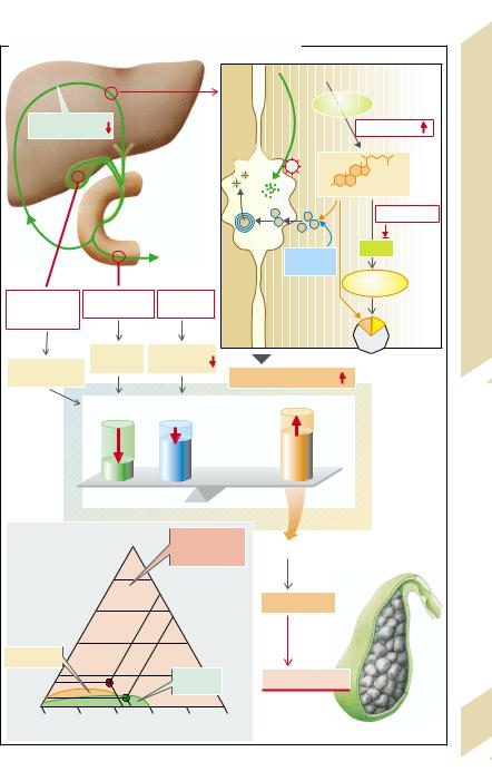
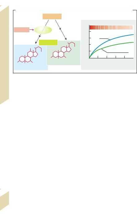
B. Cholesterol/Bile Salts: Dependence on Bile Salt Type and Bile Salt Secretion Rate


Cholesterol


–1]


h

5


12α-


kg–1·


[Cholesterol]/[Bile salts]


Estrogens


hydroxylase


·

4


[mmol


3

Cholate


secretion


Bile salts


2


OH


COO

1


Chenodeoxycholate


Intestines, Liver


12

COO


Cholesterol


HO

OH


00

10

20

30

40

50

HO


OH

Chenodeoxycholate


Bile salt secretion [mmol · kg–1· h–1]


Cholate


(after G. Paumgartner et al.)


Stomach,

!


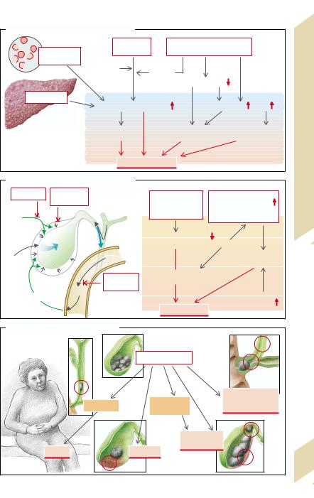
The gallbladder, in which the specific bile

In acute cholecystitis fever and leukocyto-

components (Ch, BS, Pch) are concentrated

sis are added to the symptoms listed above.

many times over by withdrawal of water,

Important causes are trauma to the gallblad-

6

also plays an important part (D) in the for-

der epithelium caused by stones. Prostaglan-


mation of gallstones (cholelithiasis after cho-

dins are liberated from the gallbladder epi-


lecystectomy is rare). Disorders of gallblad-

thelium in addition to phospholipase A2. The


der emptying can be among the causes, ei-

latter splits phosphatidylcholine to lysoleci-


ther due to insufficient CCK being liberated

thin (i.e., removal of the fatty acid at C2),


(lack of free fatty acid [FFA] release in the lu-

which in turn brings about acute cholecysti-


men in pancreatic insufficiency), so that the

tis. In some circumstances it may lead to


main stimulus for gallbladder contraction is

gallbladder perforation.


weakened, or because after nonselective va-

Bacterial cholangitis usually occurs when


gotomy the second most important contrac-

bile flow is stopped because of cholelithiasis.


tion signal, acetylcholine, is absent. Gallblad-

A rise in pressure with dilation of the bile


der contraction is also weakened in pregnan-

ducts is the result, and posthepatic cholesta-


cy. This means that not only occasional or ab-

sis and biliary pancreatitis may also develop.


sent emptying (see above) but also incom-

In relatively rare cases gallbladder cancer


plete emptying increases the duration for

develops on the basis of gallstone disease.


which bile remains in the gallbladder. As a re-


sult, there is enough time for the precipitated


crystals to form large concrements. A raised


mucus secretion (stimulated by prostaglan-


dins) can thus lead to an increased number


of nuclei of crystallization.


Possible

consequences of

cholelithiasis


are (E):


Colic. When the cystic duct or the com-


mon bile duct is transiently blocked by a


stone, pressure rises in the bile ducts and in-


creased peristaltic contraction in the region


of the blockage causes severe visceral pain


166in the epigastric area, possibly with radiation into the back, as well as vomiting (p.140).

Silbernagl/Lang, Color Atlas of Pathophysiology © 2000 Thieme

All rights reserved. Usage subject to terms and conditions of license.

C. Causes of Pigment Stone Formation


Bilirubin


Bacteria


Hemolytic

glucuronide


(cholangitis, cholecystitis)


anemia

Non-


β-gluco-


enzymatic


sidase

Micellar formation


Liver cirrhosis


Unconjugated bilirubin

Free fatty acids

pH


Calcium


Calcium palmitate

Calcium carbonate


bilirubinate


and -stearate

and -phosphate


Pigment stones


D. Role of Gall Bladder in Cholelithiasis

Vagotomy

Pregnancy

CCK deficiency,

Bile:


vagotomy,

Cholesterol/Bile salt

Acetyl-


pregnancy

(cf fig. A),


bile concentration

choline


Mucus

Gallbladder emptying


(not often enough,

Fasting


Fats

incomplete)

Cholesterol

Prosta-


crystals

glandins


Pancreatic

Bile lingers for


a long time


insufficiency


CCK

FFA


Mucus


Gall stones

secretion


Plate 6.17 Cholelithiasis II

E. Consequences of Cholelithiasis


Gall stones


Cholangitis, ob-

Spasm

Acute

structive jaundice,

cholecystitis

pancreatitis


Ulceration,


perforation


Colic

Carcinoma


167

(partly after Netter)


Silbernagl/Lang, Color Atlas of Pathophysiology © 2000 Thieme

All rights reserved. Usage subject to terms and conditions of license.

Jaundice (Icterus)

Cholestasis

6 Stomach, Intestines, Liver

Bilirubin, largely originating from hemoglobin breakdown (ca. 230 mg/d), is taken up by the liver cells and coupled by glucuronyl transferase to form bilirubin-monoglucuro- nide and bilirubin-diglucuronide. This wa- ter-soluble conjugated (direct reacting) bilirubin is secreted into the bile canaliculi and 85% is excreted in the stool. The remaining 15% is deglucuronated and absorbed in the intestine for enterohepatic recirculation.

The normal plasma concentration of bilirubin is maximally 17 µmol/L (1 mg/dL). If it rises to more than 30 µmol/L, the sclera become yellow; if the concentration rises further, the skin turns yellow as well (jaundice [icterus]). Several forms can be distinguished:

Prehepatic jaundice is the result of increased bilirubin production, for example, in hemolysis (hemolytic anemia, toxins), inadequate erythropoiesis (e.g., megaloblastic anemia), massive transfusion (transfused erythrocytes are short-lived), or absorption of large hematomas. In all these conditions unconjugated (indirect reacting) bilirubin in plasma is increased.

Intrahepatic jaundice is caused by a specific defect of bilirubin uptake in the liver cells (Gilbert syndrome Meulengracht), conjugation (neonatal jaundice, Crigler–Najjar syndrome), or secretion of bilirubin in the bile canaliculi (Dubin–Johnson syndrome, Rotor syndrome).

In the first two defects it is mainly the unconjugatedplasma bilirubin that is increased; in the secretion type it is the conjugated bilirubin that is increased. All three steps may be affected in liver diseases and disorders, for example, in viral hepatitis, alcohol abuse, drug side effects (e.g., isoniazid, phenytoin, halothane), liver congestion (e.g., right heart failure), sepsis (endotoxins), or poisoning (e.g., the Amanita phalloides mushroom).

In posthepatic jaundice the extrahepatic bile ducts are blocked, in particular by gallstones (p.164ff.), tumors (e.g., carcinoma of the head of the pancreas), or in cholangitis or pancreatitis (p.158). In these conditions

Cholestasis (A,B), i.e., blockage of bile flow, is due to either intrahepatic disorders, for example, cystic fibrosis (p.162), granulomatosis, drug side effects (e.g., allopurinol, sulfonamides), high estrogen concentration (pregnancy, contraceptive pill), graft versus host–reaction after transplantation, or, secondarily, extrahepatic bile duct occlusion

(see above).

In cholestasis the bile canaliculi are enlarged, the fluidity of the canalicular cell membrane is decreased (cholesterol embedding, bile salt effect), their brush border is deformed (or totally absent) and the function of the cytoskeleton, including canalicular motility, is disrupted. In addition, one of the two ATP-driven bile salt carriers, which are meant for the canalicular membrane, is falsely incorporated in the basolateral membrane in cholestasis. In turn, retained bile salts increase the permeability of the tight junctions and reduce mitochondrial ATP synthesis. However, it is difficult to define which of these abnormalities is the cause and which the consequence of cholestasis. Some drugs (e.g., cyclosporin A) have a cholestatic action by inhibiting the bile salt carrier, and estradiol, because it inhibits Na+-K+-ATPase and reduces membrane fluidity.

Most of the consequences of cholestasis (B) are a result of retention of bile components: bilirubin leads to jaundice (in neonates there is a danger of kernicterus), cholesterol to cholesterol deposition in skin folds and tendons, as well as in the cell membranes of liver, kidneys, and erythrocytes (echinocytes, akanthocytes). The distressing pruritus (itching) is thought to be caused by retained endorphins and/or bile salts. The absence of bile in the intestine results in fatty stools and malabsorption (p.152ff.). Finally, infection of accumulated bile leads to cholangitis, which has its own cholestatic effect.

168it is particularly conjugated bilirubin that is increased.

Silbernagl/Lang, Color Atlas of Pathophysiology © 2000 Thieme

All rights reserved. Usage subject to terms and conditions of license.

A. Types of Jaundice

Hemolysis etc.


Jaundice:


Blood


Hemoglobin etc.

Bilirubin

Production


Pre-


hepatic


· Specific syndromes

Uptake


(Icterus), Cholestasis


(Gilbert, Crigler-Najjar,


Dubin-Johnson, Rotor)

Conjugation

Conjugated


· Acute and chronic


bilirubin

Intrahepatic

liver damage


· Drug side effects


Secretion


· Estrogens, cystic


Outflow


fibrosis, etc.


= Cholestasis


Liver


Jaundice


Post-


Extrahepatic


Gall stones, tumors, etc.


hepatic

6.18


outflow


B. Mechanisms and Consequences of Cholestasis


Plate


Blood

Bile salts,

Liver cell


bilirubin,


cholesterol

Mitochondria


Enzymes,


Carrier insertion


copper,


on wrong side


endorphins


ATP

ATP synthesis


Fluidity of

impaired


cell membrane

Drugs


Retention of


bile components


Cholestasis


Deformation of


Bile salts increase

brush border


permeability of


tight junctions


Dilation


Bile salt


deficiency


in intestine


Bilirubin


Icterus


Endorphins (?)


Pruritus

Cholangitis


Bile salts (?)


Cholesterol


Cholesterol


Fat stools,


(hepatic breakdown

,

deposition


vitamin A, E and


enteral synthesis

)


K deficiency

169


Silbernagl/Lang, Color Atlas of Pathophysiology © 2000 Thieme

All rights reserved. Usage subject to terms and conditions of license.

Portal Hypertension

6 Stomach, Intestines, Liver

Venous blood from stomach, intestines, spleen, pancreas, and gallbladder passes via the portal vein to the liver where, in the sinusoids after mixture with oxygen-rich blood of the hepatic artery, it comes into close contact with the hepatocytes (A1). About 15% of cardiac output flows through the liver, yet its resistance to flow is so low that the normal portal vein pressure is only 4 –8 mmHg.

If the cross-sectional area of the liver’s vascular bed is restricted, portal vein pressure rises and portal hypertension develops. Its causes can be an increased resistance in the following vascular areas, although strict separation into three forms of intrahepatic obstructions is not always present or possible:

Prehepatic: portal vein thrombosis (A2);

Posthepatic: right heart failure, constrictive pericarditis, etc. (A2 and p. 228);

Intrahepatic (A1):

presinusoidal: chronic hepatitis, primary biliary cirrhosis, granuloma in schistosomiasis, tuberculosis, leukemia, etc.

sinusoidal: acute hepatitis, damage from alcohol (fatty liver, cirrhosis), toxins, amyloidosis, etc.

-postsinusoidal: venous occlusive disease of the venules and small veins; Budd–

Chiari syndrome (obstruction of the large hepatic veins).

Enlargement of the hepatocytes (fat deposition, cell swelling, hyperplasia) and increased production of extracellular matrix (p.172) both contribute to sinusoidal obstruction. As the extracellular matrix also impairs the exchange of substances and gases between sinusoids and hepatocytes, cell swelling is further increased. Amyloid depositions can have a similar obstructive effect. Finally, in acute hepatitis and acute liver necrosis the sinusoidal space can also be obstructed by cell debris.

Consequences of portal hypertension. Wherever the site of obstruction, an increased portal vein pressure will lead to disorders in the preceding organs (malabsorp-

from abdominal organs via vascular channels that bypass the liver. These portal bypass circuits (A3) use collateral vessels that are normally thin-walled but are now greatly dilated (formation of varices; “haemorrhoids” of the rectal venous plexus; caput medusae at the paraumbilical veins). The enlarged esophageal veins are particularly in danger of rupturing. This fact, especially together with thrombocytopenia (see above) and a deficiency in clotting factors (reduced synthesis in a damaged liver), can lead to massive bleeding that can be acutely life-threatening.

The vasodilators liberated in portal hypertension (glucagon, VIP, substance P, prostacyclins, NO, etc.) also lead to a fall in systemic blood pressure. This will cause a compensatory rise in cardiac output, resulting in hyperperfusion of the abdominal organs and the collateral (bypass) circuits.

Liver function is usually unimpaired in prehepatic and presinusoidal obstruction, because blood supply is assured through a compensatory increase in flow from the hepatic artery. Still, in sinusoidal, postsinusoidal, and posthepatic obstruction liver damage is usually the cause and then in part also the result of the obstruction. As a consequence, drainage of protein-rich hepatic lymph is impaired and the increased portal pressure, sometimes in synergy with a reduction in the plasma’s osmotic pressure due to liver damage (hypoalbuminemia), pushes a protein-rich fluid into the abdominal cavity, i.e., ascites develops. This causes secondary hyperaldosteronism (p.174) that results in an increase in extracellular volume.

As blood from the intestine bypasses the liver, toxic substances (NH3, biogenic amines, short-chain fatty acids, etc.) that are normally extracted from portal blood by the liver cells reach the central nervous system, among other organs, so that portalsystemic (“hepatic”) encephalopathy develops (p.174).

170tion, splenomegaly with anemia and thrombocytopenia) as well as to blood flowing

Silbernagl/Lang, Color Atlas of Pathophysiology © 2000 Thieme

All rights reserved. Usage subject to terms and conditions of license.

Соседние файлы в папке книги студ