

Hart C. Anthony, Shears Paul. Color Atlas of Medical Microbiology.pdf
.pdfNematoda (Roundworms) 585
Serum antibodies are present in about 85% of immunocompetent persons with S. stercoralis larvae in their stools (Table 11.5, p. 626). In infections with other helminths, especially filariae, cross-reactions occur that can be avoided by using recombinant proteins as antigens in the ELISA.
Therapy and prevention. The main drugs used for therapy are albendazole, mebendazole, and more recently ivermectin. Preventive measures resemble those taken to prevent hookworm infections. Travelers returning from tropical countries should be thoroughly examined for Strongyloides infections before any immunosuppressive measures are initiated (e.g., for a kidney transplantation).
Enterobius
Enterobius vermicularis (Pinworm)
Causative agent of enterobiosis (oxyuriosis)
Occurrence. The pinworm occurs in all parts of the world and is also a frequent parasite in temperate climate zones and developed countries. The age groups most frequently infected are fiveto nine-year-old children and adults between 30 and 50 years of age.
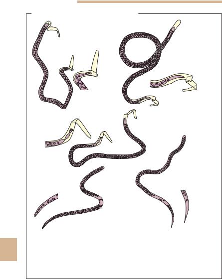
Parasite, life cycle, and epidemiology. Enterobius vermicularis which belongs to the Oxyurida has a conspicuous white color. The males are 2– 5 mm long, the females 8–13 mm. The long, pointed tail of the female gives the pinworm its name.
Sexually mature pinworms live on the mucosa of the large intestine and lower small intestine. Following copulation, the males soon die off. The females migrate to the anus, usually passing through the sphincter at night, then move about on the perianal skin, whereby each female lays about 10 000 eggs covered with a sticky proteinaceous layer enabling them to adhere to the skin. In severe infections, numerous living pinworms are often shed in stool and are easily recognizable as motile worms on the surface
of the feces. 10 The eggs (about 50 ! 30 lm in size) are slightly asymmetrical, ellipsoidal
with thin shells (Fig.10.1, p. 544). With their sticky surface they adhere to skin and other objects. Freshly laid eggs contain an embryo that develops into an infective first-stage larva at skin temperature in about two days. Eggs that become detached from the skin remain viable for two to three weeks in a moist environment.
Infection occurs mainly by peroral uptake of eggs (each containing an infective larva) that are transmitted to the mouth with the fingers from the anal region or from various objects. The sticky eggs adhere to toys and items of

586 10 Helminths
everyday use or are disseminated with dust. In the intestinal tract, larvae hatch from the ingested eggs, molt repeatedly, and develop into sexually mature pinworms in five to six weeks. “Retroinfection” is also conceivable, whereby infective larvae would be released at the anus to migrate back into the intestine.
Pathogenesis and clinical manifestations. The pinworms living on the large intestine mucosa are fairly harmless. Occasionally, different stages of the pinworm penetrate into the wall of the large intestine and the appendix or migrate into the vagina, uterus, fallopian tubes, and the abdominal cavity, where they cause inflammatory reactions.
The females of Enterobius produce in particular a very strong pruritus that may result in nervous disorders, developmental retardation, loss of weight and appetite, and other nonspecific symptoms. Scratch lesions and eczematous changes are produced in the anal area and can even spread to cover the entire skin.
Diagnosis. A tentative diagnosis based on clinical symptoms can be confirmed by detection of pinworms spontaneously excreted with feces and eggs adhering to the perianal skin (Fig. 10.1). Standard stool examination techniques are not sufficient to find the eggs. Egg detection by the “adhesive tape method” has proved most efficient (p. 622).
Therapy and prevention. The following drugs are effective: albendazole, mebendazole, and pyrantel. Reinfections are frequent, so that treatment usually should be repeated once or more times, extended to include all potential parasite carriers (e.g., family members, kindergarten members), and combined with measures, the purpose of which is to prevent egg dissemination: washing the perianal skin (especially in the morning), covering it with ointments, washing the hands, hot laundering of underwear, and cleaning contaminated objects with hot water.
10

Nematoda (Roundworms) 587
Nematodal Infections of Tissues and the Vascular System
Filarial nematodes, the Medina worm, and Trichinella are discussed in this section along with infections caused by the larvae of various nematode species.
Filarioidea (Filariae)
Causative agents of filarioses
& The nematode genera of the superfamily Filarioidea (order Spirurida) will be subsumed here under the collective term filariae, and the diseases they cause are designated as filarioses. In the life cycle of filariae infecting humans, insects (mosquitoes, blackflies, flies etc.) function as intermediate hosts and vectors. Filarioses are endemic in subtropical and tropical regions; in other regions they are observed as occasional imported cases. The most important filariosis is onchocercosis, the causative agents of which, Onchocerca volvulus, is transmitted by blackflies. Microfilariae of this species can cause severe skin lesions and eye damage, even blindness. Diagnosis of onchocercosis is based on clinical symptoms, detection of microfilariae in the skin and eyes, as well as on serum antibody detection. Other forms of filarioses include lymphatic filariosis (causative agent: Wuchereria bancrofti, Brugia species) and loaosis (causative agent: Loa loa). Dirofilaria species from animals can cause lung and skin lesions in humans (see p. 605). &
General. Filariae are threadlike (filum: thread) nematodes. The length of the adult stages (= macrofilariae) of the species that infect humans varies between 2–50 cm, whereby the females are larger than the males. The females release embryonated eggs or larvae called microfilariae. These are about 0.2–0.3 mm long, snakelike stages still surrounded by an extended eggshell (sheathed microfilariae) or they hatch out of it (unsheathed microfilar-
iae) (Fig. 10.17 p. 592). They can be detected mainly in the skin or in blood 10 (Table 10.4).
Based on the periodic appearance of microfilariae in peripheral blood, periodic filaria species are differentiated from the nonperiodic ones showing continuous presence. The periodic species produce maximum microfilaria densities either at night (nocturnal periodic) or during the day (diurnal periodic). Different insect species, active during the day or night, function as intermediate hosts accordingly to match these changing levels of microfilaremia.

588 10 Helminths
Life Cycle of Filariae
Insect: ! Ingestion of microfilaria with a blood meal ! development in thoracic musculature with two moltings to become infective larva ! migration to mouth parts and tranmission into skin of a new host through puncture wound during the next blood meal.
Human: ! Migration to definitive localizations and further development with two more moltings to reach sexual maturity.
Wuchereria bancrofti and Brugia species
Causative agents of lymphatic filariosis
Parasites and occurrence. About 120 million people in 80 countries suffer from lymphatic filariosis caused by Wuchereria bancrofti or Brugia species (one-third each in India and Africa, the rest in southern Asia, the Pacific region, and South America), and 1.1 billion people are at infection risk (WHO, 2000). (Table 10.4). Humans are the only natural final hosts of W. bancrofti and the most widely disseminated Brugia strains. There are, however, other Brugia strains using also animals as final hosts (cats, dogs, and monkeys).
Life cycle and epidemiology. The intermediate hosts of W. bancrofti and B. malayi are various diurnal or nocturnal mosquito genera (Table 10.4). The development of infective larvae in the insects is only possible at high environmental temperatures and humidity levels; in Wuchereria bancrofti the process takes about 12 days at 28 8C. Following a primary human infection, the filariae migrate into lymphatic vessels where they develop to sexual maturity. Microfilariae (Mf) do not appear in the blood until after three months at the earliest (B. malayi, B. timori) or after seven to eight months (W. bancrofti). Tables 10.4 and Fig. 10.17 show their specific characteristics. The adult parasites survive for several years.
Pathogenesis and clinical manifestations. The pathologies caused by W. bancrofti and Brugia species are very similar. The initial symptoms can appear
10as early as one month p.i. although in most cases the incubation period is five to 12 months or much longer. The different courses taken by such infections can be summarized as follows:
&Asymptomatic infection, but with microfilaremia that can persist for years.
&Acute symptomatic infection: inflammatory and allergic reactions in the lymphatic system caused by filariae ! swelling of lymph nodes, lymphangitis, intermittent recurrent febrile episodes, general malaise, swellings on legs, arms, scrotum and mammae, funiculitis, orchitis.

Nematoda (Roundworms) 589
Table 10.4 Filarial Species Commonly Infecting Humans
Species and |
Distribution |
Vector |
Localization |
Microfilariae: |
Pathology |
||
length (cm) |
|
|
of adults |
characteristics |
|
|
|
|
|
|
|
and periodicity |
|
|
|
|
|
|
|
|
|
|
|
Wuchereria |
Southeast |
Mosqui- |
Lymphatic |
244–296 lm, |
Lymphangitis |
||
bancrofti |
Asia, Pacific, |
toes: |
system |
sheathed, |
and lymph- |
||
FF: 2.4–4.0 |
trop. Africa, |
Culex, |
|
in blood, |
adenitis, ele- |
||
ff: 5.0–10.0 |
Caribbean, |
Anopheles, |
|
nocturnal, |
phantiasis |
||
|
trop. South |
Aedes |
|
diurnal or |
|
|
|
|
America |
|
|
subperiodic2 |
|
|
|
Brugia |
South and |
Mosqui- |
Lymphatic |
177–230 lm, |
Lymphangitis |
||
malayi |
East Asia |
toes: |
system |
sheathed, in |
and lymph- |
||
FF: 2.2–2.5 |
|
Anopheles, |
|
blood, nocturnal |
adenitis, ele- |
||
ff: 4.3–6.0 |
|
Aedes, |
|
or subperiodic |
phantiasis |
||
|
|
Mansonia |
|
|
|
|
|
Brugia timori |
Indonesia |
Mosqui- |
|
Nocturnal |
|
|
|
|
|
toes: |
|
periodic |
|
|
|
|
|
Anopheles |
|
|
|
|
|
|
|
|
|
|
|
|
|
Loa loa |
Tropical |
Flies: |
Subcutaneous 250–300 lm, |
Skin swel- |
|||
FF: 3.3–3.4 |
Africa |
Chrysops |
connective |
sheathed, in |
lings, infec- |
||
ff: 5.0–7.0 |
|
|
tissue |
blood, diurnal |
tion of con- |
||
|
|
|
|
periodic |
junctiva |
||
|
|
|
|
|
|
||
Onchocerca |
Africa, Central |
Black flies: Subcutaneous 221–358 lm, |
Skin no- |
||||
volvulus |
and South |
Simulium |
connective |
unsheathed, |
dules, der- |
||
FF: 2.0–4.5 |
America |
|
tissue |
in skin, not |
matitis, eye |
||
ff: 23–50 |
|
|
|
periodic |
lesions |
||
|
|
|
|
|
|
|
|
Mansonella |
Africa, South |
Midges: |
Peritoneal |
190–200 lm, |
Normally |
||
perstans |
America |
Culicoides |
and pleural |
unsheathed, in |
apathogenic |
||
FF: 4.5 |
|
|
cavities |
blood, nocturnal |
|
|
|
ff: 7.0–8.0 |
|
|
|
subperiodic |
|
|
|
|
|
|
|
|
|
|
|
Mansonella |
Tropical |
Midges: |
Subcutaneous 180–240 lm, |
Skin edema, |
|||
streptocerca |
Africa |
Culicoides |
connective |
unsheathed, |
dermatitis |
|
|
|
|||||||
FF:3 |
|
|
tissue |
in skin, |
|
10 |
|
f3 |
|
|
|
not periodic |
|
|
|
Mansonella |
Central and |
Midges: |
Peritoneal |
173–240 lm, |
Normally |
|
|
|
|||||||
ozzardi |
South America |
Culicoides |
cavity |
unsheathed, |
apathogenic |
||
FF:3 |
|
|
|
in blood (not |
|
|
|
ff: 6.5–8.1 |
|
|
|
periodic) |
|
|
|
1See Fig. 10.1 for details on differentiation of microfilariae.
2Subperiodic: periodicity is not pronounced.
3No exact data are available.

590 10 Helminths
&Chronic symptomatic infection: chronic obstructive changes in the lymphatic system ! hindrance or blockage of the flow of lymph and dilatation of the lymphatic vessels (“lymphatic varices”) ! indurated swellings caused by connective tissue proliferation in lymph nodes, extremities (especially the legs, “elephantiasis”), the scrotum, etc., thickened skin (Fig. 10.16). Lymphuria, chyluria, chylocele etc. when lymph vessels rupture. This clinical picture develops gradually in indigenous inhabitants over a period of 10–15 years after the acute phase, in immigrants usually faster.
&Tropical, pulmonary eosinophilia: syndrome with coughing, asthmatic pulmonary symptoms, high-level blood eosinophilia, lymph node swelling and high concentrations of serum antibodies (including IgE) to filarial antigens. No microfilariae are detectable in blood, but sometimes in the lymph nodes and lungs. This is an allergic reaction to filarial antigens.
Diagnosis. A diagnosis can be based on clinical symptoms (frequent eosinophilia!) and finding of microfilariae in blood (blood sampling at night for nocturnal periodic species!). Microfilariae of the various species can be differentiated morphologically in stained blood smears (Table 10.4, Fig. 10.17) and by DNA analysis. Conglomerations of adult worms are detectable by ultrasonography, particularly in the male scrotal area. Detection of serum antibodies (group-specific antibodies, specific IgE and IgG subclasses) and circulating antigens are further diagnostic tools (Table 11.5, p. 625). The recent development of a specific ELISA and a simple quick test (the ICT filariosis card test) represents a genuine diagnostic progress due to the high levels of sensitivity and specificity with which circulating filarial antigens can now be detected, even in “occult” infections in which microfilariae are not found in the blood.
Therapy. Both albendazole and diethylcarbamazine have been shown to be at least partially effective against adult filarial stages. However, optimal treatment regimens still need to be defined. Adjunctive measures against bacterial and fungal superinfection can significantly reduce pathology and suffering.
Control and prevention. In 1997, the WHO initiated a program to eradicate lymphatic filariosis. The mainstay control measure is mass treatment of pop-
10ulations in endemic areas with microfilaricides. Concurrent single doses of two active substances (albendazole with either diethylcarbamazine or iver-
Fig.10.16 a Infection with Wuchereria bancrofti: elephantiasis; b infection with Loa loa: eyelid swelling; c onchocercosis: cutaneous nodules caused by Onchocerca volvulus; d blindness caused by O. volvulus; e Trichinella spiralis; larvae in rat musculature; f larva migrans externa. (Images a, b, d: Tropeninstitut Tu¨bingen, c: Tropeninstitut Amsterdam; f: Dermatologische Klinik der Universita¨t Zu¨rich.) "

Nematoda (Roundworms) 591
Nematode Infections
10
592 10 Helminths
Microfilariae of Various Filarial Species
Wuchereria bancrofti
Loa loa
2
1
1a
2a
|
Brugia malayi |
|
3a |
|
|
|
3 |
|
|
|
5 |
Mansonella perstans |
4 |
Mansonella ozzardi |
|


 5a 4a
5a 4a 


Fig.10.17 Differential diagnosis of microfilariae in human blood: sheathed, large:
101 Loa loa: tip of tail (1a) with several nuclei; 2 Wuchereria bancrofti: tip of tail (2a) without nuclei; 3 Brugia malayi: tip of tail (3a) with single nucleus. Unsheathed, smaller: 4 Mansonella perstans: tip of tail (4a) rounded with densely packed nuclei, often in several rows reaching nearly to the tip of the tail; 5 Mansonella ozzardi: tip of tail (5a) pointed, tip free of nuclei.

Nematoda (Roundworms) 593
mectin) are 99% effective in removing microfilariae from the blood for one year after treatment. Mass-treatment with albendazole or ivermectin is also expected to have a controlling effect on intestinal nematodes (Ascaris, hookworms, Strongyloides, Trichuris). Measures to avoid mosquito bites are the same as for malaria.
Loa
Loa loa
Causative agent of loaosis (loiasis, Loa loa filariosis, African eyeworm)
Occurrence, life cycle, and epidemiology. Thirteen million people are infected with this filarial species in the tropical rainforest areas of Africa (western and central Africa, parts of Sudan) (WHO, 1995).
The adult and pre-adult parasites (Table 10.4) live in and migrate through the subcutaneous connective tissues. The microfilariae appear in a periodic pattern during the day in peripheral blood (Table 10.4, Fig.10.17). Accordingly, the intermediate hosts are diurnally active horsefly species (Tabanidae: Chrysops species). The prepatent period is five to six months. In some cases, microfilariae do not appear in the blood even in older cases of infection. The adult filariae live for several years.
Pathogenesis and clinical manifestations. Clinical symptoms can occur two to 12 months after the infection. They are probably mainly allergic in nature. The filariae migrating through the connective tissues cause edematous swellings in the limbs, face, and body (“Calabar swellings”) and itching nodules (Fig. 10.16b). The infection is often accompanied by blood eosinophilia. Migration of a parasite beneath the conjunctiva causes lacrimation, erythema, and other symptoms.
Diagnosis, therapy, and prevention. Diagnosis involves observation of typical symptoms, adult parasites in subcutis or conjunctiva and microfilariae in peripheral blood (in blood specimens sampled during the day!) (Table 10.4,
Fig. 10.17). The drug of choice is diethylcarbamazine that kills microfilariae 10 and damages macrofilariae after long-term therapy (N.B.: possible side ef-
fects).
Mansonella species
See Table 10.4 and Fig. 10.17 for Mansonella species that should be taken into account in differential diagnostic procedures.

594 10 Helminths
Onchocerca
Onchocerca volvulus
Causative agent of onchocercosis
This filarial species causes onchocercosis, a disease that manifests mainly in the form of skin alterations, lymphadenopathy, and eye damage, which latter is the reason for the special importance of the disease.
Occurrence. Onchocerca volvulus is endemic in 30 countries in tropical Africa (from the Atlantic coast to the Red Sea) and southern Arabia (Yemen) as well as in six countries in Central and South America (Mexico, Guatemala, Columbia, Venezuela, Brazil, Ecuador). About 17.6 million persons are currently infected and 267 000 are blind due to onchocercosis (WHO, 1998, 2000). The WHO has been coordinating successful control programs in 11 African countries since 1974, and in six Latin American countries since 1991 (see below).
Life cycle. The adult Onchocerca live in the connective and fatty tissues, usually tightly coiled in connective tissue nodules in the subcutis or deeper tissue layers (Fig. 10.16c). Sexually mature parasites can live as long as 15 years.
Female Onchocerca produce microfilariae that live in the tissue within the nodules or skin. Starting from the site of the female worms, the microfilariae migrate through the deep corium of the dermis into other skin regions and can also affect the eyes—especially if the nodule is located on the head or upper body. Through the lymphatics, the microfilariae can penetrate into the bloodstream and also appear in urine, sputum, and cerebrospinal fluid. The relatively large microfilariae have no sheath (Table 10.4); their lifespan in human hosts is from six to 30 months.
Simuliidae (blackflies) are the intermediate hosts and vectors. The development of the infective larvae, transmitted by a blackfly to a human host, takes many months before the nematodes reach sexual maturity. Microfilariae are usually be detected in skin after 12–15 months (seven to 24 months) (prepatent period).
10Epidemiology. Humans are the sole parasite reservoir of O. volvulus. Onchocercosis occurs in endemic foci along the rivers in which the larvae and pupae of the blackflies develop. Therefore, the blindness caused by onchocercosis is designated as “river blindness.”
Pathogenesis and clinical manifestations. Pathological reactions are provoked by adult parasites and by microfilariae. These reactions are influenced by the immune status of infected individuals.