

Hart C. Anthony, Shears Paul. Color Atlas of Medical Microbiology.pdf
.pdfEntamoeba histolytica and Other Intestinal Amebas 505
3 Fig. 9.11 |
|
|
a Giardia intestinalis (pathogenic): |
|
|
trophozoites: 9–21 ! 5–12 lm; |
|
|
cysts: 8–14 ! 8–10 lm (see also p. 478). |
|
|
b Dientamoeba fragilis (apathogenic or facultatively pathogenic): |
|
|
trophozoites: 5–15 lm, 3/4 of stages with two nuclei, the rest with one; |
|
|
karyosome consisting of four to six granules; |
|
|
cysts: none. |
|
|
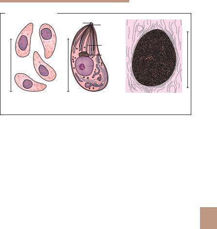
c Entamoeba histolytica (pathogenic): |
|
|
trophozoites: small form (1) 10–20 lm, with Entamoeba nucleus and small number |
|
|
of vacuoles containing bacteria; larger form (2) 20–60 lm, with phagocytosed |
|
|
erythrocytes; (3) cysts: 10–16 lm, one to four nuclei. Here binuclear cyst with gly- |
|
|
cogen vacuole and cigarshaped chromidial bodies and (4) a tetranuclear cyst. |
|
|
Entamoeba dispar (apathogenic): morphologically identical with E. histolytica |
|
|
(see text). |
|
|
Entamoeba hartmanni (5) (apathogenic): similar to E. histolytica, but smaller. |
|
|
Trophozoites and cysts approximately 3–10 lm. |
|
|
d Entamoeba coli (apathogenic): |
|
|
trophozoites: with Entamoeba nucleus, 10–50 lm, in most cases numerous va- |
|
|
cuoles containing bacteria and particles.Cysts: 15–25 lm, one to eight nuclei, |
|
|
chromidial bodies slender, splinter-shaped. |
|
|
e Iodamoeba bu¨tschlii (apathogenic): |
|
|
trophozoites: 6–20 lm, nucleus with large karyosome, either centrally located or |
|
|
contiguous with the nuclear membrane. |
|
|
Cysts: 5–18 lm, one nucleus, rarely two. |
|
|
f Endolimax nana (apathogenic): |
|
|
trophozoites: 6–15 lm, nucleus with karyosome. |
|
|
cysts: usually oval, 8–12 lm long. |
|
|
g Microsporidia (pathogenic): |
|
|
spores: very small (!), 1–3.5 lm long depending on species, oval shape; spores |
|
9 |
often not stained homogeneously by chromotropic staining according to Weber |
|
|
|
|
|
(see also Fig. 9.20c, p. 539). |
|
|
h Blastocystis hominis (facultatively pathogenic?): |
|
|
single cells: 5–20 lm. |
|
|
i Cryptosporidium species (pathogenic): |
|
|
oocysts: 4–5 lm (see also Fig. 9.14a, p. 516). |
|
|
j Cyclospora cayetanensis (pathogenic): |
|
|
oocysts: 4–5 lm, spherical, unsporulated in fresh stool; after sporulation two spor- |
|
|
ocysts with two sporozoites each (see also Fig. 9.14a, p. 516). |
|
|
k Sarcocystis species (pathogenic): |
|
|
sporocyst: 14 ! 9 lm; oocyst: 20 ! 13 lm. |
|
|
l Isospora belli (pathogenic): |
|
|
oocyst: 20–33 ! 10–19 lm. |
|
|

5069 Protozoa
—Differentiation of E. histolytica and E. dispar. A new type of PCR is now used in specialized laboratories that facilitates direct detection of these amebic species in stool specimens as well as a differential diagnosis.
—Detection of coproantigen. E. histolytica antigen can be detected in stool
specimens using an ELISA based on monoclonal antibodies with high levels of sensitivity and specificity.
—Serological antibody assay. Antibodies can be detected serologically in 95–100% of patients with amebic liver abscess. This is also frequently the case in invasive intestinal amebosis caused by E. histolytica. On the other hand, antibodies are produced far less often in E. dispar infections. When stages of the E. histolytica/E. dispar complex are detected in stool, the presence or absence of serum antibodies can be used in differential diagnosis of invasive vs. noninvasive intestinal amebosis.
& Extraintestinal amebosis. This type of amebosis is diagnosed with the help of clinical methods (ultrasound, computer tomography, etc.) and serological antibody detection (see above).
Therapy. Nitromidazole derivatives are effective against symptomatic intestinal and extraintestinal forms of amebosis. On the other hand, amebicides with only luminal activity are effective against asymptomatic intestinal amebosis (e.g., diloxanide furoate) (Table 9.5). A new drug against intestinal amebic infections is nitazoxandide. Besides chemotherapy, other measures may also be required, e.g., surgery and symptomatic treatment for liver abscesses.
Prevention. Travelers to endemic areas should decontaminate drinking water by boiling or filtering it (e.g., with Katadyn filters), not eat salads, eat only fruit they have peeled themselves and exercise caution when it comes to changing their diet. Chemoprophylactic dugs are not available.
9
Table 9.5 Chemotherapy in Amebosis (examples)
Group of amoebicides* |
Active substance |
Indication |
|
|
|
Luminal amebicides |
Diloxanide furoate |
Asymptomatic cyst excreters, |
|
Paromomycin |
follow-up treatment of invasive |
|
Nitazoxanide |
intestinal form |
Systemic amebicides |
Nitroimidazoles: |
Invasive intestinal and extrain- |
|
Metronidazole |
testinal forms |
|
Ornidazole |
|
|
Tinidazole |
|
|
|
|
*Application per os |
|
|

Naegleria, Acanthamoeba, and Balamuthia 507
Naegleria, Acanthamoeba, and Balamuthia
Causative agents of naegleriosis, acanthamebosis, and balamuthiosis
Free-living ameba of the genera Naegleria, Acanthamoeba, and Balamuthia have the potential to infect vertebrates and to cause diseases in humans. The morphological characteristics of these amebas include: nucleus with large karyosome, lack of chromatin granules at the nuclear membrane (see Entamoeba). Trophozoites: Naegleria fowleri (15–30 lm) with wide pseudopods, produces flagellated stages in water; Acanthamoeba spp. (24–56 lm) with fingerlike protrusions (“filopods”); Balamuthia mandrillaris (12–60 lm) has irregularly branched pseudopods. All of these genera produce cysts.
Naegleria. The causative agent of primary amebic meningoencephalitis (PAM) is Naegleria fowleri. This species occurs worldwide in bodies of freshwater, especially warm water in swimming pools, storage containers or thermally polluted lakes and rivers, etc.
Infection of humans occurs by the nasal route with water containing trophozoites, i.e., during a swim or shower. The amebas migrate from the olfactory epithelium along the nerve tracts into the CNS and cause, after an incubation period of two to seven (rarely as long as 15) days a hyperacute to acute meningoencephalitis that usually has a lethal outcome. The infection occurs mainly in children and youths. Sporadic occurrence is reported from all continents. Treatment with amphotericin B has been successful in a small number of cases.
Acanthamoeba. Potential human pathogens in this genus include Acanthamoeba culbertsoni and several other species. These amebas occur worldwide in soil, sand, dust (also house dust), air, and water (also in tap water). The cysts can survive for several years in a dry state and disseminate with dust. Inhalation of cysts with dust is considered to be one of the main transmission routes. Acanthamebas are frequent colonizers of human nasal mucosa and have been isolated from the oral mucosa, skin lesions, and the cornea. From the portal of entry, they can spread
hematogenously to the CNS and other organs. 9 Acanthamoeba infections in humans frequently take an asymptomatic course. Ker-
atitis is observed occasionally, especially in contact lens wearers. Diagnosis: culturing of amebas from conjunctival and contact lens rinsing liquids. Prevention: use only sterile lens rinsing liquid. In rare cases of generalized infection, Acanthamoeba can cause granulomatous amebic encephalitis (GAE) as well as granulomatous lesions in the lungs and other organs, especially in immunodeficient patients. Balamuthia mandrillaris can also cause GAE.

508 9 Protozoa
Toxoplasma gondii
Causative agent of toxoplasmosis
& Toxoplasma gondii is the causative agent of a zoonosis that occurs worldwide with high prevalences (up to 80% depending on region and age). Humans are infected by ingesting oocysts excreted by the definitive hosts (cats) or by eating unprocessed meat containing Toxoplasma cysts. If a women contracts toxoplasmosis for the first time during pregnancy, diaplacental transmission of the pathogen to the fetus is possible with potential severe consequences (for example malformations, eye damage, clinical symptoms during childhood). There is, however, no risk to the fetus from mothers who had been infected before their first pregnancy and have produced serum antibodies (about 35–45%). Latent infections can be activated by immunodeficiencies (e.g., in AIDS patients) and may result in cerebral or generalized symptomatic toxoplasmosis. Serological surveillance in pregnant women is important to prevent prenatal infections. &
Occurrence. T. gondii occurs worldwide. The low level of host specificity of this organism explains its ready ability to infect a wide spectrum of warmblooded vertebrate species (for example humans, sheep, pigs, cattle, horses, dogs, cats, wild mammals, bird species). It is estimated that approximately one-third of the world population is infected with T. gondii, although prevalences vary widely depending on age and region. According to a seroepidemiological study in Switzerland (published in 1995) of 4000 persons aged one to 70, an average of 52% was infected with seroprevalence rates in different age groups as follows: one to nine years: 24%, 20–39 years: 43%, and 40–70 years: 69%. Of 9000 pregnant women, 46% were infected. Women of childbearing age groups in other European countries showed either lower or
9higher seroprevalences. High prevalences were also observed in various animal species (see Epidemiology p. 514).
Parasite. Various strains of T. gondii differ in virulence and certain biological and genetic characteristics. The life cycle of T. gondii includes various stages:
&Tachyzoites (endozoites) (Fig. 9.12a, b) are proliferative forms that reproduce rapidly within a parasitophorous vacuole in nucleate host cells by means of endodyogeny (endodyogeny: formation of two daughter cells from a mother cell by endogenous budding). The tachyzoites are sickleshaped (toxon: bow) cells about 4–7 lm long and 2–4 mm wide. An apical complex is located at the anterior pole, consisting of serveal components, including the conoid (a conical structure of spirally arranged microtubuli), a pole ring complex, the rhoptries, and micronemes (Fig. 9.12b). The apical complex

510 9 Protozoa
within two to four days, producing two sporocysts with four sporozoites each. Sporulated oocysts are infective for humans and animals.
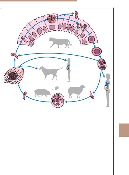
Life cycle. The developmental cycle of T. gondii involves three phases: intestinal, external, and extraintestinal (Fig. 9.13).
&The intestinal phase with production of sexual forms (gamogony) takes only place in enterocytes of definitive hosts. Only domestic cats, and several other felid species of little epidemiological significance, can function as definitive hosts for T. gondii. Only extraintestinal development is seen in intermediate hosts (pigs, sheep, and many other animal species) as well as in dead-end hosts (humans). Following primary infection of a cat with Toxoplasma cysts in raw meat, asexual reproductive forms at first develop in the small intestine epithelium, with sexually differentiated stages and oocysts following later. The oocysts are shed in feces after a prepatent period of three to nine days. When cats are infected with sporulated oocysts, the prepatent period is extended to 20–35 days because in these cases the intestinal development of Toxoplasma is preceded by an extraintestinal asexual reproduction (see below). Oocyst shedding lasts for only a few days to a maximum of three weeks, but can be highly intensive (up to 600 million oocysts per eat during the patent period!).
&External phase. Oocysts excreted in cat feces sporulate at room temperature within two to four days, rendering them infective. Kept moist, they remain infective for up to five years and are not killed by standard disinfectant agents. They die within a few minutes at temperatures exceeding 55 8C.
&Extraintestinal phase. This phase follows a peroral infection with oocysts or cysts and is observed in intermediate hosts (dogs, sheep, pigs, other vertebrates, birds) and dead-end hosts (humans), as well as in the definitive hosts (cats). Starting from the intestine, the Toxoplasma organisms travel
9in blood or lymph to various organs and multiply in nucleate host cells, especially in the reticulohistiocytic system, in musculature, and in the CNS. Repeated endodyogenic cycles produce as many as 32 individual daughter cells in the expanding host cell before it bursts. The tachyzoites thus released attack neighboring cells.
These processes result in focal necroses and inflammatory reactions in affected tissues. Generalization of the infection can lead to colonization of the placenta and, about three to four weeks later, infection of the fetus. Cysts that elicit no inflammatory reactions in the near vicinity are produced early in the course of the infection. Such cysts (tissue cysts) are found above all in the CNS, in the skeletal and heart muscles as well as in the retina, the uterine wall, and other organs. They can remain viable for years without causing noticeable damage to the host.
Toxoplasma gondii 511
Toxoplasma gondii: Life Cycle
a |
2 |
3
1 |
|
|
4 |
|
|
|
|
|
|
|
|
|
5 |
|
b |
|
|
|
|
13 |
|
|
|
|
|
|
|
6 |
|
12a |
12b |
|
|
|
|
|
|
|
|
11 |
8b |
|
|
|
|
|
|
|
|
10 |
|
|
|
|
c |
|
7 |
8a |
|
|
|
|
||
|
9 |
|
|
|
Fig. 9.13 a Development in the definitive host (cat): intestinal phase with pro- |
|
|||
duction of sexual forms: 1 Toxoplasma that has penetrated into a small intestine |
|
|||
epithelial cell; 2 asexual reproductive stage with merozoites (can continue for |
|
|||
several generations). The arrow indicates that an extraintestinal cycle precedes |
9 |
|||
the enteral cycle when a cat is infected with oocysts; 3 production of sexual forms |
||||
(gamogony) and formation of the zygote; 4 oocyst. |
|
|
|
|
b External phase with sporogony: 5 unsporulated oocyst, shed in cat feces; |
|
|||
6 sporulated oocyst with two sporocysts each containing four sporozoites. |
|
|||
c Development in an intermediate host (mammals, birds, humans): extraintestinal |
|
|||
phase with only asexual multiplication of parasites; 7 sporozoite released in |
|
|||
organism following oral ingestion of oocysts; 8a human infection; 8b infection |
|
|||
of various animal species; 9 asexual reproductive stages (tachyzoites) in a somatic |
|
|||
cell; 10 free tachyzoite; 11 cyst with bradyzoites in musculature; 12a infection of |
|
|||
a dog with Toxoplasma cysts in animal meat; 12b infection of a human with |
|
|||
Toxoplasma cysts; 13 infection of cat with infective stages of cysts in meat or |
|
|||
with oocysts. |
|
|
|
|

512 9 Protozoa
Immunity. Various Toxoplasma antigens induce humoral and cellular immune responses which, following a primary infection, result in antibody production and inhibition of tachyzoite multiplication. Toxoplasmas then “escape” from the immune defense system by encysting (immunoevasion). This enables them to persist in a latent state for many years in immunocompetent hosts, at the same time maintaining an immune status that confers protection from new infections due to the continuous presentation of antigens.
The relevant immune defense mechanisms include mainly cellular mechanisms and production of IFNc. Bradyzoites can also migrate out of cysts (without cyst rupture), are then, however, locally inactivated in immunocompetent persons, sometimes leading to formation of satellite cysts. In cases of cellular immune deficiency, this control system is lacking and the latent infection progresses to become an acute, manifest toxoplasmosis. Similarly, latent Toxoplasma infections in AIDS patients are usually activated and turn symptomatic when the CD4+ cell count falls below 200/ll.
Pathogenicity and clinical manifestations. Focal necrotic, inflammatory and immunopathological processes are the basis of the pathogenesis and varied clinical manifestations observed in toxoplasmosis. Cases are differentiated as to time of acquisition, i.e., postnatal and prenatal infections.
Forms of Postnatal Toxoplasma Infection
&Primary infection in immunocompetent persons. This is the most frequent form without clinical manifestations, recognizable by the specific serum antibodies. The infection can persist for the life of the host, and it may exacerbate in response to immunosuppression. Subacute cervical lymphadenitis occurs in about 1% of infected persons.
&Primary infection during pregnancy. This may cause prenatal infection
9of the fetus and thus become a significant threat (see prenatal toxoplasmosis p. 513).
&Primary infection in immunosuppressed persons. In cases of immune deficiency (with significant disturbance of CD4+ and CD8+ cell functions) or immunosuppressant therapies (e.g., in organ transplantations) the infection gives rise to febrile generalized illness with maculopapulous exanthema, generalized lymphadenitis, necrotizing interstitial pneumonia, hepatosplenomegaly, mycocarditis, meningoencephalitis, eye damage, and other manifestations. There is a high rate of lethality if left untreated.
&Reactivation toxoplasmosis in cases of immune deficiency. Local and generalized reactivation of a Toxoplasma infection originating from tissue cysts. Cerebral manifestations are the most frequent (up to 40% of patients

Toxoplasma gondii 513
in full-blown AIDS stage), for example, with multiple coagulative necroses, small-focus hemorrhages, and surrounding edema. Other organ systems are affected more rarely (in about 15% of cases), e.g., myocardium and lungs.
Prenatal Toxoplasmosis
Occurrence. The prenatal fetal infection occurs only in mothers who contract their primary (first!) infection with toxoplasmas during pregnancy! There is no risk of prenatal fetal infection in women having a latent infection with serum antibodies at conception.
Incidence. The rate of verified primary infections in pregnant women was estimated in Germany, Austria, and Switzerland in 1995 at 0.5–0.7%. A prenatal fetal infection is to be expected in some of these cases, whereby the infection risk for the fetus is lower in the first trimester than later in the pregnancy. The frequency of toxoplasmosis in newborns (prenatal toxoplasmosis) is between 0.1 and 0.3% in various European countries. The rate is lower in some countries, e.g., in Austria (0.01%), where monitoring examinations for pregnant women have been obligatory for a number of years (for details see Aspo¨ck, 2000, p. 660).
Possible consequences of prenatal infection:
&10% clinically severe cases, 85% of these with brain damage (e.g., hydrocephalus, intracerebral calcifications), 15% perinatal deaths,
&15% milder symptoms (99% chorioretinitis, 1% brain damage),
&75% subclinical cases (15% no damage, 85% chorioretinitis).
Children in this last group appear to be clinically normal at birth, but signs of brain and eye damage, as well as other symptoms, may manifest later in infancy and early childhood.
Diagnosis. In immunocompetent adults, toxoplasmosis is normally diag- |
9 |
nosed serologically by detection of parasite-specific IgG and IgM antibodies (Table 11.5, p. 625). IgM antibodies can be detected as early as one week after the primary infection, peak within two to four weeks, then drop to below the detection limit within a few weeks; in some cases persistence at low titers lasts longer. IgG antibodies appear somewhat later, peak after two to four months and persist for many years. A high or rising IgG titer with contemporal detection of IgM indicates an acute primary infection. The isolated cases of ocular toxoplasmosis normally cannot be diagnosed by serological methods.
Serological findings are often not reliable indicators in immunodeficient patients due to reduced antibody production. The cerebral form of the infection seen frequently in reactivated toxoplasmosis is therefore usually diagnosed by means of clinical imaging methods.

514 9 Protozoa
In prenatal diagnostics, PCR is seeing increasing use for direct detection of pathogens in the amniotic fluid. Detection of Toxoplasma DNA with this method is a reliable sign of fetal infection and demands a chemotherapeutic or other response accordingly. Diagnosis of prenatal toxoplasmosis in neonates is difficult, but highly important. Since IgG antibodies are transmitted from mother to child diaplacentally, detection of them in the child cannot serve as a definitive diagnostic indicator. IgM is only present in about 50% of prenatally infected children. In suspected cases, the blood or cerebrospinal fluid should by examined using the PCR.
Therapy. The following cases require treatment: acute or subacute, symptomatic infections in children and adults as well as symptomatic or asymptomatic primary infections in pregnant women. In an acute primary infection during pregnancy, the risk of infection for the developing fetus can be eliminated by starting chemotherapy immediately. Several different therapeutic schemes are recommended for this indication. For example, spiramycin daily for four weeks from diagnosis to the end of the 15th week of gravidity, and in the period beginning with the 16th week of gravidity pyrimethamine daily for four weeks together with sulfadiazine and folic acid. The recommendations also vary for treatment of toxoplasmosis in AIDS cases, e.g., pyrimethamine/ sulfadiazine or pyrimethamine/clindamycin.
Epidemiology and prevention. Humans become infected by peroral ingestion of raw meat containing cysts or by uptake of sporulated oocysts. T. gondii is also transmitted diaplacentally and by transplantation of infected organs to uninfected recipients.
In Europe about 1–6% of domestic cats are oocyst excreters. Sheep and goats are frequently infected with toxoplasmas; infection prevalences of pigs have decreased significantly in pig farms with high hygienic standards as demonstrated in recent studies. Cattle have always been considered rare
9carriers, but recently in Switzerland Toxoplasma DNA was found using PCR in 1–6% of the animals examined (n = 350). The epidemiological significance of these findings has not yet been clarified. Certain wild animal species (e.g., wild boars) are relatively frequently infected, not so horses and chickens. Milk and eggs are not considered sources of infection.
Toxoplasma cysts remain viable and infectious in meat for up to three weeks at 4 8C. Deep-freezing to –20 8C kills bradyzoites within three days, heating to 70 8C is lethal to them within a few minutes. Toxoplasma oocysts show considerable environmental resistance, but can be killed rapidly by heat (70 8C).
Pregnant women should eat only meat that has been thoroughly heated or deep-frozen. Close contact with cats should be avoided. Cat litter boxes in the house should be cleaned out daily and flushed with boiling water (wear rubber gloves!). Cats can be fed canned (boiled) meat to protect them from