

Gale Encyclopedia of Genetic Disorder / Gale Encyclopedia of Genetic Disorders, Two Volume Set - Volume 2 - M-Z - I
.pdfAbnormalities can also arise when the zygote begins to divide. This type of abnormality is usually severe, eventually leading to a miscarriage. If an abnormality occurs after the zygote has divided one or more times, the baby will have some normal cells and some abnormal cells. This situation is referred to as “mosaicism” and “mosaic” may be used to describe the person’s condition.
Molar pregnancies
Molar pregnancies can occur in one of two ways. Sometimes the original cell that duplicates and divides to form the fetus is completely of paternal origin. The chromosomes in a sperm duplicate themselves, then proceed to divide as if they were a normal zygote. These pregnancies are completely abnormal and miscarry. Another type of molar pregnancy occurs when two sperm fertilize one egg. The zygote is triploidy and has 69 chromosomes instead of 46. Although some fetal parts can be seen, these pregnancies normally miscarry in the first or second trimester.
Birth defects
The term birth defect describes many different types of abnormalities, including physical malformations. Abnormalities of anatomical structures may be significant or insignificant; minor variations in structure are common. Approximately 3% of newborns have major malformations. The causes are: chromosome abnormalities (6–7%), inherited genetic conditions (7-8%), environmental factors (7–10%), and multifactorial causes (20–25%). The cause of the remaining 50–60% of malformations is unknown. Multifactorial refers to causes with both genetic and environmental components. Environmental factors include exposures to drugs, chemicals, or other substances that affect the development of the fetus while he/she is in the uterus. Substances that cause birth defects are referred to as teratogens.
Artificial reproductive technology
Couples may pursue assisted reproductive technologies for a number of reasons. If a couple has artificial insemination, the sperm is inserted into the uterus when the woman in ovulating. Fertilization then occurs as it would normally. If a couple has in vitro fertilization (IVF), the egg and sperm are mixed outside the body in the laboratory. The zygote forms in a petri dish if fertilization occurs. After a number of cell divisions, the developing embryo is placed in the woman’s uterus. If the sperm are incapable of fusing with the egg themselves, the sperm may be injected into the egg. This additional step to the IVF procedure is called intracytoplasmic sperm injection (ICSI).
A human zygote. (Photo Researchers, Inc.)
In the year 2001, preimplantation diagnosis is possible for a number of genetic diseases. Couples may pursue this if they are at a significant risk for having a child with a disease that could be diagnosed prior to becoming pregnant through preimplantation diagnosis. The procedure is like that of in vitro fertilization, with an additional step. After fertilization occurs and the zygote has begun to divide, a single cell is removed. Removing the cell does not harm the other cells. The cell that is removed is tested for the genetic disease for which the couple is at risk. Multiple developing embryos are tested. Only the embryos that do not have the condition are placed in the woman’s uterus to complete development.
The development of a person from the zygote is a fascinating and amazing process. It is a difficult area to study because scientists cannot manipulate human embryos to observe the effects, and the development of the fetus cannot be directly observed. Researchers still have many unanswered questions. Following a doctor’s recommendations from prior to the pregnancy throughout pregnancy (such as folic acid intake and avoidance of alcohol and other drugs) increases the chances that the development of a zygote into a full-term infant will be normal. However, there are many babies born with severe birth defects or genetic diseases despite the parents’ efforts at doing everything in their power to prevent a problem. Most birth defects and genetic disorders occur because of an event out of control of the parents.
Zygote
GALE ENCYCLOPEDIA OF GENETIC DISORDERS |
1229 |
Zygote
Resources
BOOKS
Agnew, Connie L. Twins!: Expert Advice From Two Practicing Physicians on Pregnancy, Birth, and the First Year. New York: HarperCollins, 1997.
Brasner, Shari E. Advice From a Pregnant Obstetrician. New York: Hyperion, 1998.
Nathanielsz, P.W. Life in the Womb: The Origin of Health and Disease. Ithaca, NY: Promethean Press, 1999.
Vaughn, Christopher C. How Life Begins: The Science of Life in the Womb. New York: Times Books, 1996.
PERIODICALS
Check, Erika. “What Moms Can Do Now.” Newsweek (27
September 1999): 57–58.
Christensen, Damaris. “Sobering Work.” Science News (8 July
2000): 28–29.
Kowalski, Kathiann. “High-tech Conception in the 21st
Century.” Current Health (January 2000): 1–4.
Miller, Annetta, and Joan Raymond. “The Infertility
Challenge.” Newsweek (Spring/Summer 1999 Special
Edition): 26–28.
ORGANIZATIONS
American College of Obstetricians and Gynecologists. PO Box 96920, 409 12th St. SW, Washington, DC 20090-6920.http://www.acog.org .
American Society for Reproductive Medicine. 1209 Montgomery Highway, Birmingham, AL 35216-2809. (205) 978-5000. asrm@asrm.org. http://www.asrm
.org .
RESOLVE, The National Infertility Association. 1310 Broadway, Somerville, MA 02144-1779. (617) 623-0744. resolveinc@aol.com. http://www.resolve.org .
WEBSITES
The InterNational Council on Infertility Information Dissemination, Inc. http://www.inciid.org .
Maternal and Child Health Bureau.
http://www.mchb.hrsa.gov/ .
Organization of Teratology Information Services.
http://www.otispregnancy.org/index.html .
Michelle Queneau Bosworth, MS, CGC
1230 |
GALE ENCYCLOPEDIA OF GENETIC DISORDERS |
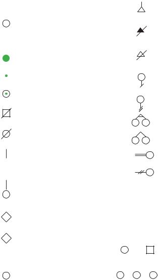
SYMBOL GUIDE FOR PEDIGREE CHARTS
Pedigree charts are a visual tool for documenting biological relationships in families and the presence of disorders. Using these charts, a medical professional such as a geneticist or genetic counselor, can analyze the genetic risk in a family for a particular trait or condition by tracking which individuals have the disorder and determining how it is inherited.
A standard set of symbols has been established for use in creating pedigree charts. Those found within the body of several entries in the encyclopedia follow the symbol guide explained on the next page. The exact style and amount of information presented on the chart varies for each family and depends on the trait or condition under investigation. Typically, only data that is directly related to the disorder being analyzed will be included.
G A L E E N C Y C L O P E D I A O F G E N E T I C D I S O R D E R S |
1231 |
Symbol Guide for Pedigree Charts
|
|
|
|
|
Symbol Guide for Pedigree Charts |
|
|||||||||
|
|
|
|
|
Male |
|
|
|
|
|
|
|
|
Miscarriage |
|
|
|
|
|
|
|
|
|
|
|
|
|
|
|||
|
|
|
|
|
Female |
|
|
|
|
|
|
|
|
Pregnancy terminated |
|
|
|
|
|
|
|
|
|
|
|
|
|
|
|||
|
|
|
|
|
|
|
|
|
|
|
|
|
|
|
|
|
|
|
|
|
|
|
|
|
|
|
|
|
|
|
due to affected condition |
|
|
|
|
|
Affected male |
|
|
|
|
|
|
|
|
|
|
|
|
|
|
|
|
|
|
|
|
|
|
|
|
||
|
|
|
|
|
|
|
|
|
|
|
|
|
|
|
Elective termination |
|
|
|
|
|
Affected female |
|
|
|
|
|
|
|
|
of pregnancy |
|
|
|
|
|
|
Carrier male |
|
|
|
|
|
|
|
|
Female with no children |
|
|
|
|
|
|
|
|
|
|
|
|
|
|
|||
|
|
|
|
|
|
|
|
|
|
|
|
|
by choice |
||
|
|
|
|
|
Carrier female |
|
|
|
|
|
|
|
|
||
|
|
|
|
|
|
|
|
|
|
|
|
|
|
||
|
|
|
|
|
|
|
|
|
|
|
|
|
|
|
Female with no children |
|
|
|
|
|
|
|
|
|
|
|
|
|
|
|
due to medical infertility |
|
|
|
|
|
Deceased male |
|
|
|
|
|
|
|
|
|
|
|
|
|
|
|
|
|
|
|
|
|
|
|
|
|
Identical twin females |
|
|
|
|
|
Deceased female |
|
|
|
|
|
|
|
|
Fraternal twin females |
|
|
|
|
|
|
|
|
|
|
|
|
|
|
|
|
|
|
|
|
|
|
|
|
|
|
|
|
|
|
|
|
Consanguineous |
|
|
|
|
|
|
|
|
|
|
|
|
|
|
|
|
|
|
|
|
|
Male adopted into |
|
|
|
|
|
|
|
|
relationship |
|
|
|
|
|
|
|
|
|
|
|
|
|
|
|
||
|
|
|
|
|
a family |
|
|
|
|
|
|
|
|
Relationship no longer |
|
|
|
|
|
|
|
|
|
|
|
|
|
||||
|
|
|
|
|
|
|
|
|
|
|
|
|
|
|
|
|
|
|
|
|
|
|
|
|
|
|
|
|
|
|
exists |
|
|
|
|
|
Female adopted into |
? |
|
|
|
||||||
|
|
|
|
|
|
|
|
Unknown family history |
|||||||
|
|
|
|
|
|
||||||||||
|
|
|
|
|
a family |
|
|
|
|
|
|
|
|
||
|
|
|
|
|
Gender not specified |
|
|
|
|
|
|
|
|||
|
|
|
|
|
|
d.79y |
Died at 79 years |
||||||||
|
|
|
|
|
|
|
|
dx.41y |
Diagnosed at 41 years |
||||||
|
|
P |
|
Pregnancy |
|
|
|
|
|
|
|
|
|
||
|
|
|
|
|
|
|
|
|
|
|
|
|
|
|
Relationship line |
|
|
|
|
|
|
|
|
|
|
|
|
|
|
|
|
|
|
4 |
|
|
Four males |
|
|
|
|
|
|
|
|
Line of descent |
|
|
|
|
|
|
|
|
|
|
|
|
|
|
|
|
Sibship line |
|
|
|
|
|
|
|
|
|
|
|
|
|
|
|
|
|
|
3 |
|
|
Three females |
|
|
|
|
|
|
|
|
Individual line |
|
|
|
|
|
|
|
|
|
|
|
|
|
|
|
|
|
1232 |
G A L E E N C Y C L O P E D I A O F G E N E T I C D I S O R D E R S |
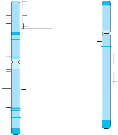
CHROMOSOME MAP
A chromosome map indicates the relative positions of the genes that code for certain characteristics. The basic format for writing a gene position is the chromosome number, arm, band, sub-band, and sub-sub-band, if known. An example is 3p22.5.
The chromosome number refers to one of the 22 autosomal chromosomes (numbered 1-22) or one of the sex-determining chromosomes known as X or Y. In the example, the gene is on chromosome 3.
Each chromosome has two arms, separated by a centromere, the pinched-in area toward the top of the chromosome. The short arm, labeled “p”, is above the centromere and the long arm, “q”, is below it. In the case of the example gene, it is found on the short arm (p) of chromosome 3, or 3p.
The arms are further divided into cytogenetic bands (regions) numbered 1, 2, 3, etc... The numbers start at the
centromere and increase to the end of the arm, known as the telomere. These bands can only be seen when stained and viewed under a microscope. Sub-bands, which are numbered the same way as bands, may be visible within bands at greater magnifications. Therefore, the exact location of the example gene is the short arm (p) of chromosome 3, band 2, sub-band region 2, and a sub-sub band 5.
The following 24 illustrations demonstrate the approximate gene location for several of the genes relating to disorders mentioned in this encyclopedia. Disorders known to be related to a specific chromosome but not necessarily at an exact location have been placed below the chromosome. These chromosome maps are in no way complete, rather, they provide an introduction to understanding relative size differences of human chromosomes and where geneticists have located the genes associated with the source of certain genetic disorders.
G A L E E N C Y C L O P E D I A O F G E N E T I C D I S O R D E R S |
1233 |

Chromosome Map
Chromosome 1
36
35
34
333
32
31
22
2
21
13
p 1 12 11
q 11
1
12
21
22
223 
24
25 
31
3
32
41
4
42
43
44 
Marshall syndrome
 GBA: Gaucher disease
PKLR: Pyruvate kinase deficiency
 Factor V deficiency HPC1: Prostate cancer
GLC1A: Glaucoma
PS2(AD4): Alzheimer disease
 CHS1: Chediak-Higashi syndrome
Chromosome 2
25
24
223 
22
21 
16
15
114
13
12
p q
11 
11 
112
13 
14
21
222
23 
24
31
32 
33 
34
35 

36
37 
ETM2: Essential tremor
Oculo-digito-esophago-duodenal syndrome
MSH2: Colon cancer
CSNU: Cystinuria
 ALMS1: Alstrom syndrome
Spastic cerebral palsy
PAX3: Waardenburg syndrome
1234 |
G A L E E N C Y C L O P E D I A O F G E N E T I C D I S O R D E R S |
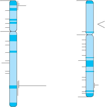
Chromosome 3
26
25 
24
223 
22
21 
14
113 
12
p |
11 |
q |
|
11
12 
1
13 
21 
22
23 
24
2
25
26
27 
28
29
VHL: von Hippei-Lindau
 BTD: Biotindase
SCLC1: Lung cancer
MLH1: Colon cancer
hMLH1: Muir-Torre syndrome
LAR1: Larsen syndrome (3p)
HGD: Alkaptonuria (3p)
Pituitary dwarfism
ETM1: Essential tremor
GM1: gangliosidosis
FBS: Faconi-Bickel syndrome
Chromosome 4
|
|
|
|
|
|
|
|
|
|
HD: Huntington disease |
|
|
16 |
|
|
|
|
|
|
|
|
MPS: Mucopolysaccharidoses |
|
|
|
|
|
|
|
|
Achondroplasia |
||||
|
|
|
|
|
|
|
|
|
|
||
|
15 |
|
|
|
|
|
|
|
|||
|
|
|
|
|
|
|
EVC: Ellis-van Creveld |
||||
|
|
|
|
|
|
|
|||||
|
1 |
|
|
|
|
|
|
|
|
|
|
|
|
|
|
|
|
|
|
|
|
|
|
|
|
14 |
|
|
|
|
|
|
|
|
|
|
13 |
|
|
|
|
|
|
|
|
|
|
|
|
|
|
|
|
|
|
|
|
|
|
p |
12 |
|
|
|
|
|
|
|
|
|
|
|
11 |
|
|
|
|
|
|
|
|
|
|
q11
112 
13
|
|
21 |
|
|
|
|
alpha-synuclein: Parkinson disease |
|
|
|
|
||||
|
|
|
|
|
|
|
|
|
|
22 |
|
|
|
|
|
2 23 |
|
|
|
|
|
|
|
|
|
|
|
|
|
||
24 |
|
|
|
|
|
||
|
|
|
|
|
|
||
25 |
|
|
|
|
RIEG: Rieger syndrome |
||
26 |
|
|
|
|
|||
|
|
|
|
LQT4: Long QT syndrome 4 |
|||
|
|
|
|
||||
|
|
|
|
|
|||
27 |
|
|
|
|
|||
28 |
|
|
|
|
|
||
31 |
|
|
|
|
|
||
|
|
|
|
|
|
|
|
|
|
|
|
|
|
|
|
|
|
|
|
|
|
|
|
|
|
|
|
|
|
|
|
3 |
32 |
|
|
|
|
|
|
|
|
33 |
|
|
|
|
|
|
|
|
|
|
|
|
|
|
|
34 |
|
|
|
|
|
|
|
|
|
|
|
|
|
|
|
35 |
|
|
|
|
|
|
|
|
|
|
|
|
|
Map Chromosome
G A L E E N C Y C L O P E D I A O F G E N E T I C D I S O R D E R S |
1235 |
Chromosome Map
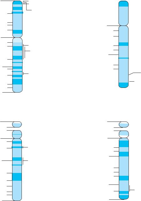
Chromosome 5
SRD51A: Steroid 4-alpha reductase 1 15 Cri du chat syndrome
14
1
13
12
p 11  CSA: Cockayne syndrome q
11
Familial adenomatous polyposis (5q)
12
113 
14
15 
21
222 
23 

 Asthma
31
Corneal dystrophy
|
|
|
|
|
|
DTD: Diastrophic dysplasia |
3 |
32 |
|
|
|||
|
|
|
|
|||
|
|
|
|
|
||
33 |
|
|
|
|
|
|
|
34 |
|
|
|
|
|
|
|
|
|
|
|
|
|
35 |
|
|
|
|
|
|
|
|
|
|
|
|
|
|
|
|
|
|
|
Chromosome 6
25 |
|
|
|
|
|
|
|
|
24 |
|
|
|
|
|
|
SCA1: Spinocerebellar atrophy |
|
|
23 |
|
|
|
|
|
||
|
|
|
|
|
||||
22 |
|
|
|
|
IDDM1: Diabetes |
|||
|
|
|
|
|||||
2 |
|
|
|
|
|
|||
21 |
|
|
|
|
|
|
COL11A2: Weissenbacher-Zweymuller syndrome |
|
|
|
|
||||||
|
|
|
|
|
|
|
|
Cleidocranial dysplasia |
|
|
|
|
|
|
|
|
|
12 |
|
|
|
|
|
|
Akylosing spondylitis |
|
|
|
|
|
|
|
|
||
p 1 11
q11 
12
113 
14
15 
16
21
22
223 
24
EPM2A: Epilepsy
25 
26
27
Major histocompatibility complex (6)
1236 |
G A L E E N C Y C L O P E D I A O F G E N E T I C D I S O R D E R S |

Chromosome 7
22 
2 21
15


 GCK: Diabetes
14
113 
12
p 11
q
1 |
11 |
|
|
|
|
|
|
ELN: Williams syndrome |
|
|
|
|
|
|
|||
|
|
|
|
|
|
|||
|
|
|
|
|
|
|
||
|
|
|
|
|
|
|
|
|
|
|
|
|
|
|
|
|
|
|
21 |
|
|
|
|
|
|
Autistic disorder (7q) |
2 |
|
|
|
|
|
|
|
|
|
|
|
|
|
|
|
|
|
|
22 |
|
|
|
|
|
|
Pendrin: Pendred syndrome |
|
|
|
|
|
|
|
|
|
|
|
|
|
|
|
|
|
|
|
31 |
|
|
|
|
|
|
CFTR: Cystic fibrosis |
|
|
|
|
|
|
|
||
|
|
|
|
|
|
|
||
|
|
|
|
|
|
|
||
3 |
|
|
|
|
|
|
OB: Obesity |
|
|
|
|
|
|
|
|
||
32 |
|
|
|
|
|
|
||
|
|
|
|
|
|
|
|
|
33 |
|
|
|
|
|
|
|
|
|
|
|
|
|
|
|
|
|
35 |
34 |
|
|
|
|
|
|
Pancreatic cancer |
|
|
|
|
|
|
|
||
|
|
|
|
|
|
|
||
|
36 |
|
|
|
|
|
|
|
Chromosome 9
2 |
|
24 |
|
|
|
|
|
|
|
|
||
22 |
|
23 |
|
|
|
|
|
|
|
|
||
|
|
|
|
|
|
|
|
|
CDKN2: Malignant melanoma |
|||
|
|
|
|
21 |
|
|
|
|
|
|
|
|
|
|
|
|
|
|
|
|
|
|
|
||
|
|
|
|
|
|
|
|
|
|
|
|
|
1 |
|
13 |
|
|
|
|
|
|
|
|
||
|
|
|
|
|
|
|
|
|
|
|||
|
12 |
|
|
|
|
|
|
|
|
|||
|
p |
|
|
|
|
|
|
|
|
|
||
|
11 |
|
|
|
|
|
|
|
|
|||
|
q |
|
11 |
|
|
|
|
|
|
|
|
|
|
|
12 |
|
|
|
|
|
|
|
FRDA: Friedreich’s ataxia |
||
|
1 |
|
|
|
|
|
|
|
|
|||
|
|
|
|
|
|
|
|
|||||
|
|
|
|
|
|
|
|
|
||||
|
|
|
|
|
|
|
|
|
|
|||
|
13 |
|
|
|
|
|
|
|
|
|||
2 |
|
21 |
|
|
|
|
|
|
|
|
||
|
|
|
|
|
|
|
|
|
||||
|
|
|
|
|
|
|
|
|||||
|
|
|
|
|
|
|
|
|
|
|||
|
|
|
|
22 |
|
|
|
|
|
|
|
Familial dysautonomia |
|
|
|
|
|
|
|
|
|
|
|
|
|
|
|
|
|
31 |
|
|
|
|
||||
|
|
|
|
|
|
|
|
|
|
|
|
|
|
|
|
|
|
|
|
|
|
|
|
|
|
|
|
|
|
32 |
|
|
|
|
|
|
|
|
3 |
|
33 |
|
|
|
|
|
|
|
OWR1: Olser-Weber-Rendu syndrome |
||
|
|
|
|
|
|
|||||||
|
|
|
|
34 |
|
|
|
|
|
|
|
|
|
|
|
|
|
|
|
|
|
|
|
|
|
|
|
|
|
|
|
|
|
|
|
|
|
|
Distal arthrogryposis syndrome (9)
|
|
|
Chromosome 8 |
Chromosome |
|||||||||||
|
|
|
|
||||||||||||
|
|
|
|
|
|
|
|
|
|
|
|
|
|
|
|
2 |
|
23 |
|
|
|
|
|
|
|
|
|
|
|
||
22 |
|
|
|
|
|
|
|
|
|
|
|
||||
|
|
|
|
|
|
|
|
|
|
|
|
|
|
|
|
|
|
|
21 |
|
|
|
|
|
|
|
|
|
|
Map |
|
|
|
|
|
|
|
|
|
|
|
|
|
|
|||
1 |
12 |
|
|
|
|
|
|
|
|
|
WRN: Werner syndrome |
||||
|
|
|
|
|
|
|
|
|
|||||||
|
|
|
|
|
|
|
|
|
|
|
|
|
|||
|
p |
11 |
|
|
|
|
|
|
|
|
|
Hereditary spherocytosis |
|
||
|
|
|
|
|
|||||||||||
|
|
|
|
|
|
|
|
|
|
||||||
|
q |
11 |
|
|
|
|
|
|
|
|
|
|
|
||
|
|
|
|
|
|
|
|
|
|
|
|||||
1 |
|
|
|
|
|
|
|
|
|
|
|
||||
|
|
|
|
|
|
|
|
|
|
|
|
|
|||
12 |
|
|
|
|
|
|
|
|
|
|
|
||||
|
|
|
|
|
|
|
|
|
|
|
|
|
|
||
|
|
|
|
|
|
|
|
|
|
|
|
|
|
|
|
|
|
|
13 |
|
|
|
|
|
|
|
|
|
|
|
|
|
|
|
|
|
|
|
|
|
|
|
|
|
|
Cohen syndrome |
|
|
|
|
21 |
|
|
|
|
|
|
|
|
|
|
||
|
|
|
|
|
|
|
|
|
|
|
|
||||
|
|
|
|
|
|
|
|
|
|
|
|
Klippel-Feil syndrome |
|
||
|
|
|
|
|
|
|
|
|
|
|
|
||||
|
|
|
|
|
|
|
|
|
|
|
|
|
|
|
|
2 |
22 |
|
|
|
|
|
|||||||||
|
|
|
|
|
|
|
|
|
|
||||||
|
|
|
|
|
|
|
|
|
|
|
|||||
|
|
|
|
|
|
|
|
|
|
||||||
|
|
|
|
|
|
|
|
|
|
|
|
|
|||
23 |
|
|
|
|
|
|
|
|
|
|
|
||||
|
|
|
|
|
|
|
|
|
|
|
|
|
|
||
|
|
|
|
|
|
|
|
|
|
|
|
MYC: Burkitt lymphoma |
|
||
|
|
|
24 |
|
|
|
|
|
|
|
LGS: Langer-Giedon syndrome |
|
|||
|
|
|
|
|
|
|
|
|
|
|
|||||
|
|
|
|
|
|
|
|
|
|
|
|||||
|
|
|
|
|
|
|
|
|
|
|
|
|
|
|
|
Chromosome 10
|
|
15 |
|
|
|
|
|
|
|
|
|
|
|
|
PAHX: Refsum disease |
|
|
14 |
|
|
|
|
|
|
|
|
|
|
|
||
|
|
|
|
|
|
|
|
|
|
13 |
|
|
|
|
|
1 |
12 |
|
|
|
|
|
|
|
|
|
|
|
|||
|
|
|
|
|
|||
|
|
|
|
|
|
|
|
|
p |
11 |
|
|
|
|
|
|
|
|
|
|
|
|
|
|
q |
|
|
|
|
|
|
111 
21
22
223
24
25
|
26 |
|
|
|
|
Apert syndrome |
OAT: Gyrate atrophy |
|
|
|
|
||||
|
|
|
|
Crouzon syndrome |
|||
|
|
|
|
|
|
|
|
|
|
|
|
|
|
|
|
G A L E E N C Y C L O P E D I A O F G E N E T I C D I S O R D E R S |
1237 |
Chromosome Map
Chromosome 11
HRAS: Harvey ras oncogene
LQT1: Long QT syndrome
15
IDDM2: Diabetes
114
13 
12
p |
11 |
q |
11 |
12DHC: Smith-Lemli-Opitz syndrome
1MEN1: Multiple endocrine neoplasia
13
 MKS2: Meckel syndrome, type 2
14
21
22
ATM: Ataxia telangiectasia
223
24
25 
Chromosome 13
13 
1 12 
p |
11 |
q |
11 |
|
12 |
|
BRCA2: Breast cancer |
13
1 

 RB1: Retinoblastoma
14
ATP7B: Wilson disease
21
2
22
31
332 
33
34 
Chromosome 12
1 |
13 |
|
|
|
|
PXR1: Zellweger syndrome |
|
|
|
|
|||
|
|
|
|
|
|
|
|
|
|
|
|
|
|
12 |
|
|
|
|
|
|
|
|
|
|
|
||
|
|
|
|
|
|
|
p |
11 |
|
|
|
|
|
|
|
|
|
|
q11 
12
113 
14
15 
221
22 
|
23 |
|
|
|
|
HOS: Holt-Oram syndrome |
|
24 |
|
|
|
|
PAH: Phenylketonuria |
|
|
|
|
|
||
|
|
|
|
|
Noonan syndrome |
|
|
|
|
|
|
||
|
|
|
|
|
||
|
|
|
|
|
|
|
|
|
|
|
|
|
|
Chromosome 14
13 
1 12  p 11
p 11 
q
11
12
1
13
21
222 
23
24

 PS1(AD3): Alzheimer disease
31
3Krabbe disease
32
1238 |
G A L E E N C Y C L O P E D I A O F G E N E T I C D I S O R D E R S |