

Gale Encyclopedia of Genetic Disorder / Gale Encyclopedia of Genetic Disorders, Two Volume Set - Volume 1 - A-L - I
.pdfAlkaptonuria
may be visible in the genital regions, the larynx (voice box), and the middle ear. Dark-stained tendons can be seen when the hand is made into a fist.
Arthritis
The symptoms of ochronotic arthritis are similar to those of other types of arthritis. However, the large, weight-bearing joints usually are the most affected in ochronotic arthritis. These include the joints of the hips, knees, and shoulders, and between the vertebrae of the spine. The joints become stiff and difficult to move. This arthritis develops at an unusually early age. In unaffected individuals, similar arthritis usually does not develop before age 55. Men with AKU develop arthritis in their 30s and 40s. Women with AKU usually develop arthritis in their 50s.
AKU can lead to osteoarthrosis, a degenerative joint disease, and ochronotic arthropathy, which is characterized by the swelling and enlargement of the bones. Ankylosis, the adhesion of bones in the joints, also may occur. The pigment deposits may cause the cartilage to become brittle and susceptible to fragmenting. Individuals with AKU may be at risk for bone fractures.
Calcium deposits can lead to painful attacks similar to those of gout. This calcification may occur in the ear cartilage and in the lumbar disks of the lower back. The disks between vertebrae may become narrowed and eventually may collapse.
Organ damage
The coronary artery of the heart can become diseased as a result of AKU. The aortic valve of the heart may harden and narrow from calcification. Similar problems may develop with the mitral or left atrioventricular valve of the heart (mitral valvulitis). Deposits of pigment can lead to the formation of hard spots of cholesterol and fat (atherosclerotic plaques) in the arteries. This can put a person at risk for a heart attack.
Complications from the deficiency of the HGD enzyme arise primarily in the kidneys and the liver. HGD normally is most active in the kidneys, liver, small intestine, colon, and prostate. The calcification of the genital and urinary tract may lead to blockages in as many as 60% of individuals with ochronosis. Kidney stones and other kidney diseases may develop. Stones in the urine may occur in middle to late adulthood. Increasingly though, this condition is seen in children with AKU under the age of 15 and even as young as two. In men, pigment deposits may lead to stones in the prostate.
The teeth, the brain and spinal cord, and the endocrine system that produces hormones also may be affected by ochronosis. Breathing may become restricted
due to the effects of ochronosis on the joints where the ribs attach to the spine. Deposits of pigment on the ear bones and on the membrane of the inner ear may lead to tinnitus, or ringing of the ears, and hearing problems.
Diagnosis
Visual diagnosis
AKU is often detected in early childhood because of the characteristic dark-staining of the urine. In adults, diagnosis usually is made on the basis of joint pain and skin discoloration. Most individuals with AKU have pigment visible in the whites of their eyes by their early 40s.
A family history of AKU helps with the diagnosis. Since many individuals with AKU have no symptoms, siblings of affected individuals should be tested for the disorder.
Identification of HGA
An individual with AKU may excrete as much as 4- 8 g of HGA per day in the urine. There are several simple methods to test for HGA in the urine: the addition of sodium hydroxide (an alkali) to the urine will turn it dark; urine with HGA turns black when reacted with iron chloride; and alkaline urine containing HGA blackens photographic paper. In the laboratory, HGA can be identified in the urine using a technique called gas chromatographymass spectroscopy. This technique separates and identifies the components of a mixture.
There are a number of methods for identifying HGA in the blood and tissues. These include procedures for separating HGA from other components of the blood and instruments that can detect the characteristic color of HGA. With AKU, the concentration of HGA in the blood is approximately 40 micromolar, or 40 micromoles of per liter.
Microscopic examination
With AKU, there usually is visible black staining of cartilage in various body regions, particularly the larynx, trachea (windpipe), and cartilage junctions. Heavy deposits of pigment also occur in the bronchi (the air passages to the lungs). Pigment on the inside and outside of the cells of these tissues can be seen with a microscope.
A skin biopsy, the removal of a small piece of skin, may be used to obtain tissue for examination. The tissue is stained with dyes to reveal the yellowish-brown pigment deposits on the outside of skin cells. Pigment deposits also occur in cells of the endothelium (the thin layer of cells that line blood vessels and other tissues), in the sweat glands, and in the membranes below the skin.
58 |
G A L E E N C Y C L O P E D I A O F G E N E T I C D I S O R D E R S |
|
|
|
|
Alkaptonuria |
|
|
|
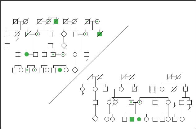
Alkaptonuria |
|
|
|
|
Autosomal Recessive |
|
|
|
|
|
1. High carrier frequency in Czechoslovakia |
|
|
|
||||
|
|
|
|
|
|
|
|
|
|
2 |
|
|
N |
|
|
|
|
|
d.71y |
|
|
Arthritis |
|
|
|
|
3 |
Heart |
3 |
|
N |
|
|
|
|
attack |
|
|
|
|
|
|||
|
|
|
|
|
|
|
|
|
|
|
40y |
38y |
|
|
|
|
|
|
2 |
|
|
2. Low carrier frequency in other countries |
|
|||
|
|
|
P |
|
|
|
|
|
|
|
12y |
7y |
5y |
|
|
? |
|
|
|
|
|
|
|
|
Stroke |
|
|
|
|
|
|
|
|
|
|
|
|
|
|
Skin |
War |
|
d.71y |
|
|
|
|
|
cancer |
|
|||
|
|
|
|
|
|
|
Breast |
|
|
|
|
|
3 |
|
|
|
cancer |
|
|
|
|
|
|
|
|
|
|
|
|
|
d.at |
43y |
41y |
|
|
|
|
|
|
birth |
|
|
|
|
|
|
|
|
N |
|
|
|
2 |
|
|
|
|
20y |
18y |
16y |
11y |
|
(Gale Group)
These pigments will not fade, even after three days in a solution of bleach.
of an organ. Lung function tests and hearing tests may be performed to assess additional complications.
Skeletal x rays |
Acquired ochronosis |
X-ray examination is used to detect calcification of the joints. Since many individuals with AKU do not have dark-staining urine, x-ray evidence of osteoarthritis may indicate a need to test for the presence of HGA in the urine. However, osteoarthritis usually affects the smaller joints; whereas ochronosis most often affects the large joints of the hips and shoulders. Spinal x rays may show dense calcification, degeneration, and fusion of the disks of the vertebrae, particularly in the lumbar region of the lower back. Chest x rays are used to assess damage to the valves of the heart.
Other procedures
Physicians may order computerized tomography (CT) scans of the brain and chest or magnetic resonance imaging (MRI) of affected joints. An electrocardiogram (ECG or EKG) may reveal signs of heart complications resulting from AKU. Kidney problems may be diagnosed by ultrasound, the use of sound waves to obtain images
In addition to being a complication of AKU, ochronosis can be acquired. In the past, ochronosis developed from the repeated use of carbolic acid dressings for treating chronic skin ulcers. The prolonged use of the drug quinacrine (atabrine) can cause ochronosis, with pigmentation occurring in many of the same sites as with AKU. Ochronosis can also result from the use of bleaching creams containing hydroquinone. Certain other substances, including phenol, trinitrophenol, quinines, and benzene, can cause ochronosis. However, these forms of ochronosis do not lead to joint disease and, unlike ochronosis from AKU, are reversible.
Treatment and management
The binding of HGA to collagen fibers is irreversible. Treatment of AKU is directed at reducing the deposition of pigment and thereby minimizing arthritis and heart problems in later life.
G A L E E N C Y C L O P E D I A O F G E N E T I C D I S O R D E R S |
59 |
Alkaptonuria
Vitamin C
Often, high doses (about 1 gm per day) of ascorbic acid (vitamin C) are administered to older children and adults with AKU. Ascorbic acid appears to slow the formation of the HGA polymer and decrease the binding of the polymer to connective tissues. Vitamin C reduces the amount of toxic benzoquinone acetic acid in the urine. However, the amount of HGA in the urine does not decrease. Furthermore, vitamin C does not appear to interrupt the progress of the disease.
Dietary restrictions
Sometimes individuals with AKU are placed on lowprotein diets. This limits the intake of phenylalanine and tyrosine from proteins. If the body has lower amounts of phenylalanine and tyrosine to break down, less HGA will be formed. However, both of these amino acids are necessary for making proteins in the body. Furthermore, phenylalanine is an essential amino acid that must be obtained from food, since the human body cannot produce it. Adult males require approximately 2 gm per day of phenylalanine. Phenylalanine also is present in some artificial sweeteners.
Restricting protein intake to no more than the daily protein requirement may be beneficial for children with AKU. Such diets appear to substantially reduce the amount of benzoquinone acetic acid in the urine. In children under the age of 12, low-protein diets significantly reduce the amount of HGA in the urine, as well. However, these diets seem to have little effect on older children and young adults with AKU, and low-protein diets are difficult to maintain. When low-protein diets are prescribed, the levels of amino acids in the blood must be monitored, to assure that there is no deficiency in phenylalanine.
Ochronosis
Most treatment of AKU is directed at the diseased joints. The treatment for ochronosis is the same as for other forms of degenerative arthritis. Treatments include painkillers, physical therapy, rehabilitation, orthopedic supports, and rest. Chiropractic manipulations and exercise regimens also are utilized.
Treatment of ochronotic arthritis eventually may require hip and/or knee joint replacements with artificial materials. In older individuals, fusion of the lumbar discs of the lower spine may be necessary. Aortic valve replacement may be necessary to treat heart disease.
Future drug treatment
The National Institutes of Health are undertaking clinical research studies to better understand the clinical, biochemical, and molecular aspects of AKU. These studies are in preparation for clinical trials of a new drug to treat AKU. It is hoped that this drug will block the production and accumulation of HGA.
Prognosis
There is no cure for AKU. Essentially all individuals with AKU eventually experience arthritic symptoms, particularly arthritis of the hips, knees, and spine. The bone and joint disease may become debilitating by the sixth to eighth decades of life. Furthermore, cardiovascular involvement and ochronotic skin abnormalities are to be expected with AKU.
Despite these difficulties, individuals with AKU have normal life expectancies. Although there is an increased risk of heart attack in later life, most individuals with AKU die of causes unrelated to the disorder.
Resources
BOOKS
La Du, B. N. “Alkaptonuria.” In The Metabolic and Molecular Bases of Inherited Disease, edited by C. R. Scriver, A. L. Beaudet, W. S. Sly, and D. Valle. New York: McGraw Hill, Inc., 1995, pp. 1371-86.
PERIODICALS
Titus, G. P., H. A. A. Mueller, S. Rodriguez de Cordoba, M. A. Penalva, and D. E. Timm. “Crystal Structure of Human Homogentisate Dioxygenase.” Nature Structural Biology 7, no. 7 (2000): 542-46.
Zatkova, A., D. B. de Bernabe, H. Polakova, M. Zvarik, E. Ferakova, V. Bosak, V. Ferak, L. Kadasi, and S. R. de Cordoba. “High Frequency of Alkaptonuria in Slovakia: Evidence for the Appearance of Multiple Mutations in HGO Involving Different Mutational Hot Spots.”
American Journal of Human Genetics 6, no. 5 (November 2000): 1333-39.
ORGANIZATIONS
AKU Hotline.http://www.goodnet.com/~ee72478/enable/hotline.htm .
National Heart, Lung, and Blood Institute. PO Box 30105, Bethesda, MD 20824-0105. (301) 592-8573. nhlbiinfo@rover.nhlbi.nih.gov. http://www.nhlbi.nih
.gov .
National Institute of Child Health and Human Development (NICHD). Patient Recruitment and Public Liaison Office, Building 61, 10 Cloister Court, Bethesda, MD 208924754. (800) 411-1222, (301) 594-9774 (TTY), (866) 4111010 (TTY). prpl@mail.cc.nih.gov. http://clinicalstudies
.info.nih.gov/detail/A_2000-CH-0141.html .
60 |
G A L E E N C Y C L O P E D I A O F G E N E T I C D I S O R D E R S |

WEBSITES
“Alkaptonuria.” AKU Database.http://www.cib.csic.es/~akudb/alkaptonuria.htm .
Burkhart, Craig G., and Craig Nathaniel Burkhart. “Ochronosis.” Dermatology/Metabolic Diseases. 25 July 2000. http://www.emedicine.com/DERM/topic476.htm .
“Clinical, Biochemical, and Molecular Investigations into Alkaptonuria.” NIH Clinical Research Studies. Protocol Number: 00-CH-0141. (March 10, 2001).http://clinicalstudies.info.nih.gov/detail/A_2000-CH- 0141.html .
Medical College of Wisconsin Physicians and Clinics. “Alkaptonuria and Ochronosis.” HealthLink. (March 18, 1999). http://healthlink.mcw.edu/content/article/ 921733488.html .
Roth, Karl S. “Alkaptonuria.” Pediatrics/Genetics and Metabolic Disease. (December 10, 2000). http://emedicine
.com/ped/topic64.htm .
Margaret Alic, PhD
I Alpha-1 antitrypsin
Definition
Alpha-1 antitrypsin is one of the most common inherited diseases in the Caucasian population. The most common symptom is lung disease (emphysema). People with alpha-1 antitrypsin may also develop liver disease and/or liver cancer. The disease is caused by a deficiency in the protein alpha-1 antitrypsin, which is why the condition is sometimes called alpha-1 antitrypsin deficiency. Other names include anti-elastase, antitrypsin, and ATT. The development of lung disease is accelerated by harmful environmental exposures, such as smoking tobacco. Alpha-1 antitrypsin is inherited. The age of onset, rate of progression, and type of symptoms vary both between and within families.
Description
The protein alpha-1 antitrypsin is a protease inhibitor, which means that it inactivates other proteins called proteases. This is an important function, as proteases themselves disable proteins. In our bodies the levels of proteases and their inhibitors are balanced so that proteases can perform their functions but not over-per- form, which leads to problems.
A protease called elastase is the most important target of alpha-1 antitrypsin. Elastase protects the lungs against bacteria and other foreign particles. However, if the action of elastase is not kept in check, elastase
destroys lung tissue. Alpha-1 antitrypsin ensures that elastase is not overactive.
Individuals with alpha-1 antitrypsin have inadequate levels of the protein alpha-1 antitrypsin. Thus, certain proteases (especially in the lungs) are overactive, which leads to emphysema and sometimes to liver disease. Alpha-1 antitrypsin is made mostly in the liver.
Some alpha-1 antitrypsin proteins are abnormal in addition to being deficient. These abnormal proteins may not move from the liver to the blood stream correctly. The build-up of the proteins in the liver may lead to liver disease. Also, the abnormal proteins may not neutralize elastase as effectively. Thus, people with alpha-1 antitrypsin have fewer proteins; those that they do have do not work as effectively.
Genetic profile
The genetics of alpha-1 antitrypsin are complicated. Scientists have identified many different forms of the gene that codes for the alpha-1 antitrypsin protein. This protein is often called Pi and the gene called PI, for protease inhibitor. One form of the gene, which scientists call Z, or PI Z, greatly reduced the amount of the active Pi protein. Because every person inherits one of each gene from his or her mother, and another copy of each gene from his or her father, everyone has two copies of every gene. People who have two copies of the PI Z gene have 85% less alpha-1 antitrypsin protein. These people have only 15% of the normal level of protein. The protein that they do have does not function as well as the normal protein. People who have one PI Z gene and one normal PI gene have about 60% of the normal level Pi protein. Other forms of the alpha-1 antitrypsin gene are associated with more or less severe deficiencies in protein.
Two other common forms of the Pi protein are called S and M. Pi M is the normal protein and PI M is the normal gene. The Pi M protein has many subtypes within the population, designated M1, M2, etc. A few abnormal alpha-1 antitrypsin genes also have unique names. The PI S gene is slightly abnormal, but not as abnormal as PI Z. Individuals with one PI S gene and one PI Z gene have approximately 38% functioning of the Pi protein (Pi SZ).
The inheritance of alpha-1 antitrypsin is autosomal recessive. This means that a person with alpha-1 antitrypsin has inherited one abnormal gene from each of his or her parents. The parents are most likely carriers, meaning they each have one normal gene and one abnormal gene. Two carriers have a one in four chance to have an affected child with each pregnancy. However, not all people with alpha-1 antitrypsin develop symptoms. Whether and when a person with two abnormal alpha-1
antitrypsin 1-Alpha
G A L E E N C Y C L O P E D I A O F G E N E T I C D I S O R D E R S |
61 |
Alpha-1 antitrypsin
antitrypsin genes develops symptoms is related to the degree of harmful exposures, such as tobacco smoke. A person who is affected with alpha-1 antitrypsin is only at risk to have an affected child if the child’s other parent is a carrier.
Although the inheritance of alpha-1 antitrypsin is autosomal recessive, the activity of the protein is equally determined by the gene inherited from either parent. For example, if a gene inherited from one parent codes for a protein with 100% activity, and the gene inherited from the other parent codes for a protein with 0% activity, the offspring would have 50% protein activity. The physical expression of the genes is autosomal recessive, but each gene has an equal effect on the protein activity—neither gene is dominant over the other gene. The gene for alpha- 1 antitrypsin is on chromosome 14. More than 90 different forms of the gene have been identified.
Demographics
Alpha-1 antitrypsin is most common in Caucasians, especially those of Northern European descent. Alpha-1 antitrypsin is less common in populations of Asian, African, and American Indian descent. Approximately one in 2,500 Caucasians have two Z genes. These individuals account for 1% of all emphysema patients. Because people with one PI Z gene and one other deleterious PI gene may also have symptoms, the number of people at risk to have alpha-1 antitrypsin associated lung disease is greater than one in 2,500. Approximately one in 20 Caucasians has one Z gene and one normal gene. The number of Caucasians with one S gene and one normal gene is even higher. Approximately one in 1,000 Caucasians of Northern European descent have two S genes (and no normal alpha-1 antitrypsin gene).
Signs and symptoms
The main symptom of alpha-1 antitrypsin is a risk for early-onset, rapidly progressive emphysema. People with alpha-1 antitrypsin who smoke tobacco are at especially high risk. Emphysema is chronic lung disease that begins with breathlessness during exertion and progresses to shortness of breath at all times, caused by destructive changes in the lung tissue. The risk for liver disease in adults is increased, as is the risk for hepatocellular carcinoma (liver cancer). Some children with alpha-1 antitrypsin develop liver disease as well. Individuals with alpha-1 antitrypsin are also at risk for chronic obstructive lung disease and reactive airway disease (asthma). Chronic obstructive lung disease is decreased breathing capacity, which may be caused by emphysema but also has other underlying causes.
Lung disease
Approximately 60–70% of the people with two PI Z genes develop chronic lung disease. Shortness of breath with exertion may begin before the age of 40 years and progress rapidly to incapacitating emphysema. Life expectancy may be reduced by 10–15 years and is reduced further if people with two PI Z genes smoke tobacco. A portion of the people with two PI Z genes never develop chronic lung disease.
The age of onset and severity of symptoms associated with alpha-1 antitrypsin are quite variable, even within the same family. Environmental exposures significantly effect whether a person will develop symptoms. Smoking puts individuals with alpha-1 antitrypsin at much greater risk to develop emphysema. The already abnormal and deficient Pi Z protein functions 1,000 times less effectively in smokers. Researcher Ronald Crystal states, “Cigarette smoking renders an already poorly defended lung completely defenseless.” People with alpha-1 antitrypsin who are not exposed to harmful environmental factors are less likely to develop emphysema. If people with two PI Z genes stop smoking before they develop lung disease, their life expectancy increases and the risk of lung disease decreases.
Individuals who have one abnormal gene with very little protein function and one gene with somewhat reduced protein function may also at risk for chronic obstructive lung disease. It is possible that people with one Z gene and one normal gene are also at risk to develop chronic lung disease if they are exposed to harmful environmental factors such as tobacco smoke. The age symptoms begin in this group would be later than that seen in people with two abnormal genes. Some researchers disagree, stating that people with PI SZ and PI MZ genes are not at significant risk for lung disease.
Liver disease
The risk of liver disease and liver cancer are increased in individuals with alpha-1 antitrypsin. Babies and children with alpha-1 antitrypsin may have abnormal liver function and inflammation. The abnormal liver function they develop is called cholestasis, which is when the liver stops secreting a digestive fluid called bile. A build-up of bile causes cholestatic jaundice (yellowing of the skin). These abnormalities sometimes progress to liver disease and liver failure, which is fatal without a liver transplant. In other babies and children, liver function returns to normal.
A small number of adults with alpha-1 antitrypsin develop liver disease, and some develop liver cancer. The age at which the liver disease begins, the rate at which it progresses, and the stage at which it is usually diagnosed
62 |
G A L E E N C Y C L O P E D I A O F G E N E T I C D I S O R D E R S |

are quite variable. Adults with alpha-1 antitrypsin who had liver abnormalities as children may be at increased risk to develop liver disease or liver cancer. People with one normal PI gene and one PI Z gene may be at increased risk for liver disease.
The likelihood that a child or adult with alpha-1 antitrypsin will develop liver disease can be predicted to some degree based on which change in the gene (mutation) they have as well as their family history. The risk that a baby with two Z genes will develop significant liver disease is approximately 10%. However if a person has a family history of alpha-1 antitrypsin with liver disease, this risk may be higher. Males (both adult and children) develop liver disease more often than females. Alpha-1 antitrypsin is the most common genetic cause of liver disease in infants and children. Researchers do not know why some people with alpha-1 antitrypsin develop progressive liver disease and many others do not. The liver disease appears to be related to abnormal antitrypsin protein remaining in the liver instead of being secreted.
Diagnosis
Alpha-1 antitrypsin may be suspected in a newborn with cholestatic jaundice, swollen abdomen, and poor feeding. In later childhood or adulthood, fatigue, poor appetite, swelling of the abdomen and legs, or abnormal liver tests may trigger the need for testing. The diagnosis of alpha-1 antitrypsin is based on measurement of antitrypsin (Pi) in the blood. If levels of Pi are deficient, genetic studies may be performed to determine which abnormal forms of the gene are present. The Pi protein can also be studied to determine which type a person has. Prenatal diagnosis is available, however, it is recommended that parental genetic studies precede prenatal testing to ensure accurate interpretation of results.
Levels of antitrypsin protein in the blood may be normal in individuals who have one PI Z gene and one normal gene, and in individuals who have one PI S gene and one PI Z gene. Studying the Pi protein will more accurately diagnose these individuals.
Lung disease in people with alpha-1 antitrypsin is diagnosed by the same methods used to diagnose lung disease in people who do not have alpha-1 antitrypsin. These studies include breathing tests such as total lung capacity and pulmonary function tests. Total lung capacity is measured with a device called a spirometer. Pulmonary function tests measure oxygen/carbon dioxide exchange by determining the amount of air exhaled, the time to exhale, and the efficiency of oxygen transport. X rays and other studies may also be performed.
K E Y T E R M S
Autosomal—Relating to any chromosome besides the X and Y sex chromosomes. Human cells contain 22 pairs of autosomes and one pair of sex chromosomes.
Emphysema—A chronic lung disease that begins with breathlessness during exertion and progresses to shortness of breath at all times, caused by destructive changes in the lungs.
Gene—A building block of inheritance, which contains the instructions for the production of a particular protein, and is made up of a molecular sequence found on a section of DNA. Each gene is found on a precise location on a chromosome.
Protein—Important building blocks of the body, composed of amino acids, involved in the formation of body structures and controlling the basic functions of the human body.
Liver disease in children and adults with alpha-1 antitrypsin is diagnosed by the same methods used to diagnose liver disease in people who do not have alpha-1 antitrypsin. Liver function studies include tests measuring two liver proteins called serum glutamic oxaloacetic transaminase (SGOT) and serum glutamic pyruvic transaminase (SGPT). SGOT is sometimes called aspartate transaminase (AST), and SGPT is sometimes called alanine aminotransferase (ALT). Studies may also be performed looking for deposits within the cells of the liver called inclusions.
Once the diagnosis of alpha-1 antitrypsin has been made, it is important to share this information with relatives related by blood, especially parents and children. These relatives may also have alpha-1 antitrypsin. If they know that they have it before they develop lung disease, they can take preventative measures such as avoiding exposure to smoke and other lung toxins. Some organizations have recommended that individuals with asthma be tested for alpha-1 antitrypsin.
Treatment and management
Although alpha-1 antitrypsin cannot be prevented, many of the condition’s consequences can be prevented. People with alpha-1 antitrypsin should not smoke cigarettes and should not be exposed to smoke or other lung
antitrypsin 1-Alpha
G A L E E N C Y C L O P E D I A O F G E N E T I C D I S O R D E R S |
63 |
Alpha-1 antitrypsin
irritants. Respiratory infections should be treated promptly because they increase the level of harmful elastase in the lungs. Some doctors recommend avoiding alcohol and oxidants; keeping hepatitis A and B vaccinations, pneumococcal vaccinations, and influenza shots up-to-date; and preventing hepatitis C exposure.
Protein augmentation
Treatment is available if individuals with alpha-1 antitrypsin develop lung disease. Infusion of alpha-1 antitrypsin protein into the bloodstream may halt or slow progression of respiratory problems. The protein is put into a blood vein weekly, biweekly, or monthly. Treatment with the replacement protein may not be effective if tissue damage to the lungs is severe. This is often called augmentation therapy. This therapy is safe and people who receive it have few adverse reactions. However, some researchers are not convinced that it is an effective treatment.
People with alpha-1 antitrypsin who have diminished lung air capacity but no other symptoms may be given prophylactic replacement antitrypsin infusions. In the year 2000, the success of prophylactic treatment has not been confirmed. The controversy over augmentation therapy may be resolved in 2001. A task force currently addressing this issue and others is scheduled to publish treatment and standard of care recommendations at that time.
Treatments in development
People who have two abnormal PI genes have reason to be hopeful that effective treatments may be available by 2010. The Pi protein may be available in an inhaled form in the first few years of the new millennium. Biotechnology based treatments such as aerosols that deliver the normal gene to lung tissue are being studied. Lung transplant may be an option in the future.
Liver disease treatments
Some doctors advocate regular monitoring of liver function in elderly patients with alpha-1 antitrypsin. In most people with alpha-1 antitrypsin, an initial liver function evaluation will be performed but it will only be repeated if the person has symptoms. Augmentation therapy (replacing the protein in the blood) does not effectively treat the liver disease. In 2001, gene therapy for liver disease is not possible.
The treatment for children with alpha-1 antitrypsin who develop liver disease is a liver transplant. Alpha-1 antitrypsin is a common reason for liver transplant in the pediatric population. If the new liver is from a donor with
normal alpha-1 antitrypsin, the new liver will have normal, functional protein after the transplant.
Prognosis
Individuals with alpha-1 antitrypsin who have never smoked nor been exposed to other respiratory irritants have the best prognosis. They may never develop lung disease. If they do develop lung disease, the age of onset is usually later than that of smokers—10 or more years later. Prognosis is improved if people with alpha-1 antitrypsin stop smoking before the onset of lung disease.
The lung disease people with alpha-1 antitrypsin develop typically progresses rapidly. Affected individuals may progress from decreased respiration during exertion to incapacitation in five years. Smoking cessation and prompt treatment are critical. Prompt treatment with replacement protein improves prognosis. Some scientists recommend delaying treatment until the affected person has quit smoking.
Prognosis of infants with liver disease is poor. If a donor is found and transplant successful, the new liver has the alpha-1 antitrypsin gene of the donor. Therefore, if the liver transplant is successful the prognosis related to alpha-1 antitrypsin is very good.
A great deal of research is done on the prevention and cure of alpha-1 antitrypsin. In 1996, the World Health Organization sponsored a meeting of experts who study the disease. The experts outlined specific topics to be researched, which included studying treatments. In 1997, 12 countries with registries of alpha-1 antitrypsin patients formed an international registry. This will make it easier for researchers to complete studies involving large numbers of patients, which are absolutely necessary to answer research questions (especially treatment questions). Pharmaceutical companies are also studying new treatment options. Researchers are hopeful about new treatments that may become available. Even with new medicines, the most important treatment for alpha-1 antitrypsin will probably be prevention.
Resources
BOOKS
Crystal, Ronald G., ed. Alpha 1-Antitrypsin Deficiency. Lung Biology in Health & Disease Series, vol. 88. New York: Marcel Dekker, Inc., 1995
ORGANIZATIONS
Alpha 1 National Association. 8120 Penn Ave. South, Suite 549, Minneapolis, MN 55431. (612) 703-9979 or (800) 521-3025. julie@alpha1.org. http://www.alpha1.org .
Alpha One Foundation. 2937 SW 27th Ave., Suite 302, Miami, FL 33133. (305) 567-9888 or (877) 228-7321. mserven @alphaone.org. http://www.alphaone.org .
64 |
G A L E E N C Y C L O P E D I A O F G E N E T I C D I S O R D E R S |

Alpha to Alpha. RR#5 Box 859, Warsaw, MO 65355. (660) 438-3045. http://www.alpha2alpha.org .
AlphaNet. (800) 557-2638. http://www.alphanet.org . American Liver Foundation. 75 Maiden Lane, Suite 603, New
York, NY 10038. (800) 465-4837 or (888) 443-7222.http://www.liverfoundation.org .
American Lung Association. 1740 Broadway, New York, NY 10019-4374. (212) 315-8700 or (800) 586-4872.http://www.lungusa.org .
WEBSITES
“Alpha1-Antitrypsin Deficiency or Inherited Emphysema.” Fact sheet. National Jewish Medical and Research Center.
http://www.nationaljewish.org/medfacts/alpha1.html .
“A1AD Related Emphysema.” Fact sheet. American Lung Association. www.lungusa.org/diseases/luna1ad.html .
Michelle Queneau Bosworth, MS, CGC
I Alzheimer disease
Definition
Alzheimer disease is a form of dementia caused by the destruction of brain cells. Dementia is the loss, usually progressive, of cognitive and intellectual functions. Alzheimer type dementia can be characterized by initial short-term memory loss, which eventually becomes more severe and finally incapacitating.
Diagnosis before death is based upon clinical findings of unexplained slowly progressive dementia and neuroimaging studies that show gross cerebral cortex atrophy (changes in the structure of the brain, usually in the form of shrinkage). Neuroimaging refers to the use of positron emission tomography (PET), magnetic resonance imaging (MRI), or computed topography (CT) scans. These are special types of pictures that allow the brain or other internal body structures to be visualized. Professor Alois Alzheimer of Germany first described the condition is 1907.
Description
Sporadic Alzheimer’s accounts for over 75% of cases of Alzheimer disease. Sporadic Alzheimer patients do not have a family history of Alzheimer disease and may develop the disease at any time during their adult life. A family history is positive for Alzheimer’s if three or more generations of a family exhibit signs of the disease. Patients are diagnosed with sporadic Alzheimer disease after all other causes of dementia are excluded.
K E Y T E R M S
Dementia—A condition of deteriorated mental ability characterized by a marked decline of intellect and often by emotional apathy.
Plaques—Abnormally deposited proteins that interfere with normal cell growth and functioning and usually progresses to cell death.
There are five common causes of dementia. If a patient has a history of strokes (blood clot in the brain) and stepwise destruction of mental capacities, multiinfarct vascular (arteries) dementia must be considered. Diffuse white matter disease is another form of vascular dementia that must be excluded as a possible cause of dementia. Diagnosis of diffuse white matter disease is made by MRI, which shows generalized death of large parts of the brain.
Parkinson disease is a brain nerve disease, which causes abnormalities in movement and functioning. Parkinson’s can be excluded by clinical presentation because most patients experience tremors and rigidity of arms and legs.
Alcoholism can also lead to dementia because patients who ingest increased quantities of alcohol over many years may have digestive problems that lead to nutritional deficiencies. These patients may experience malnutrition and possible lack of absorption of vitamins such as thiamine (B1), cobalamin (B12) and niacin (nicotinic acid). These vitamins are essential for proper function of the body and brain. Continued use of certain drugs or medications such as tranquilizers, sedatives, and pain relievers can also cause dementia. It is important to note that alcoholism and over use of medications are potentially reversible causes of dementia.
The less common causes of dementia that must be excluded as possible contributors are endocrine abnormalities (abnormalities in the hormones of the body). Thyroid dysfunction is the leading abnormality. The thyroid gland produces hormones that are essential for the basic functions of the body such as growth and metabolism. Abnormalities of the thyroid can be diagnosed by a blood test. Chronic infections, trauma or injury to the brain, tumors of the brain, psychiatric abnormalities such as depression, and degenerative disorders should also be ruled out as causes of dementia. (A degenerative disorder is a condition that causes a decrease in mental or physical processes).
Familial Alzheimer disease accounts for approximately twenty-five percent of cases of Alzheimer disease.
disease Alzheimer
G A L E E N C Y C L O P E D I A O F G E N E T I C D I S O R D E R S |
65 |

Alzheimer disease
Diseased brain tissue from a patient with Alzheimer disease showing senile plaques, seen as darker spots surrounded by lighter haloes, center and center right, located in gray matter of the brain. (Photo Researchers, Inc.)
Familial Alzheimer’s is diagnosed if other causes of dementia are ruled out and if there is a family history of the disease. Familial Alzheimer’s is further subdivided into early and late onset. Early onset indicates that the patients exhibit unexplained dementia before the age of 65. Late onset refers to the development of unexplained dementia after the age of 65. Late onset is two to four times more prevalent than early onset.
Alzheimer disease associated with Down syndrome accounts for the remaining less than one percent of Alzheimer cases. Studies have shown that Down syndrome patients over the age of forty all develop the brain cell changes that are characteristic of Alzheimer disease. Because the function of the brain is already impaired in a Down syndrome patient it is difficult to determine if changes in outward actions are related to Down syndrome or to the progression of Alzheimer disease.
Genetic profile
The gene that causes sporadic Alzheimer disease has not been identified. Currently sporadic Alzheimer’s is believed to be the result of a combination of multiple environmental influences and genetic mutations. This view is supported by research involving identical twins. Both twins develop Alzheimer disease only one third of the time. This supports the view that something besides genetic predisposition has an affect on whether sporadic Alzheimer disease develops. Females who have the Apolipoprotein E (ApoE) gene on chromosome 19 have been shown in certain cases to have an increased risk for developing sporadic Alzheimer disease. A mutation in the ApoE gene has been shown to cause an increase in the
amount of A-beta Amyloid. A-beta Amyloid is a protein that is deposited in increased amounts in the brain of patients with Alzheimer’s. Deposits of this protein in the brain are thought to interfere with another protein, which maintains nerve cell shape. A genetic test is available that detects the defect in ApoE.
Familial early onset Alzheimer’s has been associated with several genetic mutations. Identification of several genetic mutations has led to the further subdivision of early onset disease into three categories. AD3 refers to a genetic defect in the presenilin 1 (PSEN1) gene located on chromosome 14. AD1 is a genetic defect in the Amyloid precursor protein (APP) gene located on chromosome 21. AD4 is a genetic defect in the presenilin 2 (PSEN2) gene located on chromosome 1. The three genetic mutations account for approximately 50% of early onset familial Alzheimer’s. All three of these genetic mutations result in an increased amount of A-beta Amyloid. AD3 has a genetic test currently available that has been shown to detect the AD3 mutation with 20-27% accuracy. Genetic tests for AD1 and AD4 are in the research stage of development. Familial early onset Alzheimer’s is most commonly transmitted by autosomal dominant inheritance. Autosomal dominant means that either affected parent has a 50% chance of transmitting the disease to their male or female children.
The gene for familial late onset Alzheimer disease (AD2) has not been identified. An association has also been found with mutations in ApoE.
The normal person has two copies (one from each parent) of each of the 22 chromosomes. Down syndrome patients have three copies of chromosome number 21. Brain changes that are similar to those that occur in sporadic and familial Alzheimer’s patients are attributed to the gene defect in chromosome 21. Down syndrome patients also experience additional brain related changes that are similar to Alzheimer’s patients, but the gene defect for these changes has not been determined.
Demographics
Alzheimer disease is the most common form of dementia in North America and Europe. Alzheimer disease occurs most often in people over age 60 and affects 5% of individuals over the age of 70. It is estimated that four million people in the United States are afflicted with Alzheimer disease and this number is expected to increase as the estimated life expectancy of Americans increases. Females may be at greater risk than males.
66 |
G A L E E N C Y C L O P E D I A O F G E N E T I C D I S O R D E R S |

disease Alzheimer
Computer graphic compairing the brain affected by Alzheimer disease (right) to that of a normal brain (left). Due to degeneration and death of nerve cells, the affected brain is considerably smaller. (Photo Researchers, Inc.)
Signs and symptoms
Patients with Alzheimer disease progress at different rates. Progression of memory loss will vary from person to person. Impaired memory will eventually begin to interfere with daily activities. Patients may not be aware that they are experiencing failure in memory, a condition referred to as agnosognosia. Other patients are keenly aware of their memory loss and may become anxious and frustrated. Early phase manifestations of Alzheimer’s often include anxiety and frustration. Patients may also begin to experience disorientation to place and become confused by changes of environment.
During the middle phase of the disease, an individual may not be able to be left unattended. The patient can become easily confused and lost. Difficulty in many aspects of language appears at this time. Patients experience problems with comprehension and remembering the names of things in their environment. Their speech may not flow smoothly when they talk and they may experience difficulties repeating previously explained information. Simple mathematical calculations or performing tasks such as dressing or preparing a meal at the correct
time may also become impaired. Because there is individual variation in the progression of the disease, some patients may still be able to continue routine behavior and engage in a generalized type of conversation during this phase of the disease. A small number of patients may experience difficulties seeing. Changes in vision are frequently denied and only confirmed by autopsy results after death that indicate destruction in the areas of the brain, which process visual images.
If a patient remains able to get out of bed in the late phase of Alzheimer disease they may wander aimlessly. Wandering must be monitored at night because sleeping patterns may become altered. Walking may become difficult in the late phase of Alzheimer’s because some patients experience stiffening of muscles that causes their movement to be awkward and slow. Patients will require constant supervision. Rationalizing with patients becomes very difficult at this time because they experience severe mental changes. They are often unable to reason or demonstrate appropriate judgment. Patients may become uninhibited and confrontational. They may experience delusions, which are false beliefs despite ample evidence to the contrary. This can be manifested in ways
G A L E E N C Y C L O P E D I A O F G E N E T I C D I S O R D E R S |
67 |