

Gale Encyclopedia of Genetic Disorder / Gale Encyclopedia of Genetic Disorders, Two Volume Set - Volume 1 - A-L - I
.pdfCONTRIBUTORS
Christine Adamec
Medical Writer
Palm Bay, FL
Margaret Alic, PhD
Science Writer
Eastsound, WA
Lisa Andres, MS CGC
Certified Genetic Counselor
Medical Writer
San Jose, CA
Greg Annussek
Medical Writer/Editor
New York, NY
Sharon Aufox, MS CGC
Genetic Counselor
Rockford Memorial Hospital
Rockford, IL
Deepti Babu, MS
Genetic Counselor
Marshfield Clinic
Marshfield, WI
Kristin Baker Niendorf, MS CGC
Genetic Counselor
Massachusetts General Hospital
Boston, MA
Carin Lea Beltz, MS CGC
Genetic Counselor and Program Director
The Center for Genetic Counseling Indianapolis, IN
Abdel Hakim Ben Nasr, PhD
Medical Writer
Dept. of Genetics
Yale University School of
Medicine
New Haven, CT
Tanya Bivins, BS
Nursing Student
Madonna University
Livonia, MI
Bethanne Black
Medical Writer
Atlanta, GA
Jennifer Bojanowski, MS CGC
Genetic Counselor
Children’s Hospital Oakland
Oakland, CA
Shelly Q. Bosworth, MS CGC
Genetic Counselor
Eugene, OR
Michelle L. Brandt
Medical Writer
San Francisco, CA
Dawn Cardeiro, MS CGC
Genetic Counselor
Fairfield, PA
Suzanne M. Carter, MS CGC
Senior Genetic Counselor
Clinical Coordinator
Montefiore Medical Center
Bronx, NY
Pamela E. Cohen, MS CGC
Genetic Counselor
San Francisco, CA
Randy Colby, MD
Senior Medical Genetics Fellow
Greenwood Genetic Center
Greenwood, SC
Sonja Eubanks, MS CGC
Genetic Counselor
Division of Maternal-Fetal
Medicine
University of North Carolina at
Chapel Hill
Chapel Hill, NC
David B. Everman, MD
Clinical Geneticist
Greenwood Genetic Center
Greenwood, SC
L. Fleming Fallon, Jr., MD DrPH
Associate Professor of Public
Health
Bowling Green State University
Bowling Green, OH
Antonio Farina, MD PhD
Medical Writer
Dept. of Embryology
University of Bologna
Italy
Kathleen Fergus, MS
Genetic Counselor/Medical Writer
San Francisco, CA
Lisa Fratt
Medical Writer
Ashland, WI
Sallie B. Freeman, PhD
Assistant Professor
Dept. of Genetics
Emory University
Atlanta, GA
Mary E. Freivogel, MS
Account Executive
Myriad Genetic Laboratories, Inc.
Salt Lake City, UT
Rebecca Frey, PhD
Consulting Editor
East Rock Institute
Yale University
New Haven, CT
GALE ENCYCLOPEDIA OF GENETIC DISORDERS |
xv |
Contributors
Sandra Galeotti, MS
Medical Writer
Sau Paulo, Brazil
Avis L. Gibons
Genetic Counseling Intern
UCI Medical Center
Orange, CA
Taria Greenberg, MHS
Medical Writer
Houston, TX
David E. Greenberg, MD
Medicine Resident
Baylor College of Medicine
Houston, TX
Benjamin M. Greenberg
Medical Student
Baylor College of Medicine
Houston, TX
Farris Farid Gulli, MD
Plastic and Reconstructive Surgery
Farmington Hills, MI
Judy C. Hawkins, MS
Genetic Counselor
The University of Texas Medical
Branch
Galveston, TX
David Helwig
Medical Writer
London, ON, Canada
Edward J. Hollox, PhD
Medical Writer
Institute of Genetics, Queen’s
Medical Center
University of Nottingham
Nottingham, England
Katherine S. Hunt, MS
Genetic Counselor
University of New Mexico Health
Sciences Center
Albuquerque, NM
Cindy Hunter, MS CGC
Genetic Counselor
Medical Genetics Department
Indiana University School of
Medicine
Indianapolis, IN
Kevin Hwang, MD
Medical Writer
Morristown, NJ
Holly A. Ishmael, MS CGC
Genetic Counselor
The Children’s Mercy Hospital
Kansas City, MO
Dawn A. Jacob, MS
Genetic Counselor
Obstetrix Medical Group of Texas
Fort Worth, TX
Paul A. Johnson
Medical Writer
San Diego, CA
Melissa Knopper
Medical Writer
Chicago, IL
Terri A. Knutel, MS CGC
Genetic Counselor
Chicago, IL
Karen Krajewski, MS CGC
Genetic Counselor
Assistant Professor of Neurology
Wayne State University
Detroit, MI
Sonya Kunkle
Medical Writer
Baltimore, MD
Renée Laux, MS
Certified Genetic Counselor
Eastern Virginia Medical School
Norfolk, VA
Marshall Letcher, MA
Science Writer
Vancouver, BC
Christian L. Lorson, PhD
Assistant Professor
Dept. of Biology
Arizona State University
Tempe, AZ
Maureen Mahon, BSc MFS
Medical Writer
Calgary, AB
Nicole Mallory, MS
Medical Student
Wayne State University
Detroit, MI
Ron C. Michaelis, PhD FACMG
Research Scientist
Greenwood Genetic Center
Greenwood, SC
Bilal Nasser, MSc
Senior Medical Student
Universidad Iberoamericana
Santo Domingo, Domincan
Republic
Jennifer E. Neil, MS CGC
Genetic Counselor
Long Island, NY
Pamela J. Nutting, MS CGC
Senior Genetic Counselor
Phoenix Genetics Program
University of Arizona
Phoenix, AZ
Marianne F. O’Connor, MT
(ASCP) MPH
Medical Writer
Farmington Hills, MI
Barbara Pettersen, MS CGC
Genetic Counselor
Genetic Counseling of Central
Oregon
Bend, OR
Toni Pollin, MS CGC
Research Analyst
Division of Endocrinology,
Diabetes, and Nutrition
University of Maryland School of
Medicine
Baltimore, MD
Scott J. Polzin, MS CGC
Medical Writer
Buffalo Grove, IL
Nada Quercia, Msc CCGC CGC
Genetic Counselor
Division of Clinical and Metabolic Genetics
The Hospital for Sick Children Toronto, ON, Canada
Robert Ramirez, BS
Medical Student
University of Medicine & Dentistry
of New Jersey
Stratford, NJ
Julianne Remington
Medical Writer
Portland, OR
Jennifer Roggenbuck, MS CGC
Genetic Counselor
Hennepin County Medical Center
Minneapolis, MN
xvi |
GALE ENCYCLOPEDIA OF GENETIC DISORDERS |
Edward R. Rosick, DO MPH MS
University Physician/Clinical
Assistant Professor
The Pennsylvania State University
University Park, PA
Judyth Sassoon, ARCS PhD
Medical Writer
Dept. of Chemistry and
Biochemistry
University of Bern
Bern, Switzerland
Jason S. Schliesser, DC
Chiropractor
Holland Chiropractic, Inc.
Holland, OH
Charles E. Schwartz, PhD
Director of Center for Molecular Studies
JC Self Research Center Greenwood Genetic Center Greenwood, SC
Laurie H. Seaver, MD
Clinical Geneticist
Greenwood Genetic Center
Greenwood, SC
Nina B. Sherak, MS CHES
Health Educator/Medical Writer
Wilmington, DE
Genevieve Slomski, PhD
Medical Writer
New Britain, CT
Java O. Solis, MS
Medical Writer
Decatur, GA
Amie Stanley, MS
Genetic Counselor
University of Florida
Gainesville, FL
Constance K. Stein, PhD
Director of Cytogenetics
Assistant Director of Molecular
Diagnostics
SUNY Upstate Medical University
Syracuse, NY
Kevin M. Sweet, MS CGC
Cancer Genetic Counselor
James Cancer Hospital
Ohio State University
Columbus, OH
Catherine Tesla, MS CGC
Senior Associate, Faculty
Dept. of Pediatrics, Division of
Medical Genetics
Emory University School of
Medicine
Atlanta, GA
Oren Traub, MD PhD
Resident Physician
Dept. of Internal Medicine
University of Washington Affiliated
Hospitals
Seattle, WA
Amy Vance, MS CGC
Genetic Counselor
GeneSage, Inc.
San Francisco, CA
Brian Veillette, BS
Medical Writer
Auburn Hills, MI
Linnea E. Wahl, MS
Medical Writer
Berkeley, CA
Ken R. Wells
Freelance Writer
Laguna Hills, CA
Jennifer F. Wilson, MS
Science Writer
Haddonfield, NJ
Philip J. Young, PhD
Research Fellow
Dept. of Biology
Arizona State University
Tempe, AZ
Michael V. Zuck, PhD
Medical Writer
Boulder, CO
Contributors
GALE ENCYCLOPEDIA OF GENETIC DISORDERS |
xvii |

A
4p minus syndrome see Wolf-Hirschhorn syndrome
5p deletion syndrome see Cri du chat syndrome
5p minus syndrome see Cri du chat syndrome
22q1 deletion syndrome see Deletion 22q1 syndrome
47,XXY syndrome see Klinefelter syndrome
I Aarskog syndrome
Definition
Aarskog syndrome is an inherited disorder that causes a distinctive appearance of the face, skeleton, hands and feet, and genitals. First described in a Norwegian family in 1970 by the pediatrician Dagfinn Aarskog, the disorder has been recognized worldwide in most ethnic and racial groups. Because the responsible gene is located on the X chromosome, Aarskog syndrome is manifest almost exclusively in males. The prevalence is not known.
Description
Aarskog syndrome is among the genetic disorders with distinctive patterns of physical findings and is confused with few others. Manifestations are present at birth allowing for early identification. The facial appearance and findings in the skeletal system and genitals combine to make a recognizable pattern. The diagnosis is almost exclusively based on recognition of these findings.
Although the responsible gene has been identified, testing for gene mutations is available only in research laboratories. Aarskog syndrome is also called Faciogenital dysplasia, Faciogenitodigital syndrome, and AarskogScott syndrome.
Genetic profile
Aarskog syndrome is caused by mutations in the FGD1 gene, located on the short arm of the X chromosome (Xp11.2). In most cases, the altered gene in affected males is inherited from a carrier mother. Since males have a single X chromosome, mutations in the FGD1 gene produces full expression in males. Females who carry a mutation of the FGD1 gene on one of their two X chromosomes are usually unaffected, but may have subtle facial differences and less height than other females in the family.
Female carriers have a 50/50 chance of transmitting the altered gene to daughters and each son. Affected males are fully capable of reproduction. They transmit their single X chromosome to all daughters who, therefore, are carriers. Since males do not transmit their single X chromosome to sons, all sons are unaffected.
The gene affected in Aarskog FGD1 codes for a Rho/Rac guanine exchange factor. While the gene product is complex and the details of its function are incompletely understood, it appears responsible for conveying messages within cells that influence their internal architecture and the activity of specific signal pathways. However, the precise way in which mutations in FGD1 produce changes in facial appearance and in the skeletal and genital systems is not yet known.
Demographics
Only males are affected with Aarskog syndrome, although carrier females may have subtle changes of the facial structures and be shorter than noncarrier sisters. There are no high risk racial or ethnic groups.
G A L E E N C Y C L O P E D I A O F G E N E T I C D I S O R D E R S |
1 |

Aarskog syndrome
K E Y T E R M S
Rho/Rac guanine exchange factor—Member of a class of proteins that appear to convey signals important in the structure and biochemical activity of cells.
Signs and symptoms
Manifestations of Aarskog syndrome are present from birth. The facial appearance is distinctive and in most cases is diagnostic. Changes are present in the upper, middle, and lower portion of the face. Increased width of the forehead, growth of scalp hair into the middle of the forehead (widow’s peak), increased space between the eyes (ocular hypertelorism), a downward slant to the eye openings, and drooping of the upper eyelids (ptosis) are the major features in the upper part of the face. A short nose with forward-directed nostrils and simply formed small ears that may protrude are the major findings in the mid-part of the face. The mouth is wide and the chin small. As the face elongates in adult life, the prominence of the forehead and the increased space between the eyes becomes less apparent. Dental abnormalities include slow eruption, missing teeth, and broad upper incisors.
The fingers are often held in a distinctive position with flexion at the joint between the hand and the fingers, over extension at the first joint of the finger and flexion at the second joint. This hand posturing becomes more obvious when there is an attempt to spread the fingers. There may also be some mild webbing between the fingers. The fingers are short and there is often only a single crease across the middle of the palm. The toes are also short and the foot is often bent inward at its middle portion. All of the joints may be unusually loose. Excessive movement of the cervical spine may lead to impingement on the spinal cord. In some cases, the sternum (breastbone) may appear depressed (pectus excavatum).
Changes in the appearance of the genitals may also be helpful in diagnosis. One or both testes may remain in the abdomen, rather than descending into the scrotal sac. The scrotum tends to surround the penis giving a socalled “shawl scrotum” appearance. Hernias may appear in the genital and umbilical regions. Linear growth in childhood and adult height are generally less than in unaffected brothers. The head size is usually normal.
Although most affected males have normal intellectual function, some individuals will have mild impairments. There does not appear to be any particular
2
association with behavioral disturbances. However, attention deficit occurs among some boys with learning difficulties.
Diagnosis
The diagnosis of Aarskog syndrome is made on the basis of clinical findings, primarily analysis of the family history and characteristic facial, skeletal, and genital findings. There are no laboratory or radiographic changes that are specific. Although the diagnosis can be confirmed by finding a mutation in the FGD1 gene, this type of testing is available only in research laboratories.
In families with a prior occurrence of Aarskog syndrome, prenatal diagnosis might be possible through ultrasound examination of the face, hands, and feet, or by testing the FGD1 gene. However, this is not generally sought since the condition is not considered medically severe.
Few other conditions are confused with Aarskog syndrome. Noonan syndrome, another single gene disorder that has short stature, ocular hypertelorism, downslanting eye openings, and depression of the lower chest, poses the greatest diagnostic confusion. Patients with Noonan syndrome often have wide necks and heart defects, which is helpful in distinguishing them from patients with Aarskog syndrome.
The older patient may pose greater difficulty due to loss of facial findings and obscuring of shawl scrotum by pubic hair.
As in many disorders, there is a range of severity of the clinical appearance even within the same family. In these cases, examination of several affected family members and attention to family history may be helpful.
Treatment and management
Since there are no major malformations or major mental disabilities in Aarskog syndrome, the diagnosis may be reassuring. Developmental milestones and school progress should be monitored, as there may be impairment of intellectual function in some individuals.
The X-linked inheritance pattern should be described to the family.
Prognosis
Short-term and long-term prognosis is favorable. Life threatening malformations or other health concerns rarely occur. Special educational attention may be necessary for those with learning difficulties. A minority of affected persons will have spinal cord compression, usu-
G A L E E N C Y C L O P E D I A O F G E N E T I C D I S O R D E R S
|
|
|
|
|
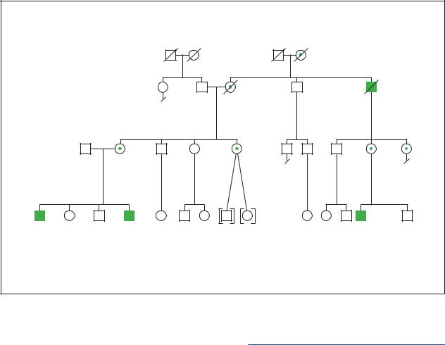
Aarskog Syndrome |
|
Aase |
|
|
|
|
|
|
X-Linked Recessive |
|
||
|
|
|
|
|
|
syndrome |
||
|
|
|
|
|
|
|
|
|
|
|
|
|
|
67y |
d.55y |
d.34y in accident |
|
|
|
|
|
|
5'11" |
Lung cancer |
"slow" |
|
|
|
|
|
|
|
5' 2" |
|
|
|
|
|
|
|
|
Webbed fingers |
|
|
|
|
|
|
|
|
Ptosis |
|
|
|
44y |
|
43y |
40y |
39y |
37y |
|
|
|
|
5'4" |
|
|
||||
|
6'1" |
5'3" |
5'10" |
5'7" |
|
|
||
|
Widows peak |
|
|
|||||
|
|
Webbed fingers |
|
|
|
|
||
|
|
|
|
Short fingers |
|
|
||
|
|
Broad thumbs |
|
|
|
|
||
|
|
|
|
|
|
|
||
19y |
15y |
14y |
9y |
|
|
|
2y |
2mos |
5'5" |
5'9" |
5'4" |
4'6" |
|
|
|
Shawl scrotum |
|
Learning |
|
Attention deficit |
|
|
|
|
||
|
|
|
|
Wide spaced eyes |
|
|||
disabilities |
|
Undescended |
|
|
|
Broad forehead |
|
|
Shawl scrotum |
testes at birth |
|
|
|
|
|
||
Inguinal hernia |
|
|
|
|
|
|
|
|
(repaired) |
|
|
|
|
|
|
|
|
(Gale Group)
ally in the neck, causing pain or injury to peripheral nerves. Neurosurgical intervention is necessary in some cases. Hernias in the umbilical and groin areas may be surgically repaired.
Resources
PERIODICALS
Aarskog, D. “A familial syndrome of short stature associated with facial dysplasia and genital anomalies.” Journal of
Pediatric Medicine 77 (1971): 856.
Pasteris, N. G., et al. “Isolated and characterization of the faciogenital dysplasia (Aarskog-Scott syndrome) gene: A putative Rho/Rac guanine nucleotide exchange factor.” Cell 79 (1994): 669.
ORGANIZATIONS
Alliance of Genetic Support Groups. 4301 Connecticut Ave. NW, Suite 404, Washington, DC 20008. (202) 966-5557. Fax: (202) 966-8553. http://www.geneticalliance.org .
National Organization for Rare Disorders (NORD). PO Box 8923, New Fairfield, CT 06812-8923. (203) 746-6518 or (800) 999-6673. Fax: (203) 746-6481. http://www
.rarediseases.org .
Roger E. Stevenson, MD
I Aase syndrome
Definition
Aase syndrome is a rare, autosomal recessive genetic disorder characterized by congenital hypoplastic anemia (CHA) and triphalangeal thumbs (TPT). People with Aase syndrome may have one or more physical abnormalities. Poor growth in childhood is common, but mental retardation and other neurological problems are not associated with Aase syndrome.
Description
Aase syndrome is sometimes also called Aase–Smith syndrome, or Congenital Anemia–Triphalangeal Thumb syndrome. It is a very rare hereditary syndrome involving multiple birth defects. The two symptoms that must be present to consider the diagnosis of Aase syndrome are CHA and TPT. CHA is a significant reduction from birth in the number of red cells in the blood. TPT means that one or both thumbs have three bones (phalanges) rather than the normal two.
G A L E E N C Y C L O P E D I A O F G E N E T I C D I S O R D E R S |
3 |

Aase syndrome
K E Y T E R M S
Blackfan-Diamond syndrome (BDS)—A disorder with congenital hypoplastic anemia. Some researchers believe that some or all individuals with Aase syndrome actually have BDS, that Aase syndrome and BDS are not separate disorders.
Congenital hypoplastic anemia (CHA)—A significant reduction in the number of red blood cells present at birth, usually referring to deficient production of these cells in the bone marrow. Also sometimes called congenital aplastic anemia.
Fontanelle—One of several “soft spots” on the skull where the developing bones of the skull have yet to fuse.
Hypoplastic radius—Underdevelopment of the radius, the outer, shorter bone of the forearm.
Triphalangeal thumb (TPT)—A thumb that has three bones rather than two.
Several other physical abnormalities have been described in individuals with Aase syndrome, including narrow shoulders, hypoplastic radius (underdevelopment of one of the bones of the lower arm), heart defect, cleft lip/palate, and late closure of the fontanelles (soft spots on an infant’s skull where the bones have not yet fused). The specific cause of Aase syndrome is not known, but recurrence of the condition in siblings implies an abnormal gene is responsible.
Genetic profile
The available evidence suggests Aase syndrome is inherited in an autosomal recessive fashion meaning that an affected person has two copies of an abnormal gene. Parents of an affected individual carry one abnormal copy of that particular gene, but their other gene of the pair is normal. One copy of the normal gene is sufficient for the parent to be unaffected. If both parents are carriers of a gene for the same autosomal recessive condition, there is a one in four chance in each pregnancy that they will both pass on the abnormal gene and have an affected child.
Autosomal recessive inheritance is suspected for Aase syndrome based on the pattern seen in the families that have been described. An autosomal recessive pattern requires that only siblings are affected by the condition (parents are unaffected gene carriers), and the disorder occurs equally in males and females. As of 2000, an abnor-
mal gene proven to cause Aase syndrome had not been discovered.
Demographics
Aase syndrome is quite rare, with possibly no more than two dozen cases reported in the medical literature.
Signs and symptoms
CHA and TPT are the two classic signs of Aase syndrome. The anemia may require treatment with steroids, or possibly blood transfusions, but tends to improve over time. TPT may cause a person with Aase syndrome to have difficulty grasping and manipulating objects with their hands. A hypoplastic radius may complicate problems with appearance and movement of the hands and arms. Narrow and sloping shoulders are caused by abnormal development of the bones in that area of the body.
Slow growth in children with Aase syndrome may be partly related to their anemia, but is more likely to be genetically predetermined due to the syndrome. Ventricular septal defect (VSD), a hole between the bottom two chambers of the heart, is the cardiac defect reported most often, and several cases of cleft lip and palate have occurred as well.
Diagnosis
The diagnosis of Aase syndrome is made when an infant has CHA and TPT, and one or more of the other symptoms. Children with another more common congenital anemia syndrome, Blackfan–Diamond syndrome (BDS), sometimes have abnormalities of their thumbs. Since the syndromes have overlapping symptoms, there is some question about whether Aase syndrome and BDS are contiguous gene syndromes or even identical conditions. Further genetic research may resolve this issue.
Treatment and management
Anemia associated with Aase syndrome is often helped by the use of a steroid medication. For serious anemia that does not respond to medications, blood transfusions from a matched donor might be necessary. Management of problems related to the skeletal abnormalities should be treated by orthopedic surgery as well as physical and occupational therapy. Heart defects and cleft lip and palate are nearly always correctable, but both require surgery and long–term follow up. A genetic evaluation and counseling should be offered to any individual
4 |
G A L E E N C Y C L O P E D I A O F G E N E T I C D I S O R D E R S |

or couple whose child is suspected of having Aase syndrome.
Prognosis
While major medical procedures such as blood transfusions and corrective surgeries might be needed for a child with Aase syndrome, the long–term prognosis seems to be good. Discovery of the specific genetic defect is not likely to immediately change the prognosis. Development of a reliable genetic test, however, might allow for carrier testing for other family members, and prenatal diagnosis for couples who already have an affected child.
Resources
ORGANIZATIONS
Aicardi Syndrome Awareness and Support Group. 29 Delavan Ave., Toronto, ON M5P 1T2 Canada. (416) 481-4095.
March of Dimes Birth Defects Foundation. 1275 Mamaroneck Ave., White Plains, NY 10605. (888) 663-4637. resourcecenter@modimes.org. http://www.modimes
.org .
National Heart, Lung, and Blood Institute. PO Box 30105, Bethesda, MD 20824-0105. (301) 592-8573. nhlbiinfo @rover.nhlbi.nih.gov. http://www.nhlbi.nih.gov .
National Organization for Rare Disorders (NORD). PO Box 8923, New Fairfield, CT 06812-8923. (203) 746-6518 or (800) 999-6673. Fax: (203) 746-6481. http://www
.rarediseases.org .
National Society of Genetic Counselors. 233 Canterbury Dr., Wallingford, PA 19086-6617. (610) 872-1192. http://www
.nsgc.org/GeneticCounselingYou.asp .
Scott J. Polzin, MS, CGC
Aase-Smith syndrome see Aase syndrome
I Abetalipoproteinemia
Definition
Abetalipoproteinemia (ABL) is a rare inherited disorder characterized by difficulty in absorbing fat during digestion. The result is absence of betalipoproteins in the blood, abnormally shaped red blood cells, and deficiencies of vitamins A, E, and K. Symptoms include intestinal, neurological, muscular, skeletal, and ocular
problems, along with anemia and prolonged bleeding in some cases.
Description
An unusual sign first described in ABL is the presence of star-shaped red blood cells, which were dubbed “acanthocytes” (literally, thorny cells). Thus, ABL is also known by the name acanthocytosis. Less commonly, ABL may be referred to as Bassen-Kornzweig syndrome.
The underlying problem in ABL is a difficulty in absorbing fats (lipids) in the intestine. Most people with ABL first develop chronic digestive problems, and then progress to neurological, muscular, skeletal, and ocular disease. Disorders of the blood may also be present. Severe vitamin deficiency causes many of the symptoms in ABL. Treatments include restricting fat intake in the diet and vitamin supplementation. Even with early diagnosis and treatment, though, ABL is progressive and cannot be cured.
Genetic profile
Fats are important components of a normal diet, and their processing, transport, and use by the body are critical to normal functioning. Lipids bind to protein (lipoprotein) so they can be absorbed in the intestine, transferred through the blood, and taken up by cells and tissues throughout the body. There are many different lipoprotein complexes in the body. One group, the betalipoproteins, must combine with another protein, microsomal triglyceride transfer protein (MTP). ABL is caused by abnormalities in the gene that codes for MTP. When MTP is nonfunctional or missing, then betalipoproteins will also be decreased or absent. The MTP gene has been localized to chromosome 4.
ABL is an autosomal recessive genetic disorder. This means that both copies of the MTP gene are abnormal in a person affected with the disorder. Since all genes are present at conception, a person cannot “acquire” ABL. Each parent of an affected child carries the abnormal MTP gene but also has a normally functioning gene of that pair. Enough functional MTP is produced by the normal gene so that the parent is unaffected (carrier). When both parents are carriers of the same recessive gene, there is a one in four chance in each pregnancy that they will have an affected child.
Demographics
ABL is rare, and the true incidence of the disorder is unknown. Prior to the description of ABL in 1950, it is
Abetalipoproteinemia
G A L E E N C Y C L O P E D I A O F G E N E T I C D I S O R D E R S |
5 |

Abetalipoproteinemia
K E Y T E R M S
Acanthocytosis—The presence of acanthocytes in the blood. Acanthocytes are red blood cells that have the appearance of thorns on their outer surface.
Ataxia—A deficiency of muscular coordination, especially when voluntary movements are attempted, such as grasping or walking.
Chylomicron—A type of lipoprotein made in the small intestine and used for transporting fats to other tissues in the body. MTP is necessary for the production of chylomicrons.
Clubfoot—Abnormal permanent bending of the ankle and foot. Also called talipes equinovarus.
Consanguinity—A mating between two people who are related to one another by blood.
Lipoprotein—A lipid and protein chemically bound together, which aids in transfer of the lipid in and out of cells, across the wall of the intestine, and through the blood stream.
Low density lipoproteins (LDL)—A cholesterol carrying substance that can remain in the blood stream for a long period of time.
Neuromuscular—Involving both the muscles and the nerves that control them.
Ocular—A broad term that refers to structure and function of the eye.
Retinitis pigmentosa—Progressive deterioration of the retina, often leading to vision loss and blindness.
Triglycerides—Certain combinations of fatty acids (types of lipids) and glycerol.
Vitamin deficiency—Abnormally low levels of a vitamin in the body.
believed that people with ABL were diagnosed as having either Friedreich ataxia (a more common form of hereditary ataxia) or some other neurologic disorder. Misdiagnosis may still occur if all of the symptoms are not present, or if they do not occur in a typical fashion. Most of the reported cases of ABL have been in the Jewish population, but individuals from other ethnic backgrounds have been described as well. As many as onethird of people with ABL have had genetically related (consanguineous) parents. Higher rates of consanguinity are often seen in rare autosomal recessive disorders.
6
Signs and symptoms
Too much fat left unabsorbed in the intestine results in the symptoms that are often noticed first in ABL, such as chronic diarrhea, loss of appetite, vomiting, and slow weight gain and growth due to reduced uptake of nutrients.
Various lipids, such as cholesterol and its components, are important in the development and normal functioning of nerve and muscle cells. Decreased lipid levels in the bloodstream, and thus elsewhere in the body, are partly responsible for the neuromuscular and ocular problems encountered in ABL. Neurological symptoms include ataxia (poor muscle coordination), loss of deep tendon reflexes, and decreased sensation to touch, pain, and temperature.
Muscular atrophy, the weakening and loss of muscle tissue, is caused by the decreased ability of nerves to control those muscles, as well as lack of nutrients for the muscles themselves. Weakened heart muscle (cardiomyopathy) may occur, and several severe cases have been reported that resulted in early death.
Retinitis pigmentosa is progressive, especially without treatment, and the typical symptoms are loss of night vision and reduced field of vision. Loss of clear vision, nystagmus (involuntary movement of the eyes), and eventual paralysis of the muscles that control the eye may also occur.
Skeletal problems associated with ABL include various types of curvature of the spine and clubfeet. The abnormalities of the spine and feet are thought to result from muscle strength imbalances in those areas during bone growth.
Severe anemia sometimes occurs in ABL, and may be partly due to deficiencies of iron and folic acid (a B vitamin) from poor absorption of nutrients. In addition, because of their abnormal shape, acanthocytes are prematurely destroyed in the blood stream.
Vitamins A, E, and K are fat soluble, meaning they dissolve in lipids in order to be used by the body. Low lipid levels in the blood means that people with ABL have chronic deficiencies of vitamins A, E, and K. Much of the neuromuscular disease seen in ABL is thought to be caused by deficiencies of these vitamins, especially vitamin E.
Approximately one-third of all individuals with ABL develop mental retardation. However, since the proportion of cases involving consanguinity is also reported to be about one-third, it is difficult to determine if mental retardation in individuals with ABL is due to the disease itself or to other effects of consanguinity. Consanguinity may also be responsible for other birth defects seen infrequently in ABL.
G A L E E N C Y C L O P E D I A O F G E N E T I C D I S O R D E R S

Diagnosis
The diagnosis of ABL is suspected from the intestinal, neuromuscular, and ocular symptoms, and is confirmed by laboratory tests showing acanthocytes in the blood and absence of betalipoproteins and chylomicrons in the blood. Other diseases resulting in similar intestinal or neurological symptoms, and those associated with symptoms related to malnutrition and vitamin deficiency must be excluded. As of 2000, there was no direct test of the MTP gene available for routine diagnostic testing. Accurate carrier testing and prenatal diagnosis are therefore not yet available. However, this could change at any time. Any couple whose child is diagnosed with ABL should be referred for genetic counseling to obtain the most up-to-date information.
Treatment and management
The recommended treatments for ABL include diet restrictions and vitamin supplementation. Reduced triglyceride content in the diet is suggested if intestinal symptoms require it. Large supplemental doses of vitamin E (tocopherol) have been shown to lessen or even reverse the neurological, muscular, and retinal symptoms in many cases. Supplementation with a water-soluble form of vitamin A is also suggested. Vitamin K therapy should be considered if blood clotting problems occur.
Occupational and physical therapy can assist with any muscular and skeletal problems that arise. Physicians that specialize in orthopedics, digestive disorders, and eye disease should be involved. Support groups and specialty clinics for individuals with multisystem disorders such as ABL are available in nearly all metropolitan areas.
Prognosis
ABL is rare, which means there have been few individuals on which to base prognostic information. The effectiveness of vitamin supplementation and diet restrictions will vary from person to person and family to family. Life span may be near normal with mild to moderate disability in some, but others may have more serious and even life-threatening complications. Arriving at the correct diagnosis as early as possible is important. However, this is often difficult in rare conditions such as ABL. Future therapies, if any, will likely focus on improving lipid absorption in the digestive tract. Further study of the MTP gene may lead to the availability of accurate carrier testing and prenatal diagnosis for some families.
Resources
ORGANIZATIONS
March of Dimes Birth Defects Foundation. 1275 Mamaroneck Ave., White Plains, NY 10605. (888) 663-4637. resourcecenter@modimes.org. http://www.modimes
.org .
National Foundation for Jewish Genetic Diseases, Inc. 250 Park Ave., Suite 1000, New York, NY 10017. (212) 371-1030.http://www.nfjgd.org .
National Organization for Rare Disorders (NORD). PO Box 8923, New Fairfield, CT 06812-8923. (203) 746-6518 or (800) 999-6673. Fax: (203) 746-6481. http://www
.rarediseases.org .
National Society of Genetic Counselors. 233 Canterbury Dr., Wallingford, PA 19086-6617. (610) 872-1192. http://www
.nsgc.org/GeneticCounselingYou.asp .
National Tay-Sachs and Allied Diseases Association. 2001 Beacon St., Suite 204, Brighton, MA 02135. (800) 9068723. ntasd-Boston@worldnet.att.net. http://www.ntsad
.org .
Scott J. Polzin, MS, CGC
Acanthocytosis see Abetalipoproteinemia
I Acardia
Definition
Acardia is a very rare, serious malformation that occurs almost exclusively in monozygous twins (twins developing from a single egg). This condition results from artery to artery connections in the placenta causing a physically normal fetus to circulate blood for both itself and a severely malformed fetus whose heart regresses or is overtaken by the pump twin’s heart.
Description
Acardia was first described in the sixteenth century. Early references refer to acardia as chorioangiopagus parasiticus. It is now also called twin reversed arterial perfusion sequence, or TRAP sequence.
Mechanism
Acardia is the most extreme form of twin-twin transfusion syndrome. Twin-twin transfusion syndrome is a pregnancy complication in which twins abnormally share blood flow from the umbilical artery of one twin to the umbilical vein of the other. This abnormal connection can cause serious complications including loss of the pregnancy.
Acardia
G A L E E N C Y C L O P E D I A O F G E N E T I C D I S O R D E R S |
7 |